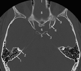
The skull base is made of the paired frontal and temporal bones as well as the ethmoid and occipital bones and these bones form the floors of the anterior middle and posterior cranial fossa. A coronal ct image.
 The Radiology Assistant Brain Anatomy
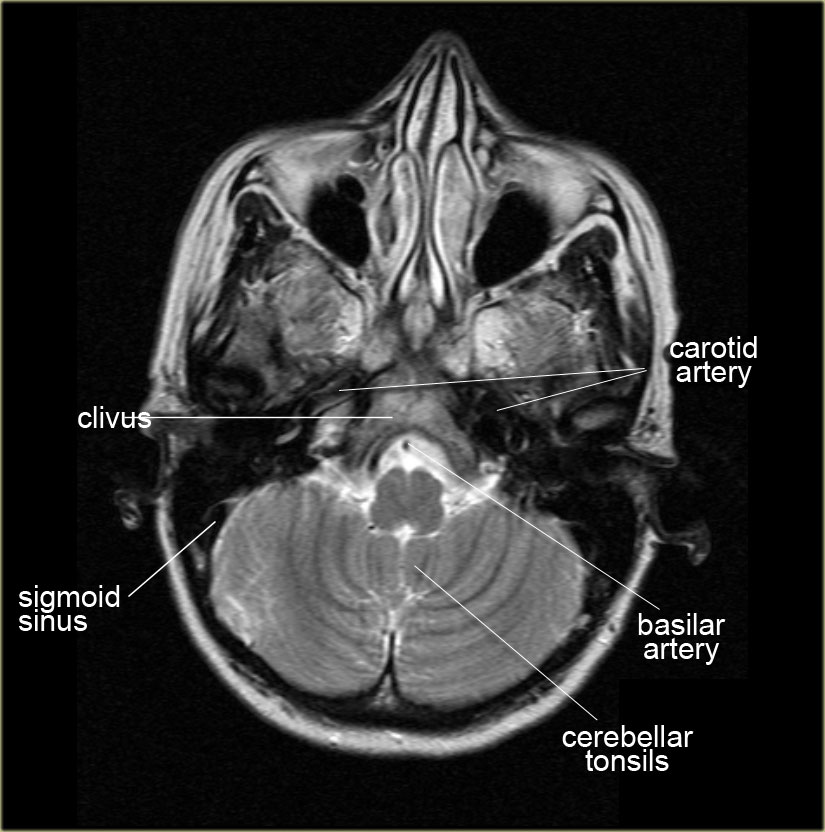
Gross anatomy the base of the skull is a bony diaphragm composed of a number of bones from anterior.

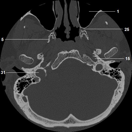
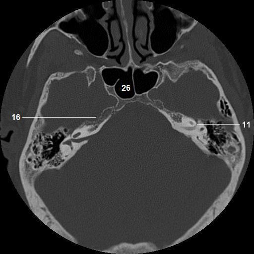
Base of skull anatomy ct. Detailed anatomy enter this module for a more detailed review of skull base anatomy. 1 anterior clinoid process 2 optic canal 3 planum sphenoidale 4 sphenoid sinus 5 foramen rotundum 6 vidian canal 7 middle cranial fossa mcf 8 nasal cavity. Ct demonstrates the bony anatomy best while mri has superior soft tissue resolution.
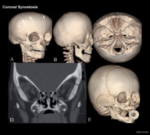
The coronal suture is the line where the parietal bone frontal bone and are in contact. Detailed skull base anatomy. Ct anatomy of skull base.
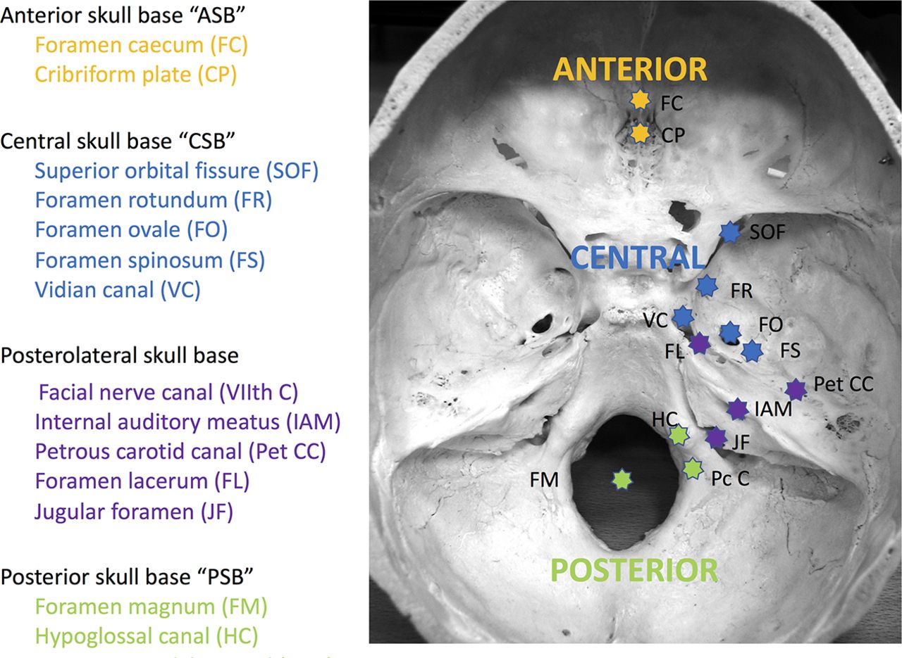
Basic anatomy review the bones sutures and fissures that comprise the skull base. Navigating the skull base identify the petro occipital fissure to navigate the major structures of the skull base. Radiologic anatomy of the skull base.
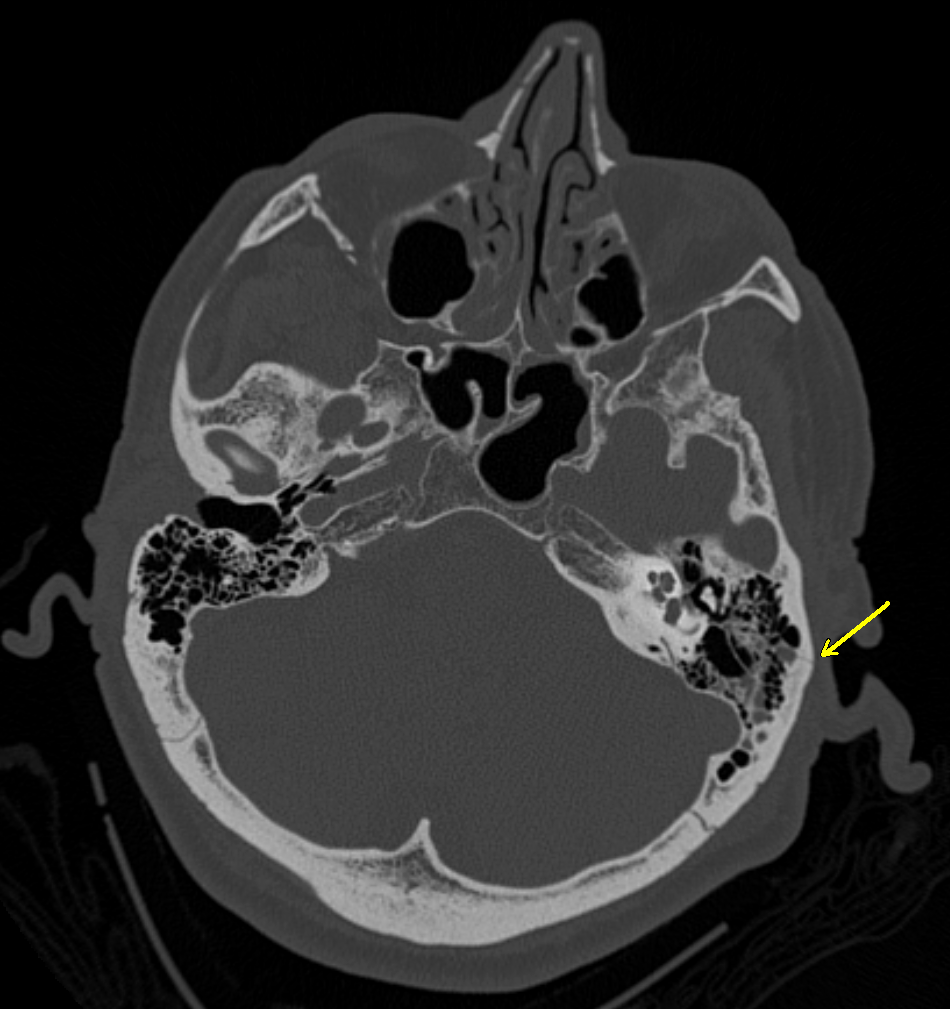
Skull ct anatomy the sagittal suture is the line where the right and left parietal bone are in contact. The skull base can be evaluated by computed tomography ct which will demonstrate the bony structures of the skull base with its foramina and fissures for vessels and cranial nerves the temporal bone and sinonasal cavities. The base of the skull or skull base forms the floor of the cranial cavity and separates the brain from the structures of the neck and face.
 Figure 1 From Lesions Of The Skull Base Imaging For
 Radiologic Anatomy Of The Skull Base Radiology Key
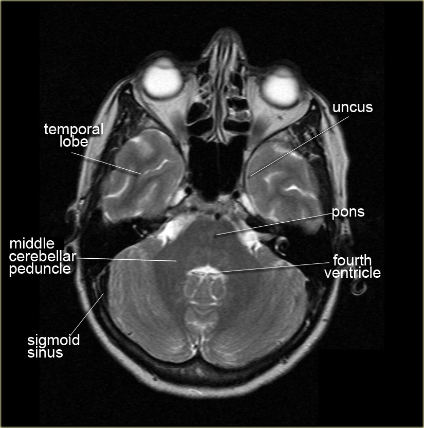 The Radiology Assistant Brain Anatomy
 Axial Ct Bone Window Of Skull Base From Inferior To Superior
 The Anatomy Brain Trauma Concussion And Coma Brainline
 Base Of The Skull Radiology Reference Article
Ct Anatomy Of The Pterygopalatine Fossa Asterisk A An

 The Skull Anatomy And Physiology I
 Ecr 2015 C 0264 Imaging Of The Anterior And Central
 Headneckbrainspine

 Brain And Face Ct Interactive Anatomy Atlas

![]() Superior View Of The Base Of The Skull Anatomy Kenhub
Superior View Of The Base Of The Skull Anatomy Kenhub

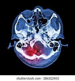 Fotos Imagenes Y Otros Productos Fotograficos De Stock
 Figure 3 From Imaging Of Paranasal Sinuses And Anterior
 Base Of Skull Wikipedia

 On Radiology Normal Anatomy Of Ct Brain At Skull Base
 Basilar Skull Fracture Wikipedia
 Skull Base Tumors Uci Head And Neck Surgery Uci Ent
 Skull Base Imaging Anatomy Pathology And Protocols
 North Jersey Brain Spine Center Computerized Tomography
Anatomy And Pathology Of The Skull Base Ct And Mri Imaging
 Radiologic Anatomy Of The Skull Base Radiology Key
 A Ct Scan At Admission Showing Intracranial Pneumatosisa

 Musculoskeletal System Skull Development Embryology
 Base Of Skull Wikipedia


Posting Komentar
Posting Komentar