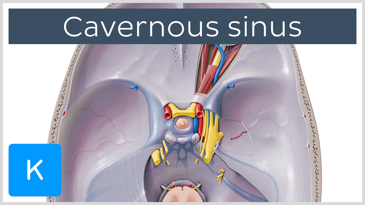
The cavernous sinuses are 1 cm wide cavities that extend a distance. Roof meningeal layer of the dura mater.

The cavernous sinus contains the internal carotid artery and several cranial nerves.

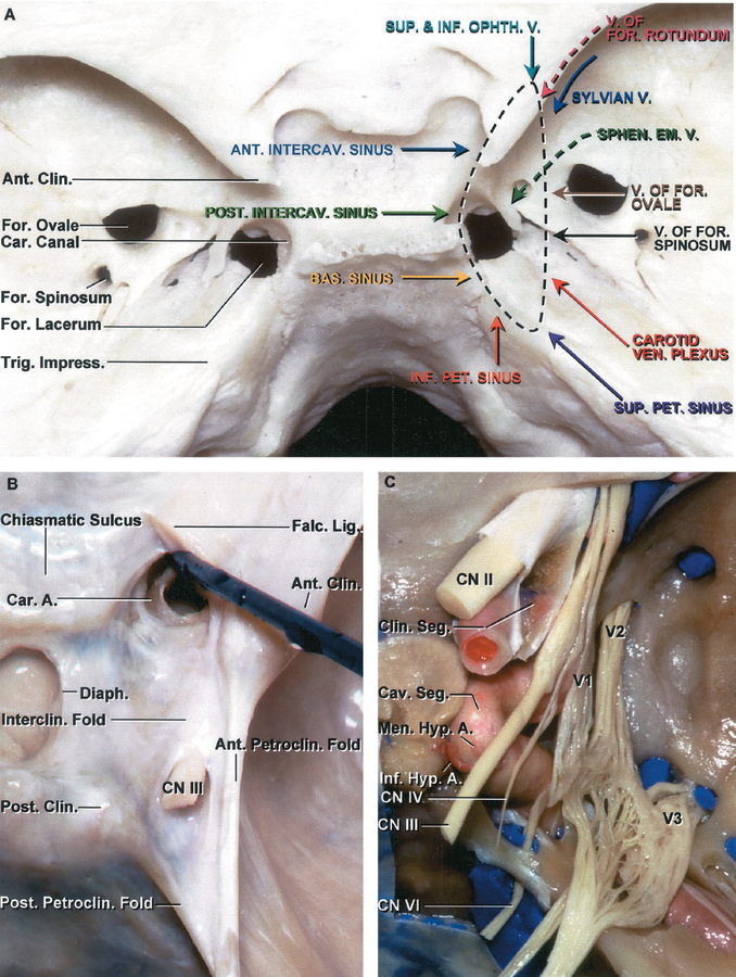
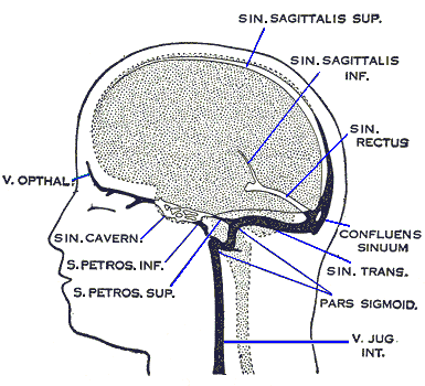
Cavernous sinus anatomy. Inferior petrosal sinus directly to the jugular bulb. Emissary veins passing through the. Lateral meningeal layer of the dura mater running from the roof to the floor.
Medial body of the sphenoid bone. Posterior petrous part of the temporal bone. It is a network of veins that sit in a cavity approximately 1 x 2 cm in size in an adult.
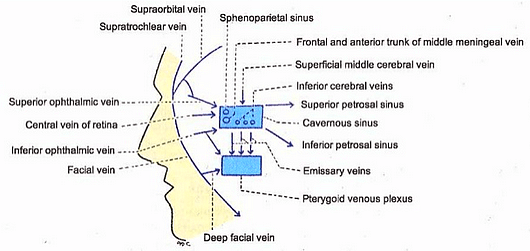
The flow of blood in all the tributaries and communications of the cavernous sinus is reversible because they possess no valves spread of infection to the cavernous sinus leads to its thrombosis the cavernous sinus communicates with the veins draining the middle area of the face dangerous area of the face. Drainage of the cavernous sinus is via. The cavernous sinus is made up of very thin walled veins that make up a venous plexus.
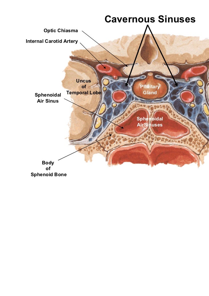
Venous plexus on the internal carotid artery ica to the clival basilar venous plexuses. The cavernous sinus is one of the dural venous sinuses of the head. The cavernous sinus is located on either side of the sella turcica and superior to the sphenoid bone.
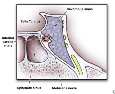
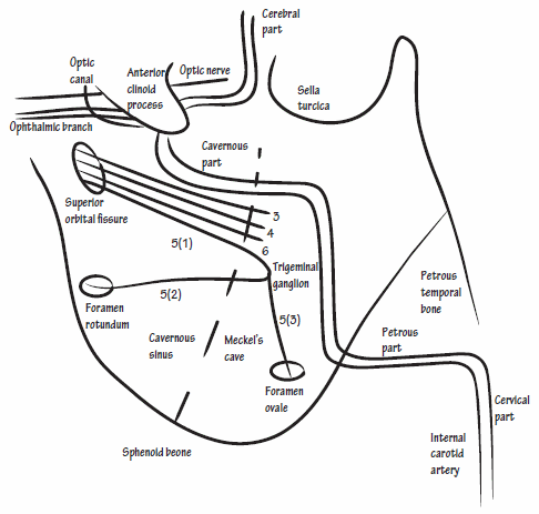
There are numerous structures surrounding the cavernous sinus that are noteworthy. 2 the carotid siphon of the internal carotid artery and cranial nerves iii iv v branches v 1 and v 2 and vi all pass through this blood filled space. Anterior superior orbital fissure.
The cavernous sinus is a relatively large venous channel formed by a splitting of the dura mater on each side of the body of the sphenoid bone. Superior petrosal sinus to the transverse sinus. Cavernous sinus syndromes refer to constellations of clinical signs and symptoms referable to pathology within or adjacent to the cavernous sinus.
The cavernous sinus extends from the medial end of the superior orbital fissure to the petrous portion of the temporal bone. The borders of the cavernous sinus are as follows.
 Cavernous Sinus An Overview Sciencedirect Topics
 Cavernous Sinus Anatomy Cavernous Sinus Syndrome
 Cavernous Sinus Thrombosis Background Pathophysiology
 No More Fear Of The Cavernous Sinuses Sciencedirect
 Instant Anatomy Head And Neck Areas Organs Meninges
 Cavernous Sinus Sagittal Anatomy Optic Nerve Brain
 Inferior Ophthalmic Vein An Overview Sciencedirect Topics
 Cavernous Sinus Anatomy
 Cavernous Sinus Location Drainage Function Human Anatomy Kenhub
 Cavernous Sinus Wikipedia
 Anatomy Of The Cavernous Sinus In Journal Of Neurosurgery
 Cavernous Sinus Anatomy
 Figure Anatomy Of The Cavernous Sinus Contributed By Okkes
 Cavernous Sinus Anatomy
 The Cavernous Sinus Drainage System Great Image Medical
 The Cavernous Sinus Contents Borders Thrombosis
 Applied Anatomy Of Cavernous Sinus Epomedicine
 Cavernous Sinuses Neurology Medbullets Step 1
 Cavernous Sinus Location Drainage Function Human
 Schematic Diagram Of The Normal Left Cavernous Sinus Anatomy
 Applied Anatomy Of Cavernous Sinus Epomedicine
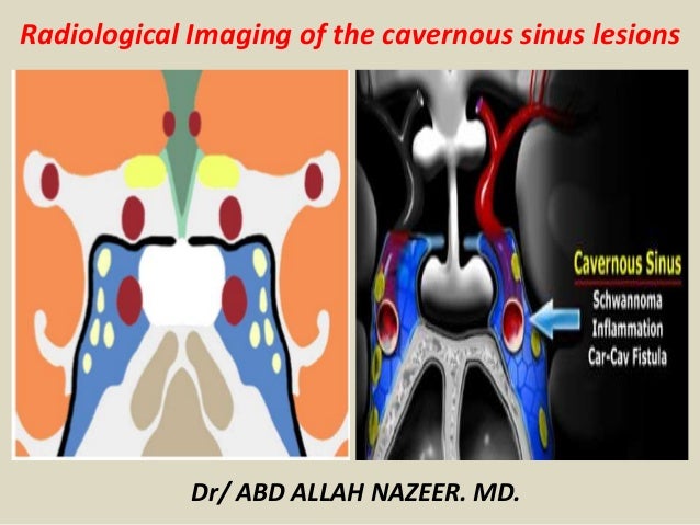 Presentation1 Radiological Imaging Of Cavernous Sinus Lesions
 Cavernous Sinus Thrombosis Wikipedia
 Anatomy Of The Cavernous Sinus In Journal Of Neurosurgery
Anatomy Of The Cavernous Sinus And Surrounding Structures


Posting Komentar
Posting Komentar