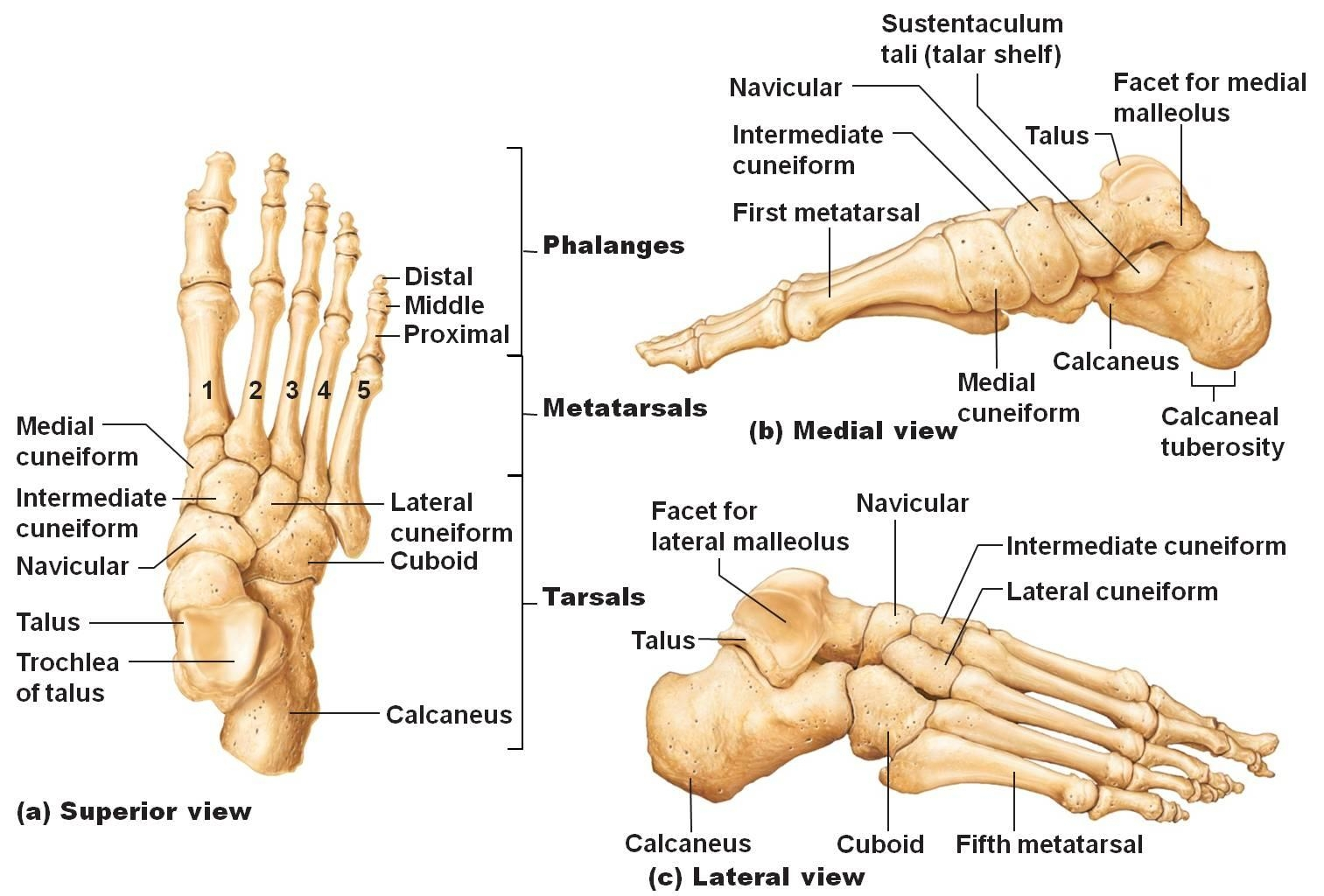
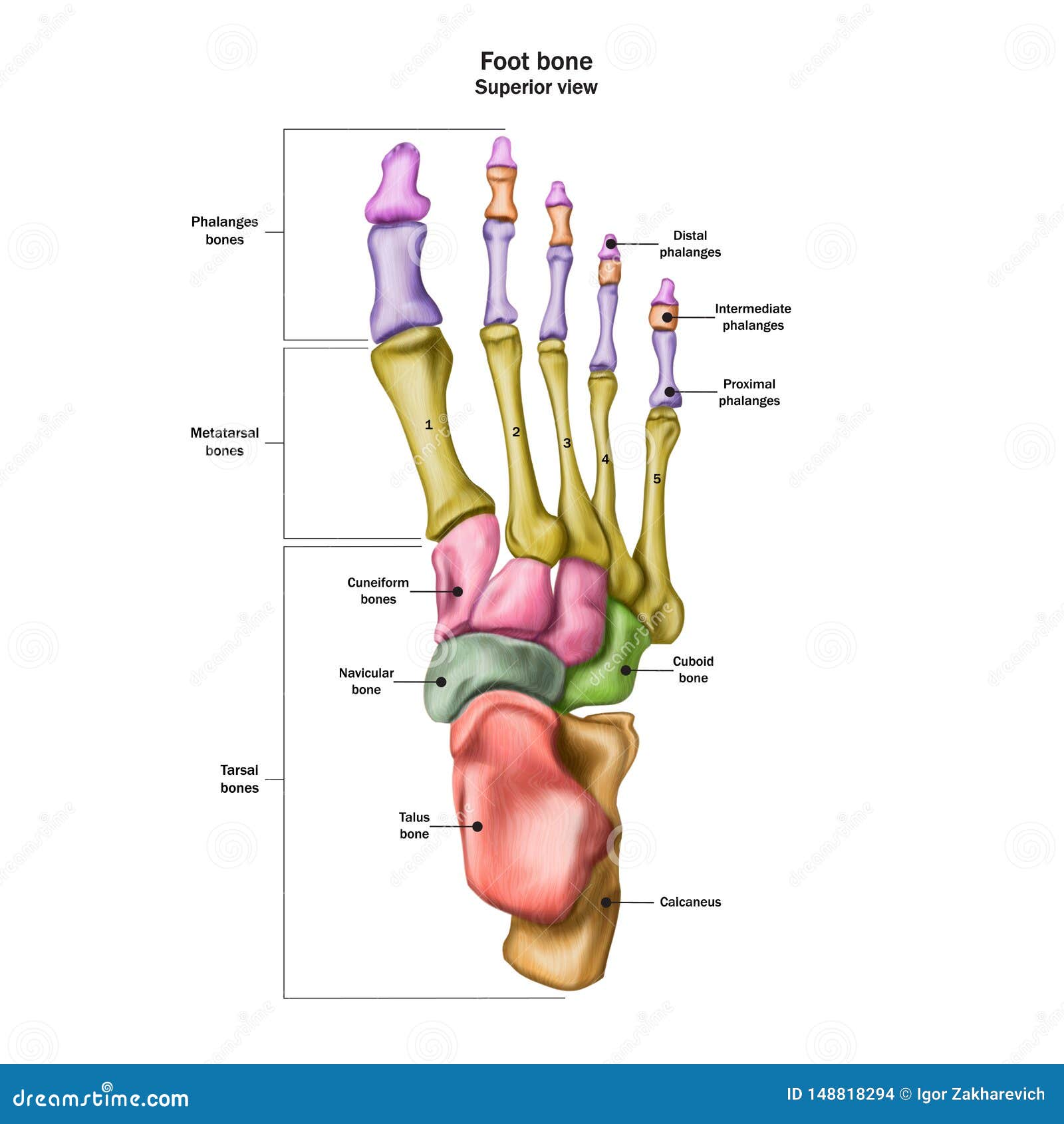
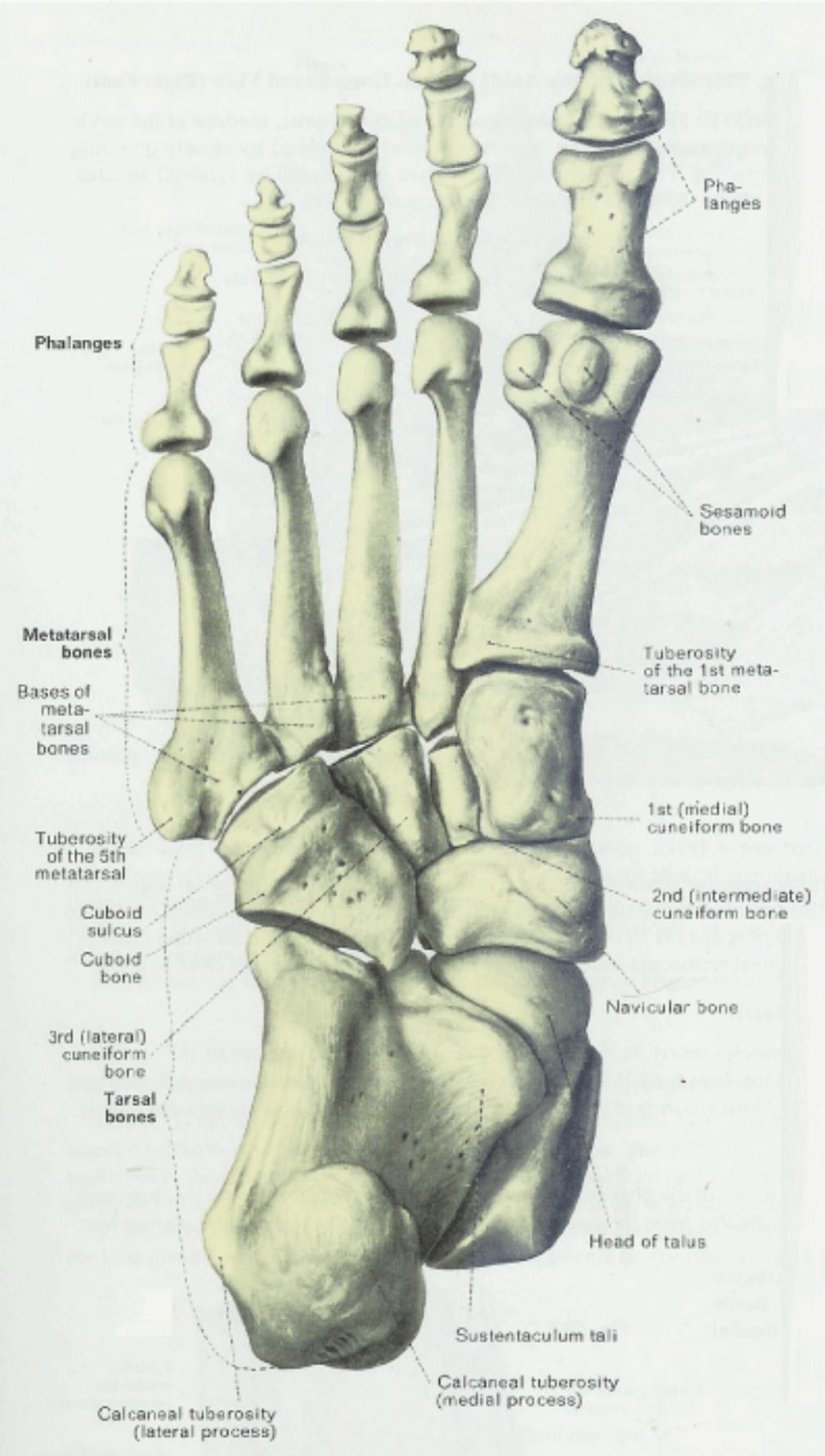
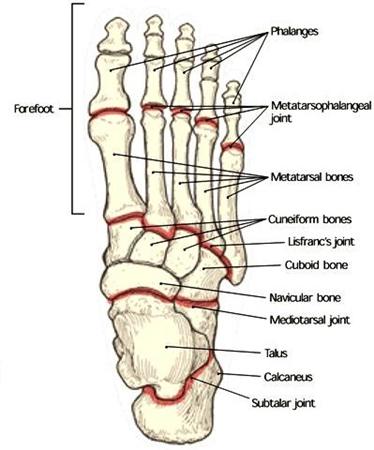
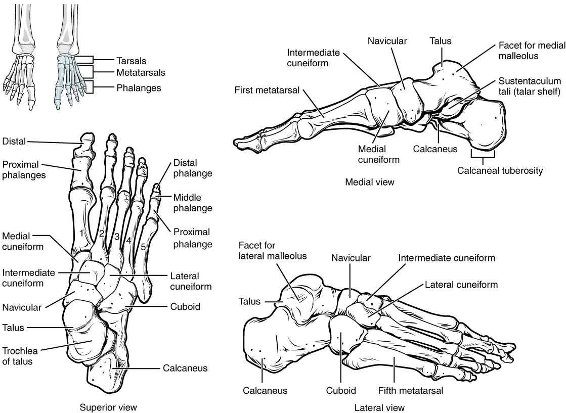
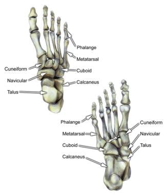
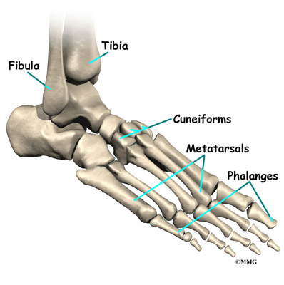
The cuneiform bones the navicularis and the cuboid all of which function to give your foot. The metatarsals which run through the flat part of your foot.
 Talus Bone Wikipedia
The phalanges which are the bones in your toes.
Foot bony anatomy. The midfoot is a pyramid like collection of bones that form the arches of the feet. Talus the bone on top of the foot that forms a joint with the two bones of the lower leg. Can you identify all the bones of the human foot.
The talus bone is the second largest bone in the entire foot and unlike the rest bones there is no attachment of muscles. Learn this topic now at kenhub. It forms the lower part of the ankle formed collectively by the tibia fibular and talus bones.
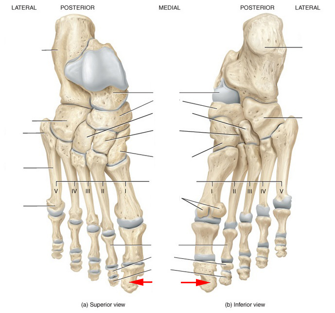
Tarsal bones gross anatomy. The feet are divided into three sections. The ankle joint is both a synovial joint and a hinge joint.
Bones and main ligaments of the foot. In many animals with feet the foot is a separate organ at the terminal part of the leg made up of one or more segments or bones generally including claws or nails. Tarsals five irregularly shaped bones of the midfoot that form the foots arch.
The talus bone supports the leg bones. Calcaneus the largest bone of the foot which lies beneath the talus to form the heel bone. The hindfoot forms the heel and ankle.
The bones of the feet are. The talus which is the. It is the terminal portion of a limb which bears weight and allows locomotion.
The calcaneus is the largest of the tarsal bones located in the heel of the foot and bears the weight of the body as the heel hits the ground. They are named the calcaneus talus cuboid navicular and the medial middle and lateral cuneiforms. Hinge joints typically allow for only one direction of motion much like a door hinge.
The tarsal bones are 7 in number. This is an article covering the muscle attachments blood supply innervation and ossification of the phalanges of the foot. The foot is an anatomical structure found in many vertebrates.
Find out with this free multiple choice picture quiz and learn more about the anatomy of the human body. The calcaneus which is the bone in your heel. The ankle joint or tibiotalar joint is formed where the top of the talus the uppermost bone in the foot and the tibia shin bone and fibula meet.
The forefoot contains the five toes phalanges and the five longer bones metatarsals. This is an article covering the articular surfaces ligaments and muscles that produce movement at the joints of the feet.


 Foot Bone Anatomy
 Anatomy Moment 52 Foot Bones Corpo Kinetic Pilates Rehab
 How To Draw Feet With Structure Foot Bone Anatomy Proko
 1 Bony Anatomy Of The Foot And Ankle Download Scientific
 Foot Bone Tarsal Bone Anatomy

 Foot And Ankle Anatomical Poster Size 12wx17t
 Racgp The Challenge Of Managing Mid Foot Pain
 Duke Anatomy Lab 2 Pre Lab Exercise
 How To Draw Feet With Structure Foot Bone Anatomy
 Bones Of The Human Foot With The Name And Description Of All
 Foot Anatomy And Biomechanics Foot Ankle Orthobullets
 The Bones In The Foot Inferior View Picture Illustrated
 Foot Bone Anatomy Vector Illustration Stock Vector Art
 8 4 Bones Of The Lower Limb Anatomy And Physiology
 Medivisuals Right Medial Foot Anatomy Medical Illustration
 Artstation Bony Anatomy Of The Human Foot Multiple Views
 The Landmarks Of The Body Noella Roos Art
 The Anatomy Of Ankle Bones What You Should Know
 Foot Bone Anatomy Overview Tarsal Bones Gross Anatomy


 Bones Of The Foot Illustrations Foot Anatomy Illustrations
 Human Foot Bone Anatomy Vector
 Sm2 Foot Bony Anatomy Diagram Quizlet
 Skeletal Anatomy Of The Tibia Fibula Ankle And Foot


Posting Komentar
Posting Komentar