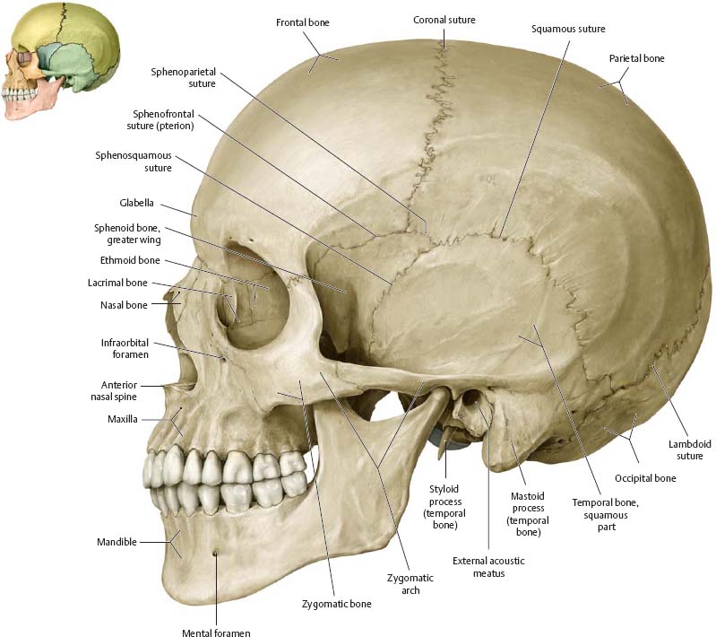
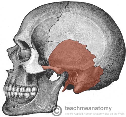
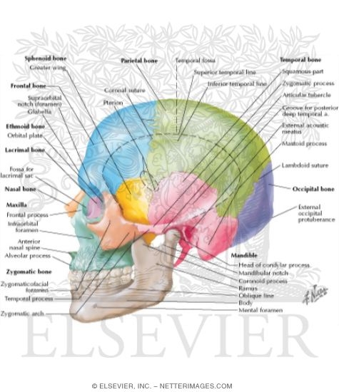
Lateral skull base anatomy surgical approaches for the neurosurgeon 6 episodes the course goal is to provide a breadth of knowledge of microsurgical anatomy of the skull base and provide an opportunity to practice surgery at this difficult location the orbit cavernous sinus middle fossa and temporal bone. The zygomatic arch is formed jointly by the zygomatic process of the temporal bone and the temporal process of the zygomatic bone.
 Procedures 3 Skull Anatomy Flashcards Quizlet
The shallow space above the zygomatic arch is the temporal fossa.

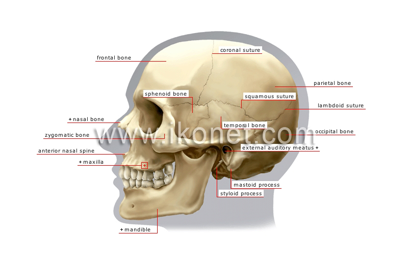
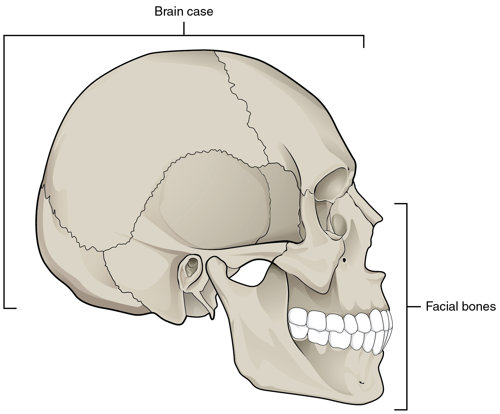
Lateral skull anatomy. Dealing with surgical approaches to the lateral skull base and describing them in a simple way is a challenging matter. The complexity of the anatomy the need for lengthy surgical training and the large number of approaches described over the years have contributed to make this subject more difficult. The skull is divided into the braincase cerebral cranium and the face visceral cranium.
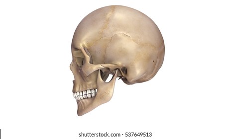
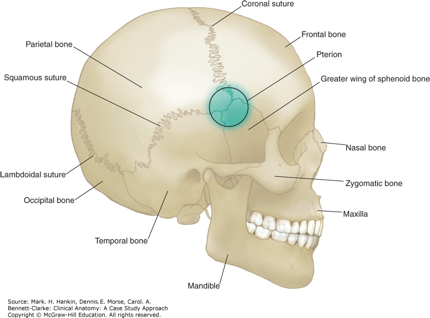
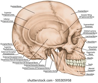
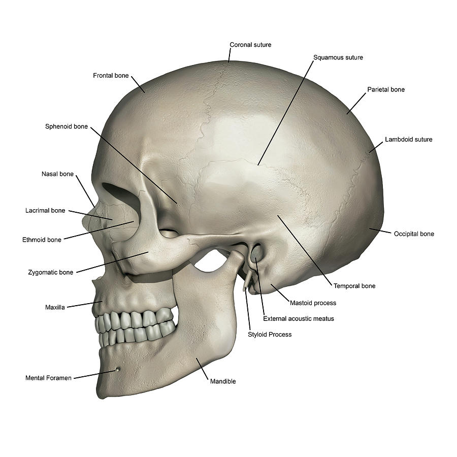
The lateral skull shows the large rounded brain case zygomatic arch and the upper and lower jaws. The posterior surface protects the region of the brain that contains the occipital lobes and cerebellum. Anterior and lateral views of the skull the human skull consists of about 22 to 30 single bones which are mostly connected together by ossified joints so called sutures.
The shallow space above the zygomatic arch is the temporal fossa. The posterior and lateral views of the skull show us important bones that maintain the integrity of the skull. The zygomatic arch is formed jointly by the zygomatic process of the temporal bone and the temporal process of the zygomatic bone.
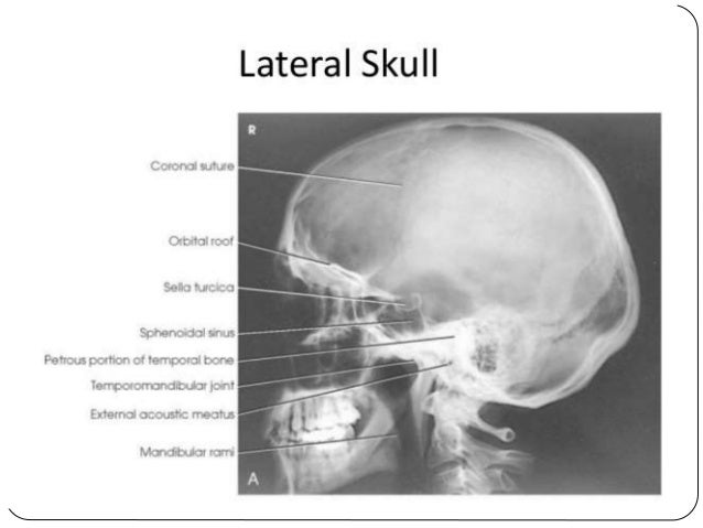
The shallow space above the zygomatic arch is the temporal fossa. The lateral skull base includes the far lateral aspect of the greater sphenoid wing the lateral temporal bone and the temporomandibular joint. The lateral skull shows the large rounded brain case zygomatic arch and the upper and lower jaws.
The zygomatic arch is formed jointly by the zygomatic process of the temporal bone and the temporal process of the zygomatic bone. When tumors affect the clival dura mater they may also spread to the cavernous sinus gasserian ganglion sphenoidal sinus the sella tentorial incisura the porus and the ventral edge of the foramen magnum. The lateral skull shows the large rounded brain case zygomatic arch and the upper and lower jaws.
 Amazon Com Surgical Anatomy A Series Of Video Cassettes
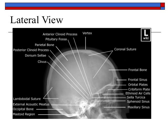
 The Skull Lateral View
 Lateral Skull Anatomy Images Stock Photos Vectors
 7 2 The Skull Anatomy And Physiology
 7 2 The Skull Anatomy And Physiology
![]() Skull Anatomy Structure Bones Quizzes Kenhub
Skull Anatomy Structure Bones Quizzes Kenhub
 Bones Of The Head Atlas Of Anatomy
 Skull Lateral View
 The Temporal Bone Parts Fractures Teachmeanatomy
 Basic Anatomy Views Importance And Positioning
 The Skull Anatomy And Physiology

![]() Biorender Life Science Icons
Biorender Life Science Icons
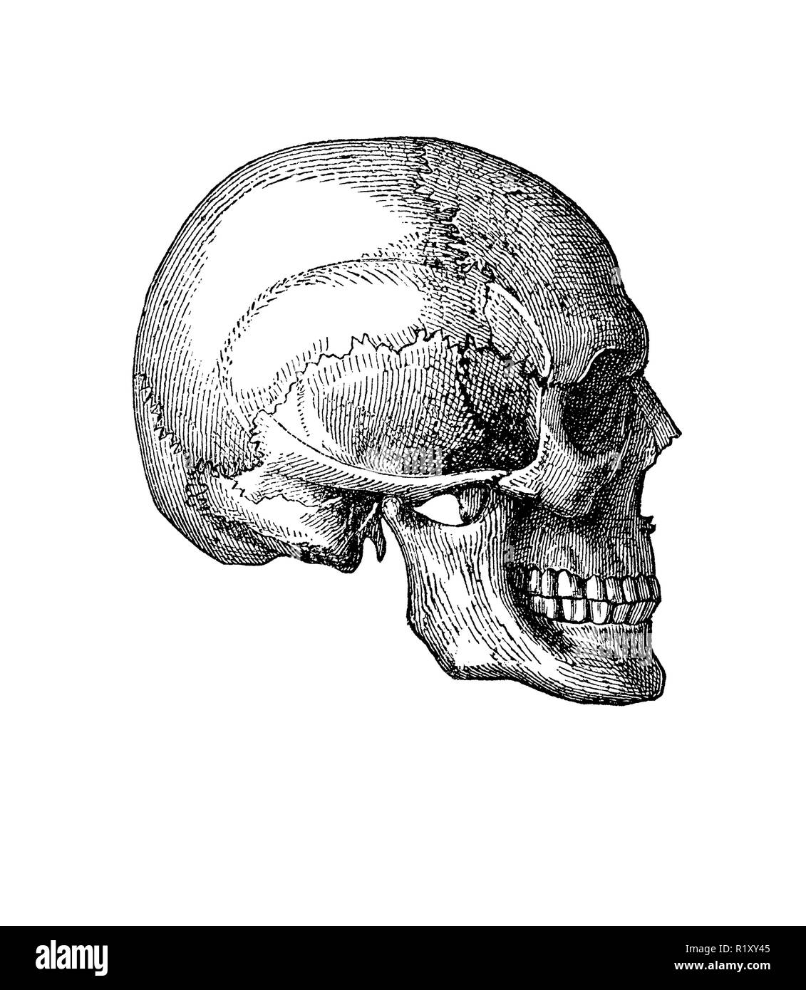 Vintage Illustration Of Anatomy Human Skull Lateral View
 Benign Tumors Of The Skull Base History Of The Procedure
 Lateral View And Maxilla Of The Skull Anatomy
 Head Clinical Anatomy A Case Study Approach
 Skull Anatomy Right Lateral View Diagram Quizlet
 Skull Anatomy And Positioning
 Bones Of Skull Lateral Purposegames
 Skull Lateral View Images Stock Photos Vectors Shutterstock
 7 2 The Skull Anatomy And Physiology
 Lateral Skull Bone Markings
 Anatomy Skull Hand Drawing Vintage Lateral Front View Human
![]() Skull Anatomy Structure Bones Quizzes Kenhub
Skull Anatomy Structure Bones Quizzes Kenhub
 7 2 The Skull Anatomy And Physiology
 Skull Lateral View
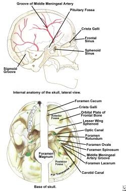
 Anatomy And Physiology Chapter 7 Internal Lateral Skull
 Lateral View Of Human Skull Anatomy
 Lateral View Of The Skeletal Anatomy Of The Skull Stock
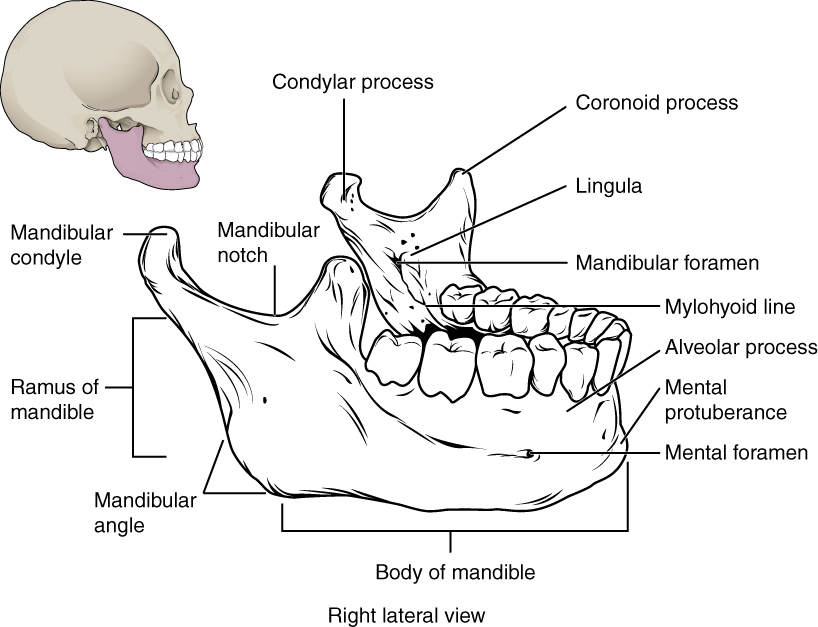
 Angle Of The Mandible Wikipedia


Posting Komentar
Posting Komentar