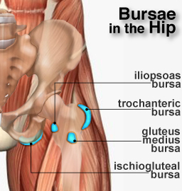
The other bursa on the inside of the hip area is called the iliopsoas bursa. In the adult it is about 1 cm lower than the head.
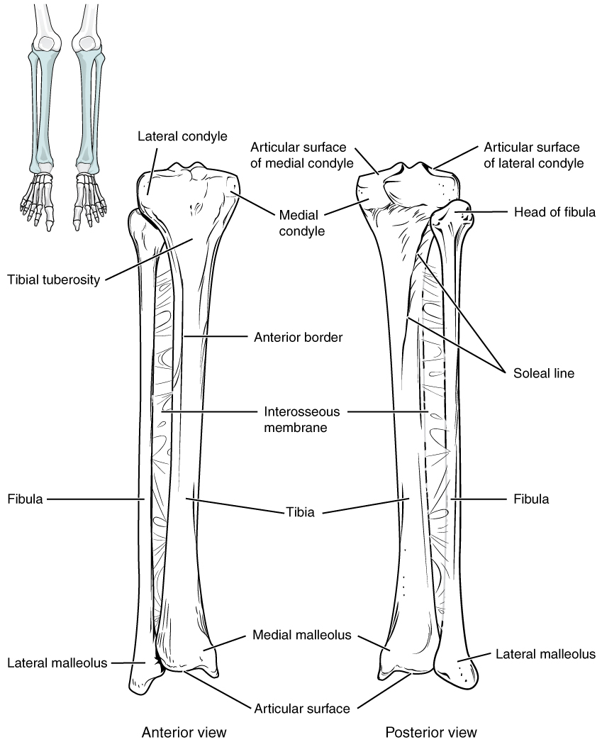 8 4 Bones Of The Lower Limb Anatomy And Physiology
A rough prominence at the upper part of the femur of many vertebrates serving usually for the attachment of muscles.

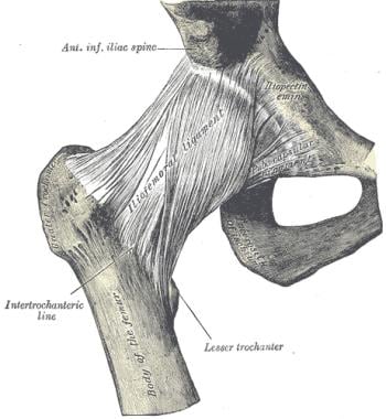
Trochanter anatomy. Each hip has two major bursae. The lesser trochanter a pyramidal prominence that projects from the proximal near and medial inside part of the shaft of the femur. These fluid filled sacs are found around the body and serve as cushions between bones and soft tissues such muscles tendons and skin.
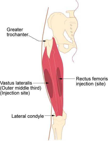
In humans and most mammals the trochanters serve as important muscle attachment sites. Because the pelvic outlet in the female is larger than in the male. The outside point of the hip which is called the greater trochanter has a bursa called the trochanteric bursa.
The outside point of the hip which is called the greater trochanter has a bursa called the trochanteric bursa. The second segment of an insects leg adjacent to the coxa. There are two trochanters.
The greater trochanter a powerful protrusion located at the proximal near and lateral outside part of the shaft of the femur. It is directed lateral and medially and slightly posterior. The greater trochanter has two surfaces.
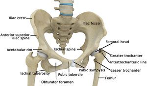
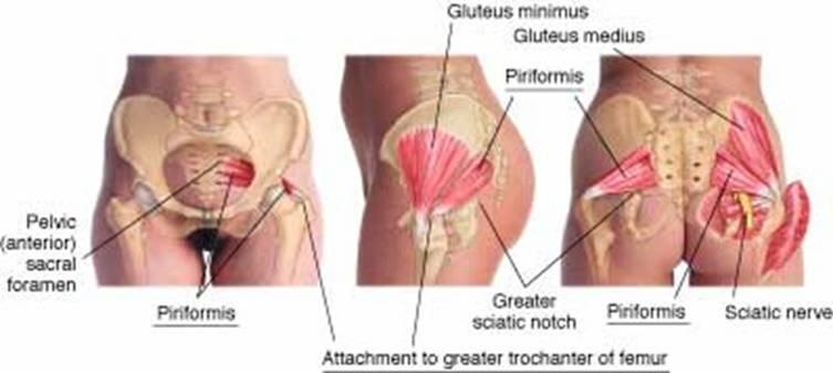
Anatomy in the greater trochanter the piriformis muscle the superior gyrus muscle the lower gyrus muscle the internal obturator muscle the external obturator muscle the gluteus maximus muscle and the gluteus medius muscle are inserted. Trochanter definition either of two knobs at the top of the femur the greater on the outside and the lesser on the inside serving for the attachment of muscles between the thigh and pelvis. The second proximal segment of the leg of an insect.
Any of several bony processes on the upper part of the femur of many vertebrates. The greater trochanter great trochanter of the femur is a large irregular quadrilateral eminence and a part of the skeletal system. A trochanter is tubercle of the femur near its joint with the hip bone.
In humans and most mammals the trochanters serve as important muscle attachment sites.
 Greater Trochanter Location Functions Anatomy Diagrams
 Issues Around The Hip From Tendonitis To Bursitis Beacon
 Hip Joint Anatomy Overview Gross Anatomy
 Trochanteric Bursitis Hip Bursitis Physioadvisor

 Hip Anatomy Recon Orthobullets
 Hip Arthroscopy Austin Tx Hip Surgery Austin Tx Hip
Snapping Hip Orthoinfo Aaos

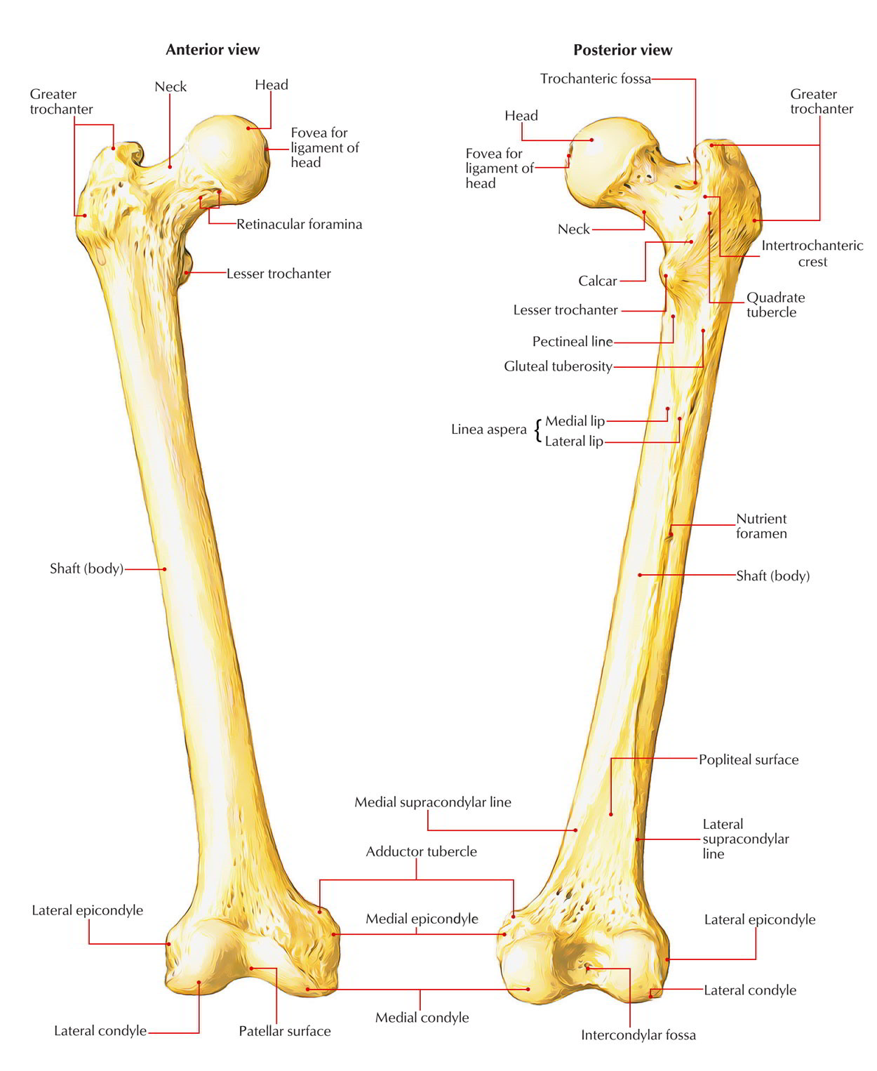 Easy Notes On Femur Learn In Just 4 Minutes Earth S Lab
 Greater Trochanter Photos 27 Greater Stock Image Results
 Pdf Greater Trochanteric Pain Syndrome A Review Article
 Anatomy Of Greater Trochanter With Tendinous Insertion Sites
 The Femur Consists Of Four Parts The Head Greater
 Greater Trochanter
 Femur Bone Anatomy Landmarks And Muscle Attachments
 Femur Labelled Leg Bones Greater Trochanter Anatomy
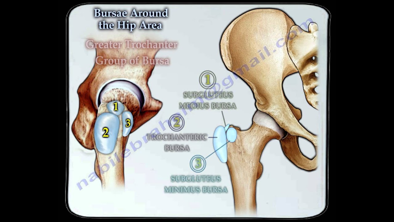 Bursitis Around The Hip Everything You Need To Know Dr Nabil Ebraheim
 Hip Anatomy Pictures Function Problems Treatment
 Dr Stephen Maclean On Twitter Today I Have An Anatomy
 Lesser Trochanter Wikipedia


Posting Komentar
Posting Komentar