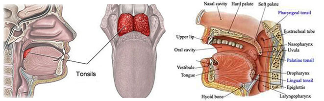
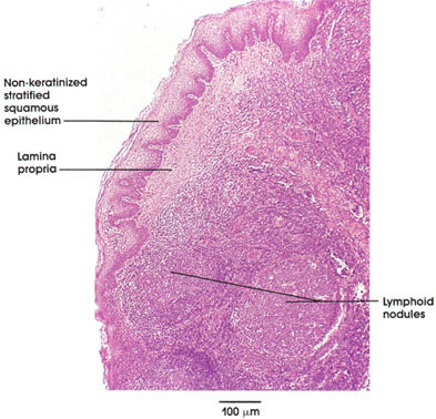
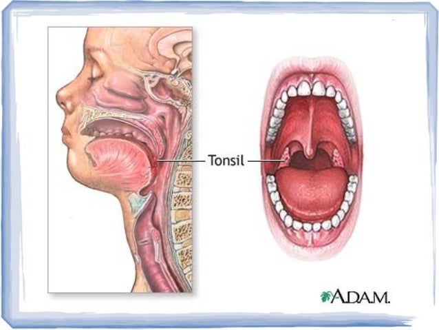
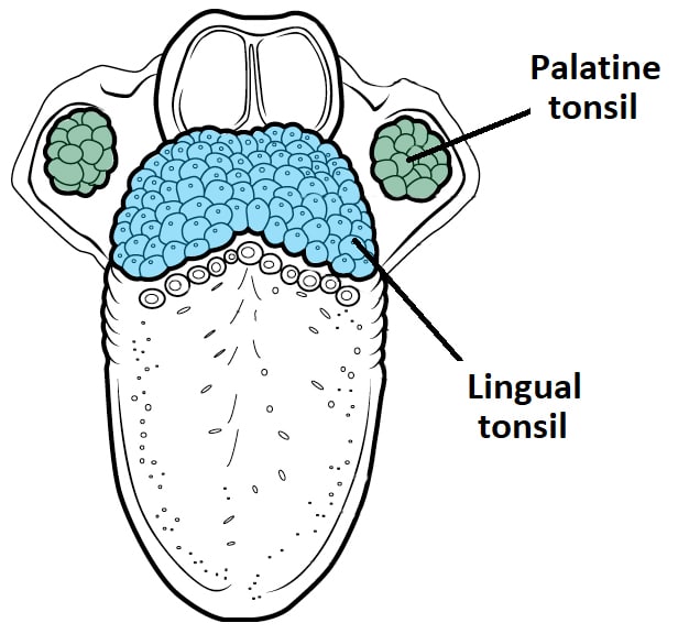
These tonsils are mainly constituted by mucous membranes nerves veins and tiny lumps of lymphoid tissue. They form the lateral part of the waldeyers ring.
 Tonsil Disease Dearborn Ent Livonia Otolaryngology
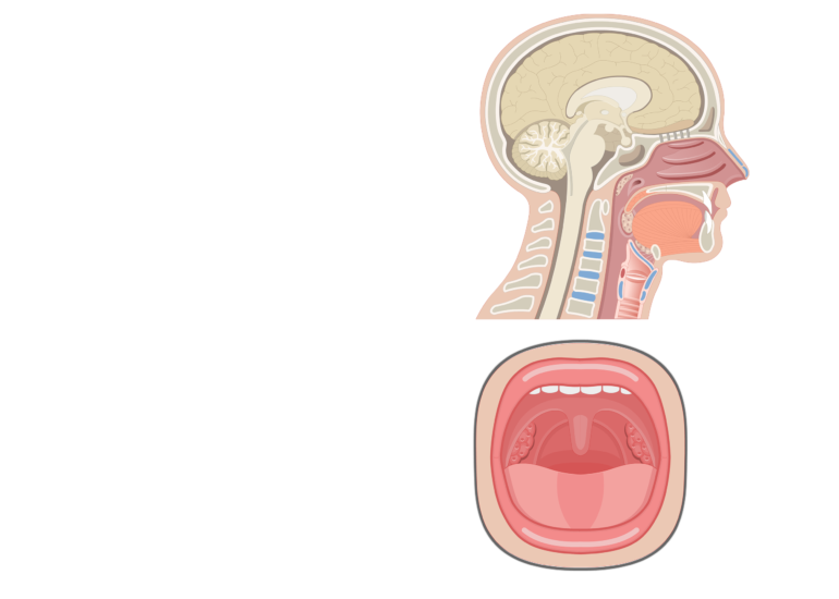
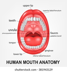
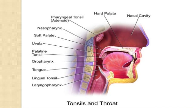
Palatine tonsils commonly called the tonsils and occasionally called the faucial tonsils are tonsils located on the left and right sides at the back of the throat which can often be seen as flesh colored pinkish lumps.

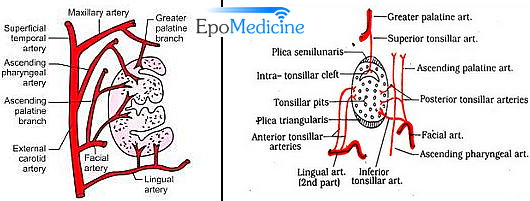
Palatine tonsil anatomy. Usually this is done to check for bacteria such as streptococcus. Ascending palatine a branch of facial artery. The tonsils contain b cells a type of white blood cell that fights infections.
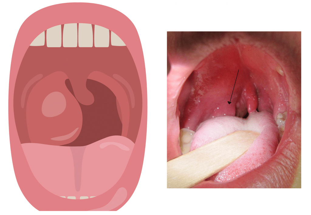
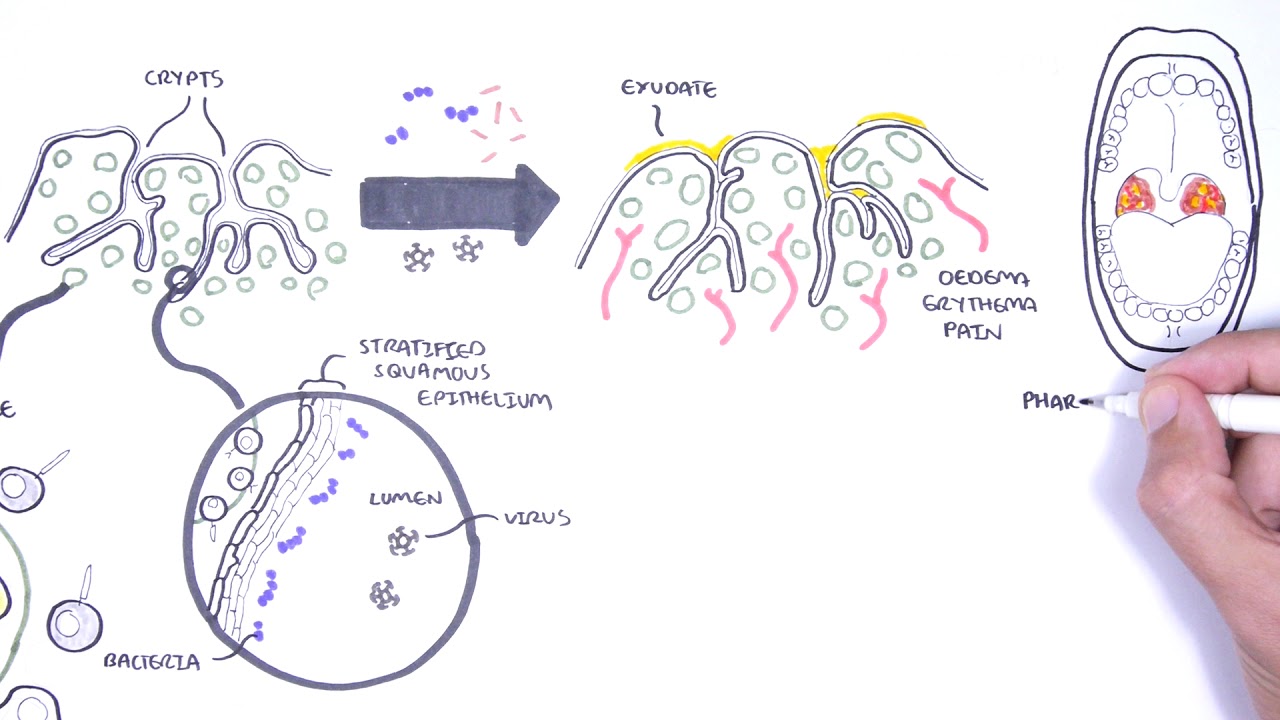
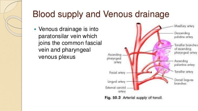
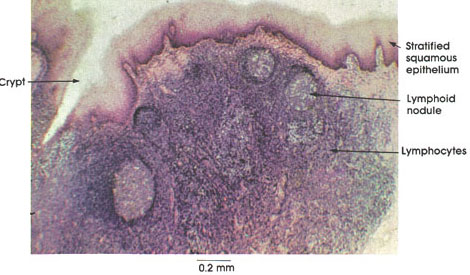
Ascending pharyngeal a branch of external carotid artery. Debris frequently lodges in the pits and. The exposed surface of each tonsil is marked by numerous pits that lead to deeper lymphatic tissue.
Ascending pharyngeal branch of the external carotid. A blood test can detect certain antibodies which can help confirm that a persons symptoms are due to mononucleosis. Palatine tonsil its location gross features relations arterial supply venous drainage and lymphatic draniageclinical anatomy of paltine tonsil and waldeyers ring.
Dorsal lingual branch of the lingual artery. In tonsil of tonsils most commonly the palatine tonsils. The following arteries supply the palatine tonsils.
The palatine tonsils are commonly referred to as the tonsils. Simple easy notes for quick revision of important questions of exams. A doctor rubs a cotton swab on the tonsils and throat and sends the swab for tests.
Each tonsil has free medial surface which projects into the pharynx. The palatine tonsils receive their blood supply from the tonsillar branches of five arteries. They are located within the tonsillar bed of the lateral oropharynx wall between the palatoglossal arch anteriorly and palatopharyngeal arch posteriorly.
Each tonsil consists of a network of crypts pits that store cells used to fight infection. Ascending palatine branch of the facial artery. These are a pair of oval shaped masses protruding from each side of the oral pharynx behind the mouth cavity.
Palatine tonsil anatomy it is round to oval in shape and is actually a lump of lymphoid tissue. Tonsillar branch of facial artery its the main artery and enters the lower pole. Tonsils only present as white lumps if they are inflamed or infected with symptoms of exudates pus drainage and severe swelling.
Dorsalis linguae branches of lingual artery. Tonsillar branch of the facial artery. Palatine tonsil gross anatomy.
They also produce antibodies against polio streptococcal pneumonia influenza and numerous other infections.
Palatine And Pharyngeal Tonsils Google Search Lingual
 Solved Are The Lingual Tonsils Located In Both The Laryng
 Oral Pharynx With Special Reference To The Palatine Tonsil
 Q Where Are The Lingual Tonsils Located Studentrdh Blog
 The Tonsils Waldeyer S Ring Lingual Pharyngeal
 Applied Anatomy Of Palatine Tonsils Epomedicine
 Anatomy Atlases Atlas Of Microscopic Anatomy Section 1 Cells
Palatine Tonsil
 Tonsils Adenoids Lymphoid Tissue Of The Pharynx

![]() Tonsils Anatomy Histology And Clinical Points Kenhub
Tonsils Anatomy Histology And Clinical Points Kenhub
 Tonsils Clinical Anatomy Palatine Lingual Tubal Adenoids
 Anatomy And Physiology Of The Palatine Tonsil
 Applied Anatomy And Diseases Of Tonsil
 Palatine Tonsils Images Stock Photos Vectors Shutterstock
 The Tonsils Waldeyer S Ring Lingual Pharyngeal
![]() Tonsils Anatomy Histology And Clinical Points Kenhub
Tonsils Anatomy Histology And Clinical Points Kenhub
 Tonsil Wikipedia
 The Tonsils Revisited Review Of The Anatomical Localization
![]() Tonsils Anatomy Histology And Clinical Points Kenhub
Tonsils Anatomy Histology And Clinical Points Kenhub
 Lab Practical 3 Flashcards Quizlet
 Anatomy Atlases Atlas Of Microscopic Anatomy Section 1 Cells
 The Tonsils Waldeyer S Ring Lingual Pharyngeal
 Lingual Tonsils Tonsillitis And Pictures
 Tonsils Anatomy Exhibits

 Applied Anatomy And Diseases Of Tonsil
 Anatomical Localization And Histological Characteristics Of


Posting Komentar
Posting Komentar