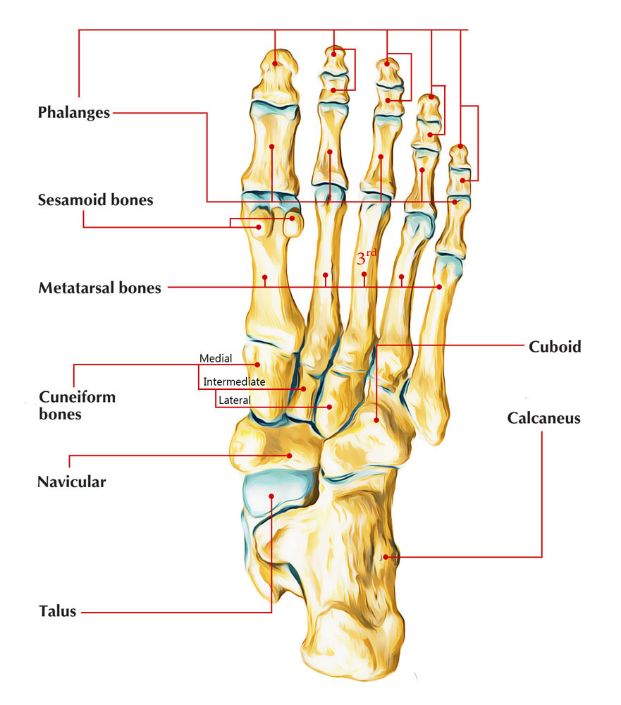
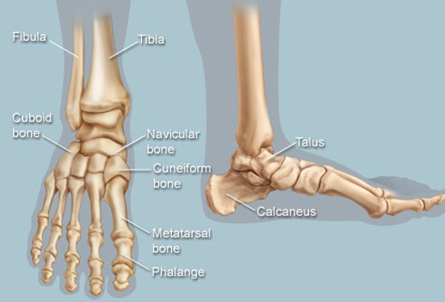
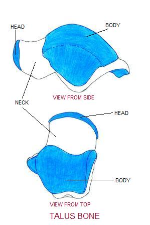
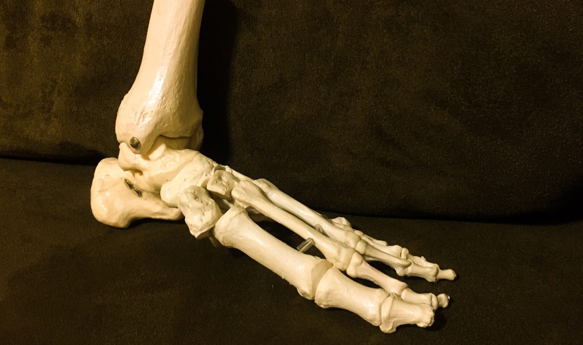
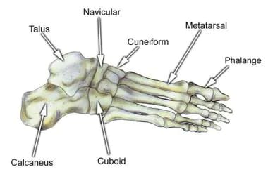
The talus is a frequent site of pathology and fracture and therefore a detailed understanding its complex anatomy is critical for accurate assessment on imaging. The talus is one in a group of seven bones of the foot which are collectively referred to as the tarsus.
 Talus Bone Images Stock Photos Vectors Shutterstock
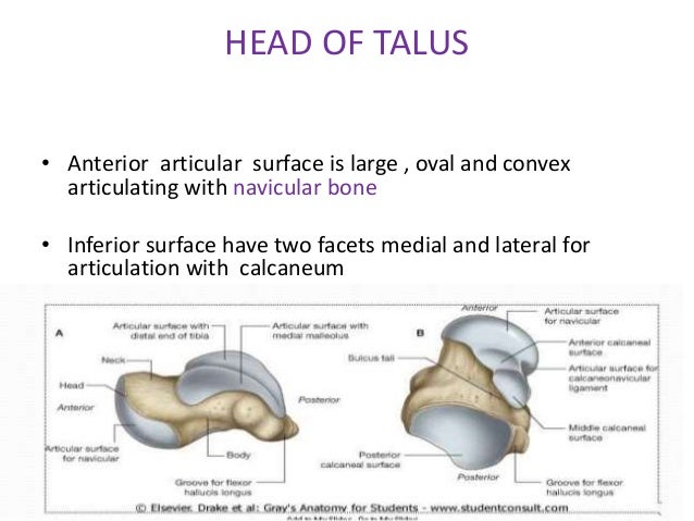
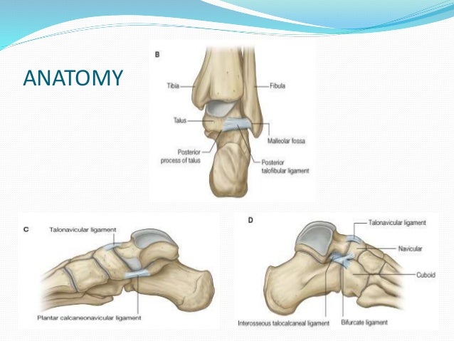
The groove on the posterior surface lodges the tendon of the flexor hallucis longus.
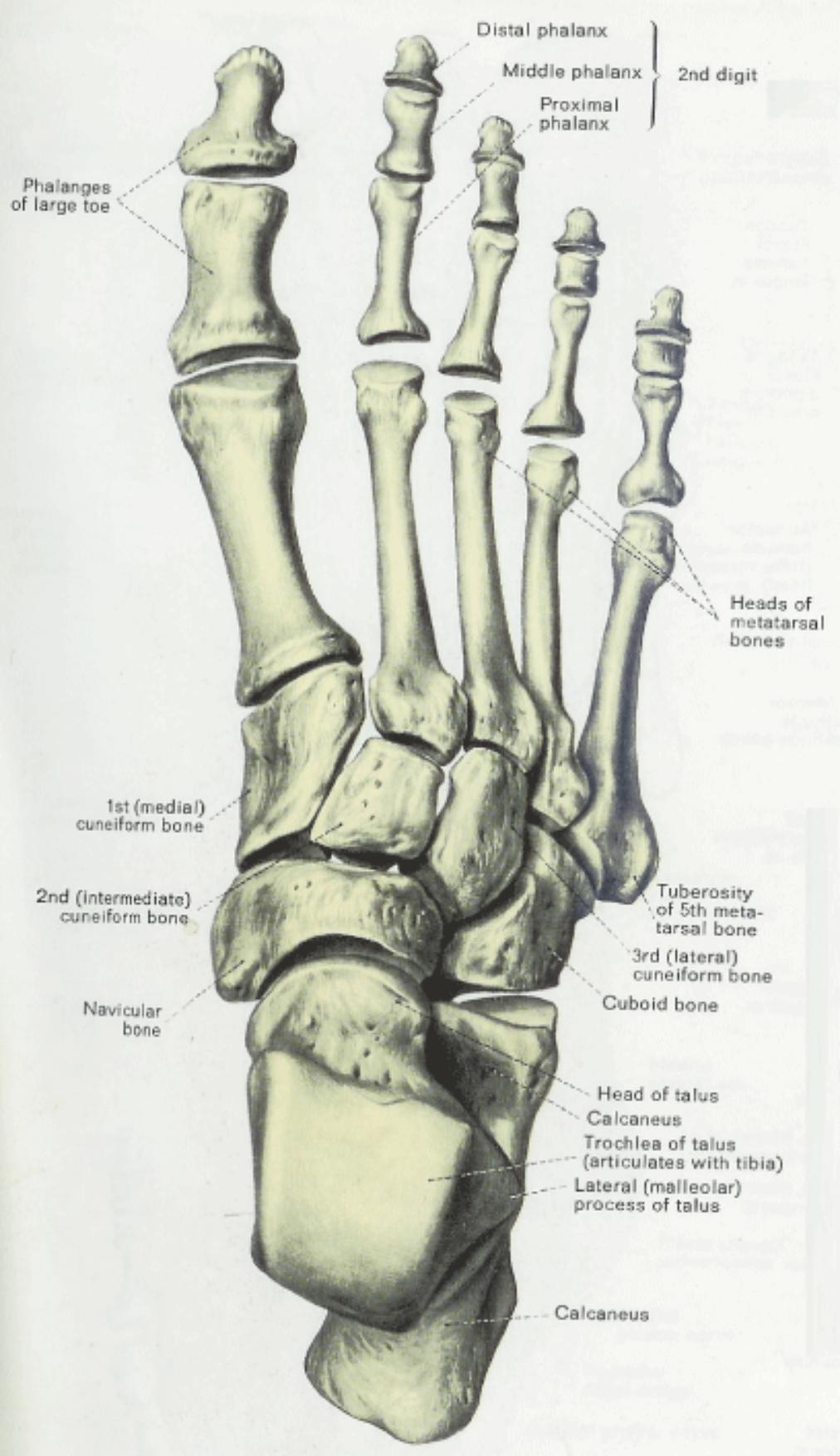
Talus bone anatomy. The talus is an important bone of the ankle joint that is located between the calcaneus heel bone and the fibula and tibia in the lower leg. The shape of the bone is irregular somewhat comparable to a turtles hump. The head of the talus has a convex surface and carries the articular surface of the navicular bone.
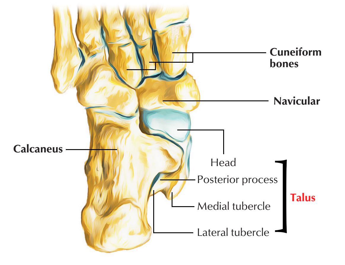
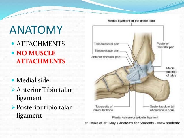
Body of talus the lower non articular part of the medial surface of the body gives attachment to the deep fibers of the deltoid ligament. No muscles are attached. Head a neck and a body.
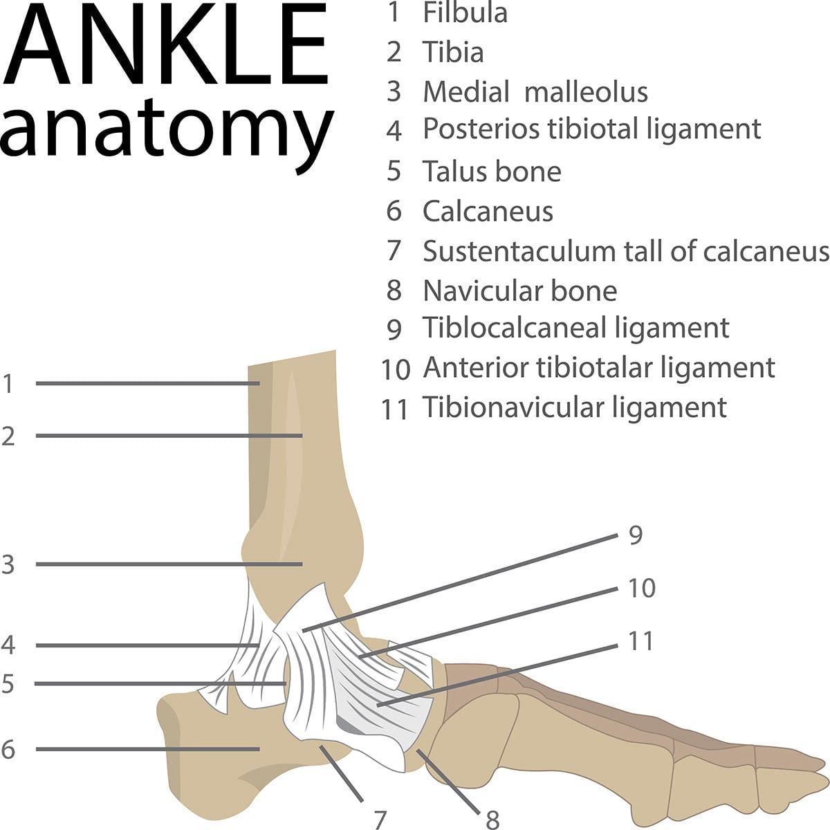
The talus shares this space with the calcaneus the cuboid the lateral cuneiform the. Snowboarders ankle if the. The talus bone is the bone that connects the lower leg bones to the foot.
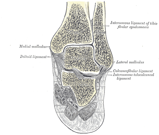
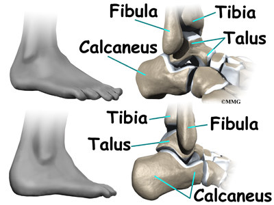
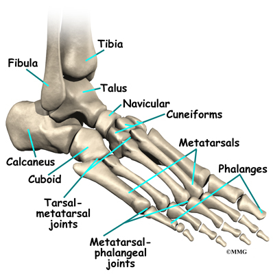
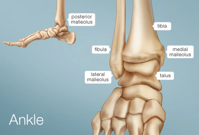
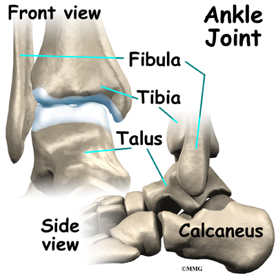
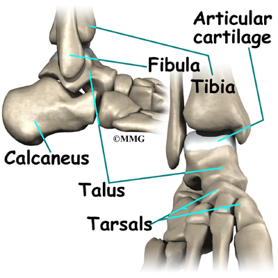
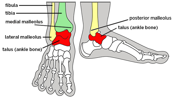
The tarsus forms the lower part of the ankle joint through its articulations with the lateral and medial malleoli of the two bones of the lower leg the tibia and fibula. Within the tarsus it articulates with the calcaneus below and navicular in front within the talocalcaneonavicular joint. The top of the talus contains round cradle like depressions that the lower leg bones fit into.
The key function of this bone is to form a connection between the leg and the foot so that body weight may be transferred from. The talus is part of a group of bones in the foot which are collectively referred to as the tarsus. Educational video describing anatomy and fracture types of the talus.
Anatomy the talus is a very compact and hard bone making up a part of the ankle joint where the tibia shin bone and fibula meet the foot. It is found at the top of the foot and is one of seven tarsal bones. Muscle and ligamentous attachments.
The vascular supply to the talus is considered tenuous due to. The talar body has a curved smooth trochlear surface also termed the talar dome which is covered with cartilage. The medial tubercle provides attachment to the superficial fibers of the.
Fracture of the talar neck can lead to avascular necrosis and arthritis of the subtalar joint. The talus has been described as having three main components. The talus is a frequent site of pathology and fracture and therefore a detailed understanding its complex anatomy is critical for accurate assessment on imaging.
From the case.
 Ankle Anatomy Orthogate
 The Radiology Assistant Ankle Mri Examination
 Tarsus Anatomy Of The Dog On Ct
 Talus Bone Articulations And Landmarks Preview Human Anatomy Kenhub

 The Radiology Assistant Ankle Mri Examination
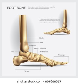
 Foot Bones Anatomy Conditions And More
 Flat Feet Eorthopod Com
Broken Ankle Or A Sprain How Do You Know Beacon
 Talus Bone Wikipedia
 Talus
Talus
 Easy Notes On Talus Learn In Just 4 Minutes Earth S Lab
 Ankle Human Anatomy Image Function Conditions More
 Fractures Of The Talus Anatomy Evaluation And Management

 Osteoarthritis Of The Ankle Orthogate
 Easy Notes On Talus Learn In Just 4 Minutes Earth S Lab
 Foot Bones Anatomy And Mnemonic Tarsals Metatarsals
 Bones Of The Lower Limb Anatomy And Physiology I
 Talus Fracture
 Feet Human Anatomy Bones Tendons Ligaments And More
 Talus Radiology Reference Article Radiopaedia Org
 The Talus Bone
 Talus Fracture
 Talus Bone Anatomy Bone And Spine
 Mr Miles Callahan Anatomy Of The Foot And Ankle

![]() Ankle Joint Anatomy Bones Ligaments And Movements Kenhub
Ankle Joint Anatomy Bones Ligaments And Movements Kenhub
 Ankle Fusion Information Florida Orthopaedic Institute
 The Talus Bone Moving Into Harmony
 Foot Bone Anatomy Overview Tarsal Bones Gross Anatomy
 Broken Ankle Types Of Fractures Diagnosis Treatments


Posting Komentar
Posting Komentar