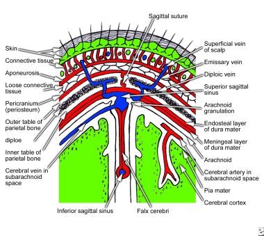
The dura mater contacts the endosteum that lines bones of the cranial cavity. The outer layer is called the periosteum.
 Scalp Anatomy Structure Nerve Supply Arterial Supply
The falx cerebri which separates the two hemispheres of the brain is located in the longitudinal cerebral fissure between the.

Dura anatomy. At its cephalad origin it fuses with the periosteum of the skull at the foramen magnum and it continues caudally in a circumferential manner to its termination. It is separated from the wall of the vertebral canal by the epidural space. The dura is pierced with a needle during a lumbar puncture spinal tap.
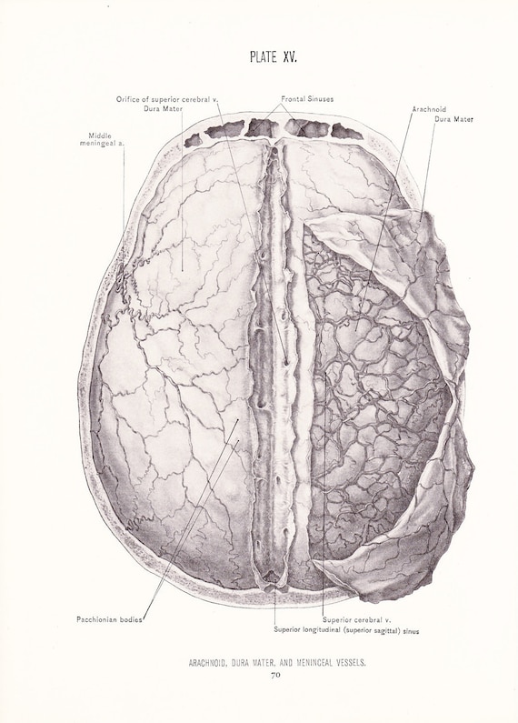
In the brain the dura mater is made up of two layers of whitish nonelastic film or membrane. These coverings differentiate from local neighbouring mesoderm. The other two layers are arachnoid mater and pia mater.
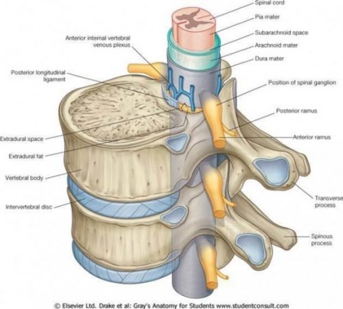
It is the space within the thecal sac which extends from below the end of the spinal cord the conus medularis typically at the level of the first to second lumbar vertebrae down to tapering of the dura at the level of the second sacral vertebra. The dura mater is described as composed of two layers. The outer layer is called the periosteum.
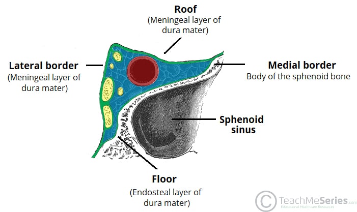
It is thick tough and inextensible. The dura mater is the top layer of the meninges lying beneath the bone tissue. This material at times opens into sinus cavities spaces located around the skull.
The endosteal layer and the meningeal layer. An inner layer the dura lines the inside of the entire skull and creates little folds or compartments in which parts of the brain are protected and secured. This is particularly notable with.
Dura mater simply called the dura is the outermost of the three meninges surrounding the brain and the spinal cord. Spinal cord with a fibrous covering the dura mater and a vascular membrane the pia arachnoid. Anatomy the outermost layer the dura mater provides a thicker and tougher layer of protection.
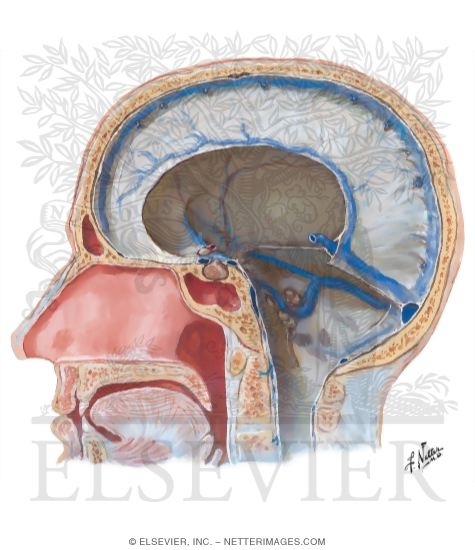
Meningitis is an infection of the meninges caused by viruses or bacteria. It is a thick fibroelastic membrane composed of collagen and elastin fibers that is a continuation of the cranial dura. The tentorium cerebelli exists between and separates the cerebellum and brainstem from the occipital lobes of the cerebrum.
The spinal dura mater is a fibrous non adherent tough layer surrounding the spinal cord. This space contains loose areolar tissue and a network of internal vertebral venous plexuses. The dura mater is the outermost of the spinal meninges.
The dura mater is a strong fibrous membrane that surrounds the brain and is continuous with the dura that covers the spinal cord. The subdural space separates the spinal dura mater from the arachnoid mater. There are two main dural reflections.
It is thick tough and inextensible. Within the cranial cavity the dura contains two connective tissue sheets. The dura mater is the outermost layer of the meninges lying directly underneath the bones of the skull and vertebral column.
Clinical anatomy for dummies.
 Anatomy And Cell Biology 3319 Lecture 2 Anatomy 3319
 Image From Page 211 Of An Atlas Of Human Anatomy For Stud
 Brain Anatomy Dura Mater Superficial Fuses Brain To Skull
/HEAD%20AND%20NECK%20(SEGMENT%20I%20-%20FACE,%20CRANIAL%20CAVITY,%20ORBIT%20DISSECTIONS)_files/Netter98.jpg) Electronic Dissection Manual Head And Neck Part 1
Electronic Dissection Manual Head And Neck Part 1
 Overview Spinal Csf Leak Foundation
![]() The Meninges
The Meninges
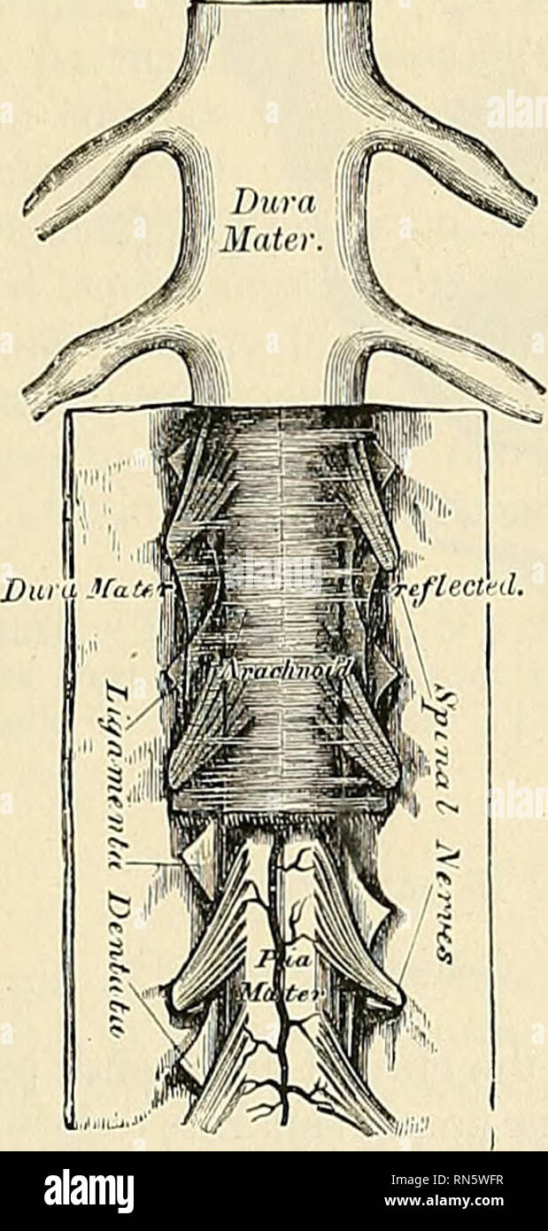 Anatomy Descriptive And Applied Anatomy The Spinal Dura

 More Evidence That Riskiest Part Of Chiari Surgery May Not
 Pin By Tom Bennett On Psychology Brain Models Gross
Dural Venous Sinuses Anatomy Qa
 Pediagenosis
 Anatomical Pia Dura Arachnoid Model Google Search Taj
 The Cavernous Sinus Contents Borders Thrombosis
:max_bytes(150000):strip_icc()/meninges-56f99a4f5f9b5829866fe6a7.png) Meninges Function And Layers And Health Problems
Meninges Function And Layers And Health Problems
 A Graphic Representation Of Dural Anatomy And The Intradural
 The Meninges Of The Brain And Medulla Spinalis Human Anatomy
 Human Brain Anatomy Flashcards Quizlet
 Dural Venous Sinuses
 1926 Human Anatomy Print Arachnoid Dura Mater Meninceal Vessles Brain Vintage Antique Medical Art Illustration Doctor Hospital Office
 The Anatomy And Immunology Of Vasculature In The Central

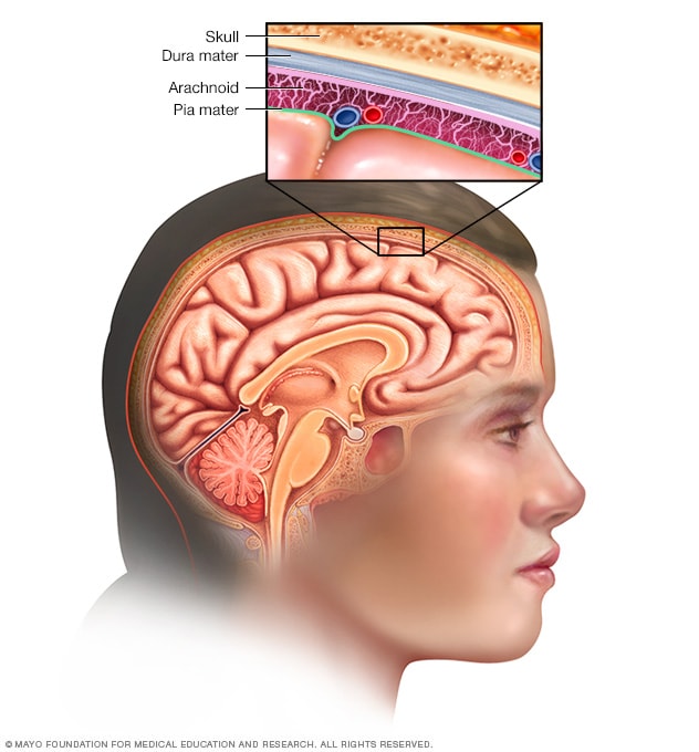 Meninges Mayo Clinic
 Dura Mater Wikipedia
 Science Source Illustration Of The Dura Mater
 Pediagenosis
 Dura And Meninges


Posting Komentar
Posting Komentar