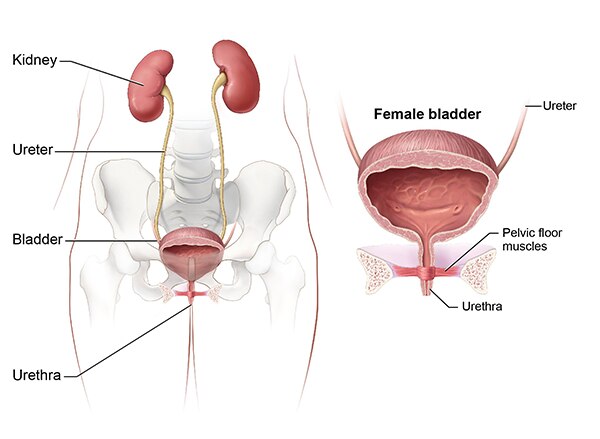
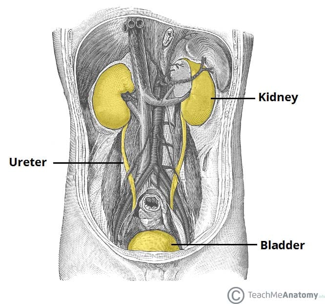
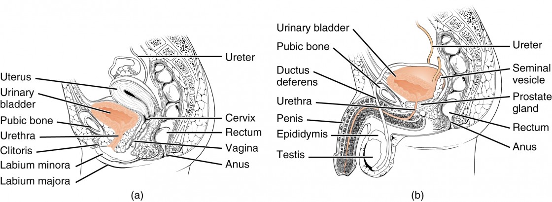
Anatomy and function of the female urethra. Shows the right and left kidneys the ureters the bladder filled with urine and the urethra.
 The Urinary System Ureter And Urinary Bladder
The urethra carries urine from the bladder out of the body.
Female urinary anatomy. The inside of the left kidney shows the renal pelvis. The bladder holds urine until youre ready to release it. In both men and women the urology system is the part of the body that deals with urination.
The main sphincter muscle circles the mid urethra. Anatomy of the female urinary system showing the kidneys ureters bladder and urethra. Female urinary organ anatomy overview.
The urethra is the tube through which urine passes from the bladder to the exterior of the body. Urine is made in the kidneys and travels. The kidneys collect chemicals and water your body doesnt need.
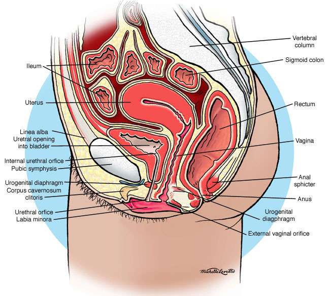
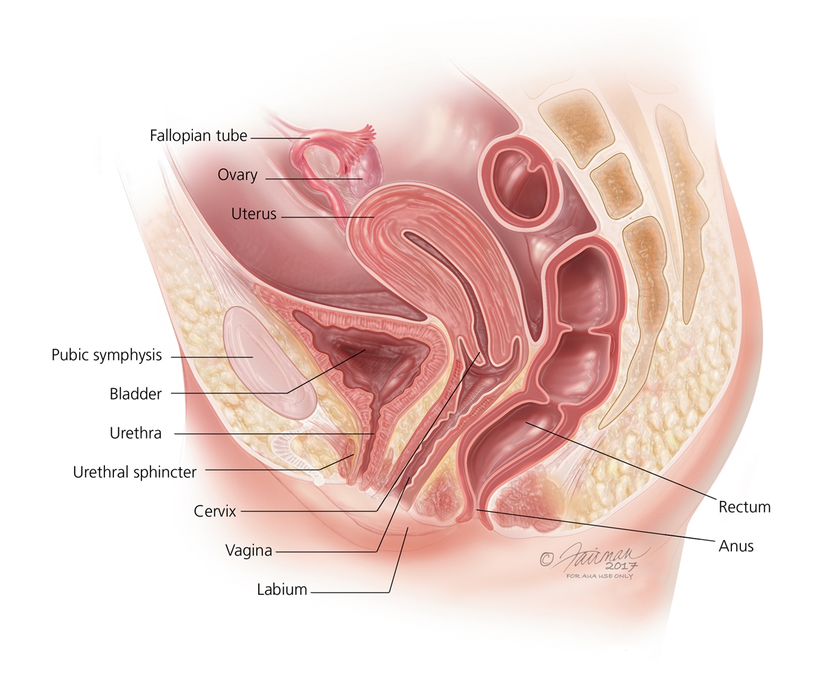
The bony female pelvis is formed by two paired hip bones. The female urinary system has similar parts and functions to the male urinary system. The kidneys collect chemicals and water your body doesnt need.
The urethra carries urine from the bladder out of the body. It extends downward through the muscular area of the pelvic floor. The female urethra begins at the bottom of the bladder known as the neck.
The female urethra is around 2 inches long and ends inferior to the clitoris and superior to the vaginal opening. Anatomy of the female urinary tract. An inset shows the renal tubules and urine.
Before reaching the urethral opening urine passes through the urethral sphincter. When empty the bladder is about the size and shape of a pear. Anatomy of the female urinary tract.
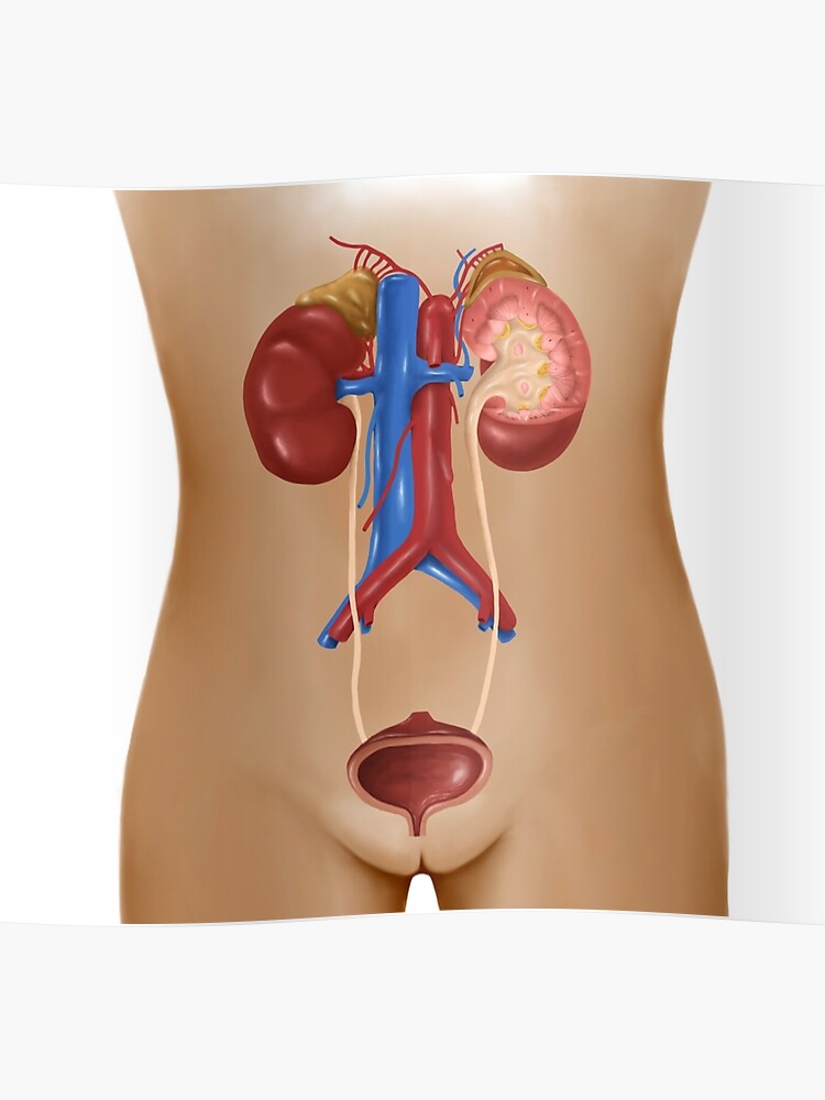
The ovary is divided into a cortex and a medulla. Urine travels out of the kidneys through the ureters to the bladder. It doesnt take a doctor to know that the urology related anatomy of men and women look very different at least from the outside.
It includes the kidneys ureters bladder urethra and the organs of reproduction uterus ovaries fallopian tubes and vagina. Urine travels out of the kidneys through the ureters to the bladder. This is turned into urine.
However internally they are similar the kidneys of both men and women. Anatomy of the female urinary system. Female urology and external sexual anatomy.
The urinary bladder is a muscular sac in the pelvis just above and behind the pubic bone. The female pelvis is accustomed to a wide range of natural and pathologic conditions. The main sphincter muscle circles the mid urethra.
In males the urethra is around 8 to 10 inches long and ends at the tip of the penis. This is turned into urine. The bladder holds urine until youre ready to release it.
The female urogenital tract consists of all the organs involved in reproduction and the formation and release of urine. It expels excess body fluids via the kidneys ureter urinary bladder and the urethra. The kidneys are located in the abdomen at the retroperitoneal area.
The uterus is also shown.
 Urinary Retention Female Articles Mount Nittany Health
Search Female Urinary Bladder
 Body Scientific International Post It Anatomy Of Urinary
 Anatomy Of Female Urinary System Poster
 Anatomy Atlases Anatomy Of First Aid A Case Study Approach
Urinary System Female Anatomy Image Details Nci Visuals
 Urinary System Female Interstitial Cystitis Cystitis


 Symptoms Causes Of Bladder Control Problems Urinary

![]() Anatomy Of Female Urinary System Art Print
Anatomy Of Female Urinary System Art Print
 Female Urinary System Art Print Poster
 Vaginal Abnormalities Urogenital Sinus Symptoms Diagnosis
 Female Urinary Anatomy
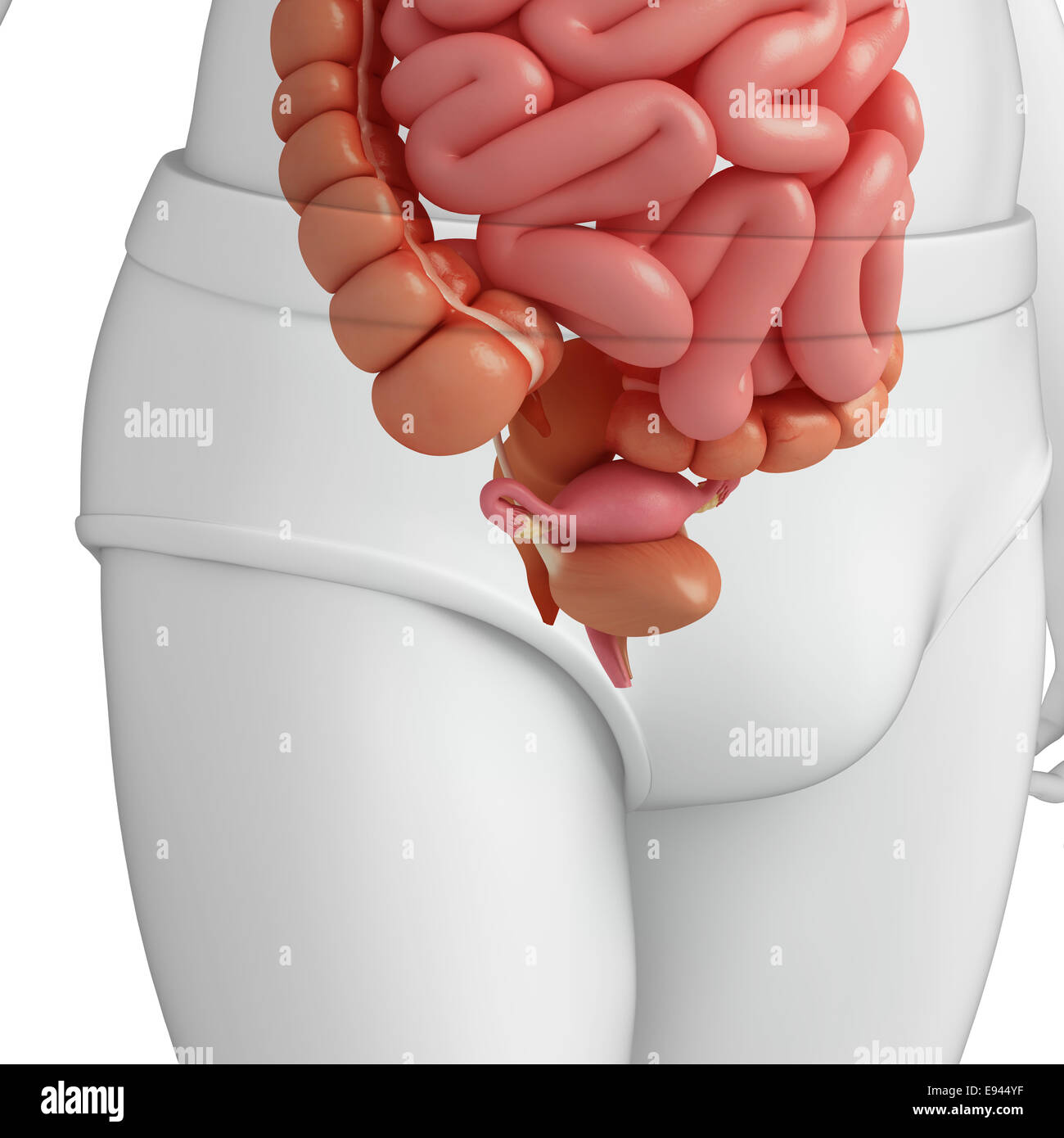 Illustration Of Female Urinary System Stock Photo 74465091
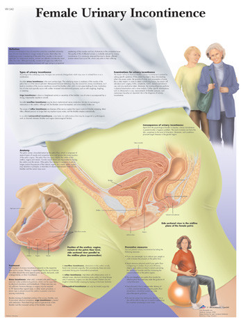 Female Urinary Incontinence
 The Urinary System Of Dogs Dog Owners Merck Veterinary
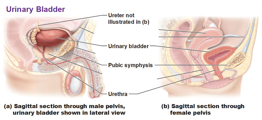
 The Urinary Bladder Structure Function Nerves
 Anatomy Of The Female Urinary Tract
 Figure Anatomy Of The Male Urinary Pdq Cancer
![]() Urinary Bladder Anatomy Function And Clinical Notes Kenhub
Urinary Bladder Anatomy Function And Clinical Notes Kenhub
 Gross Anatomy Of The Urinary System And Urine Transport


Posting Komentar
Posting Komentar