During each step with each leg a horse completes four movements. Inflammation in the joint causes excessive fluid production.
 Habitat For Horses Kids Corral
It also includes the joints of the hip stifle hock fetlock pastern and coffin.

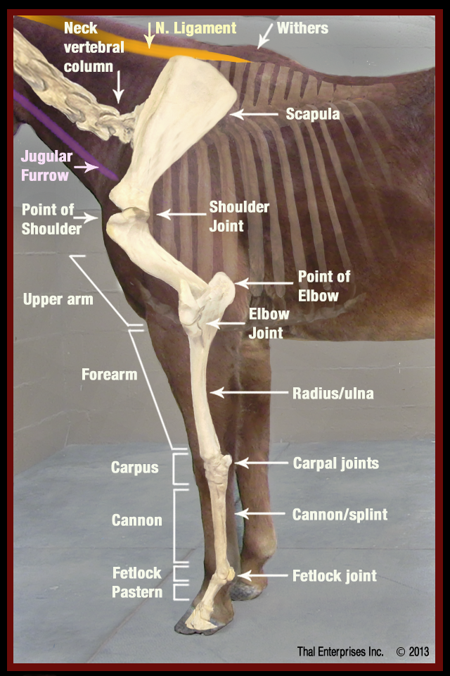
Horse leg anatomy. There are various disease processes that affect the nature of the synovial fluid because of inflammation and disease in the synovial membrane. While the horse uses muscles throughout its body to move the legs perform the functions of absorbing impact bearing weight and providing thrust. Offering anatomy charts and freeze dried and skeletal models of horse limbs.
Equine anatomy refers to the gross and microscopic anatomy of horses and other equids including donkeys and zebras. The top part of the hind limbs consists of three fused. Joint connecting the hind leg to the horse hip.
While all anatomical features of equids are described in the same terms as for other animals by the international committee on veterinary gross anatomical nomenclature in the book nomina anatomica veterinaria there are many horse specific colloquial terms used by equestrians. Is the patella and corresponds to the human kneecap. A horse with good conformation is going to have well formed.
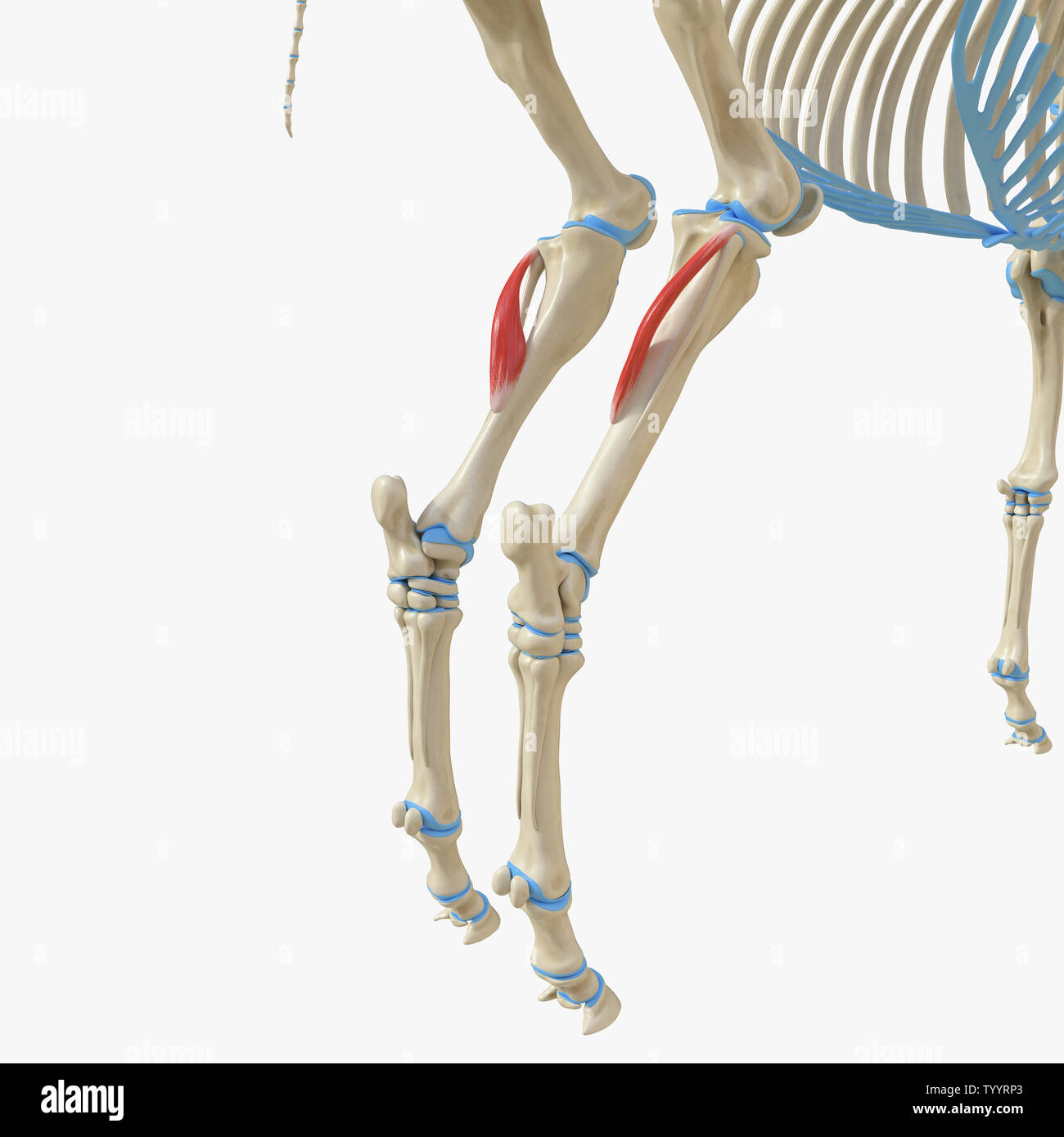
Equine rear leg bones and function the horse leg anatomy in the rear includes the bones of the pelvis the ilium ischium and pubic bones femur tibia fibula metatarsus and the phalanxes. The horses hind limbs. The horse does not have a collarbone.
The swing phase the grounding or impact the support period and the thrust. Horse seriously injured after being hit by dublin bound train commuters warned to expect delays irish examiner posted on december 12 2019 0 comments global veterinary headlights market 2015 2026 dre veterinary accesia harltons equine specialties jupiter veterinary products luxtel health 365 news. The synovial membrane secretes the synovial fluid which provides lubrication within the joint.
Important parts of the horses forelimbs. Home 3d radiographic projection select a body part and angle on the left then select the type of image from the top menu. The horse leg anatomy in the rear includes the bones of the pelvis the ilium ischium and pubic bones femur tibia fibula metatarsus and the phalanxes.
It also includes the joints of the hip stifle hock fetlock pastern and coffin. Horse leg anatomy form and function conformation and a horses legs.
Torn Horse Tendon The Long Road Back From This Equine
 Horse Leg Anatomy Stock Photos Horse Leg Anatomy Stock
 Horse Leg Anatomy Learn Everything You Did Not Know Medrego
 Novobrace Tendonitis Desmitis And Soft Tissue Injury
 Stringhalt The Marionette Horse Irongate Equine Clinic
 Horse S Lower Front Leg Horses Horse Anatomy Horse
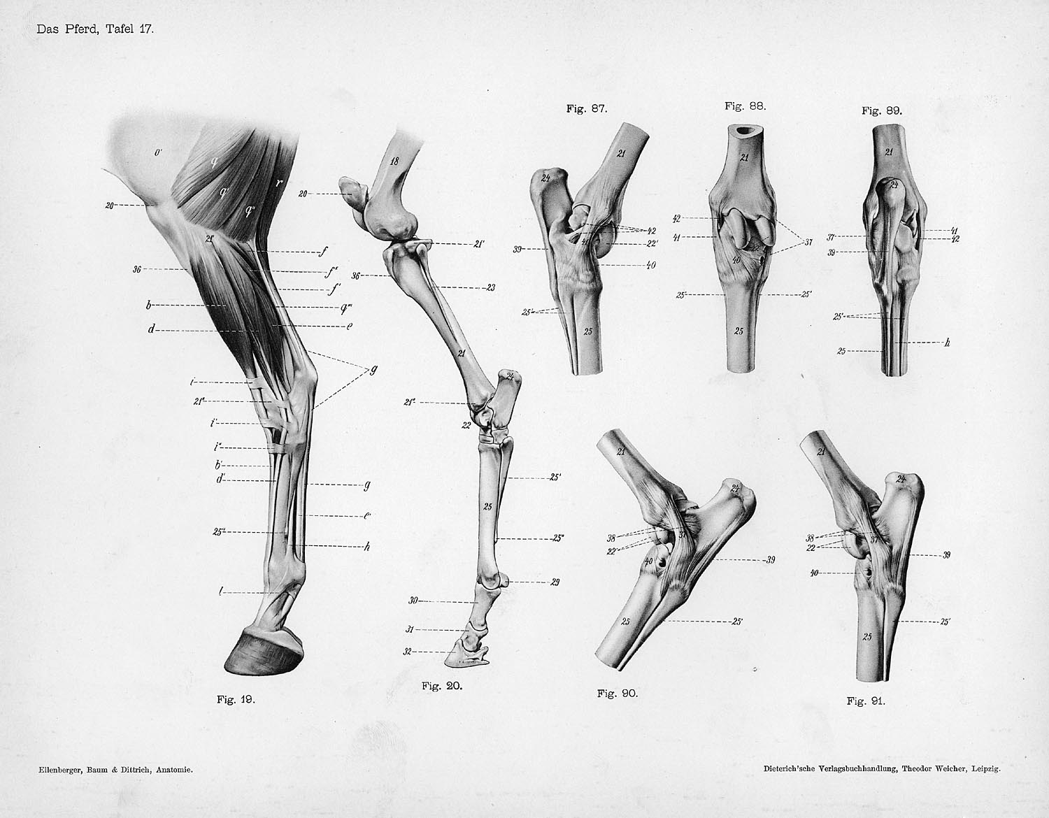 Horse Anatomy By Herman Dittrich Hind Legs Shoestring Stable
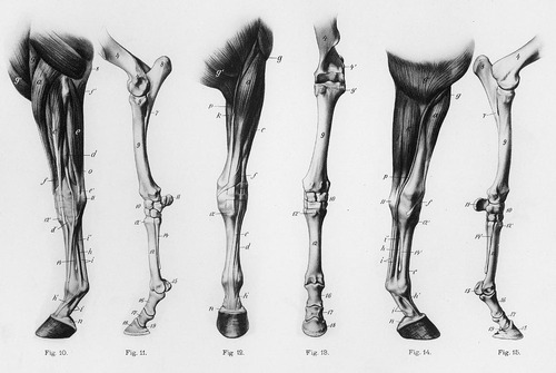
 Horse S Anatomy Legs Muscles By Silviasdesires On Deviantart
 Causes Of Equine Lameness Equimed Horse Health Matters
 Chestnut Horse Anatomy Wikivisually
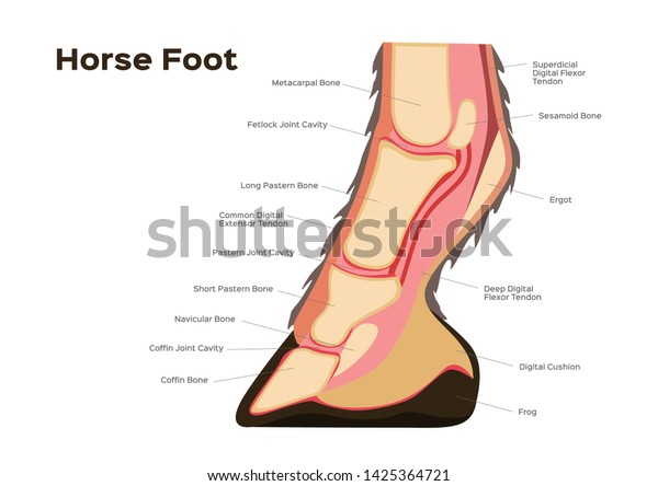 Horse Foot Leg Anatomy Infographic Chart Stock Vector
 Pin By Valerij Martynyuk On Animalistika In 2019 Horse
 Front Leg Locking Mechanisms Horse Anatomy Horses Horse
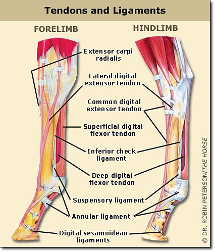
 Musculoskeletal Anatomy Of Your Horse Classic Equine
 Horse Front Leg Anatomy Oneyplays
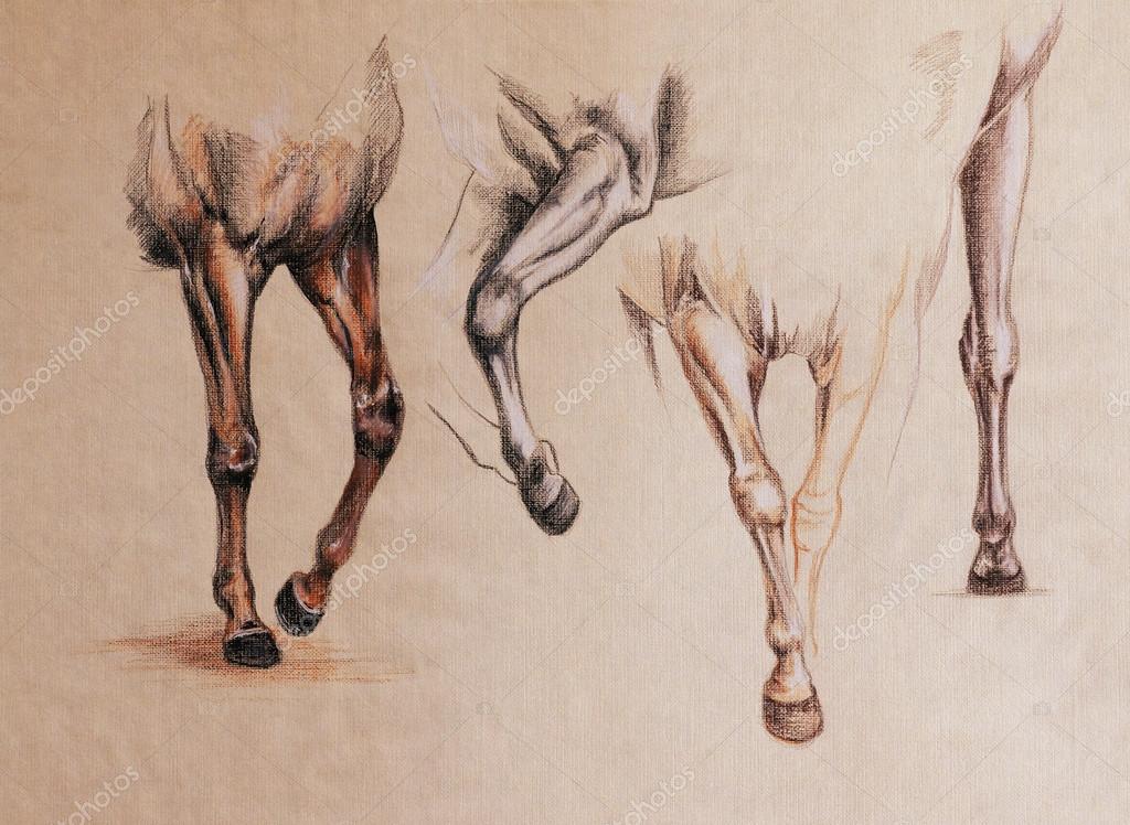 Horse Legs Study Stock Photo C Cattallina 92466600
 Limbs Of The Horse Wikipedia
 Horse Leg Anatomy Form And Function Equimed Horse
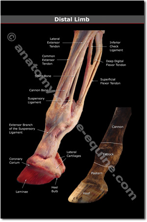
 Hoof Anatomy What Horse Hooves Are Made Of
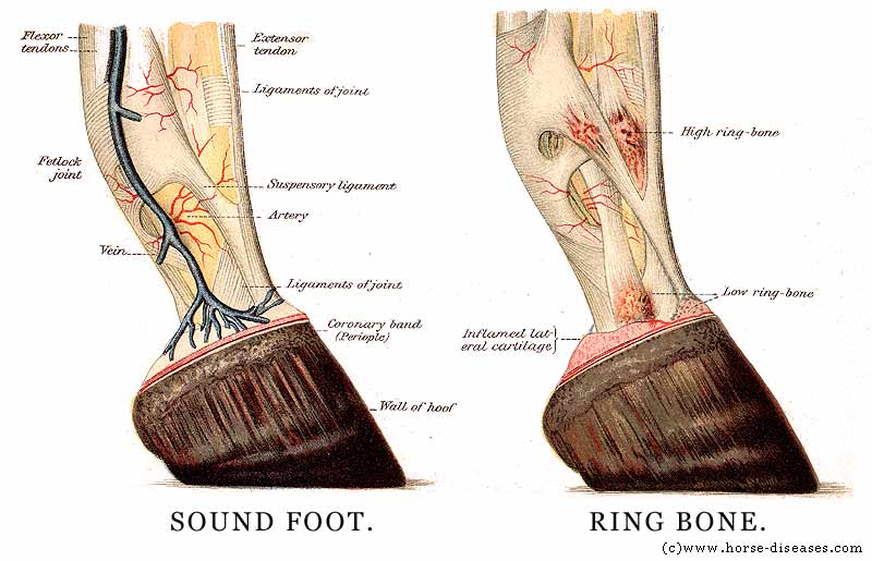
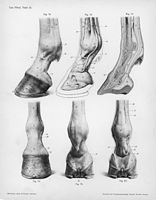
 Section Of Horse S Leg And Foot
 Equine Limb Anatomy Horse Leg Anatomy Diagram Horse
 Vitals Anatomy Horse Side Vet Guide
 Horsehoofandleganatomy Pdf Horse Hoof And Leg Anatomy A
 Horse Leg Muscles And Skeleton Structure Diagram
 Limbs Of The Horse Wikiwand
 Novobrace Tendonitis Desmitis And Soft Tissue Injury
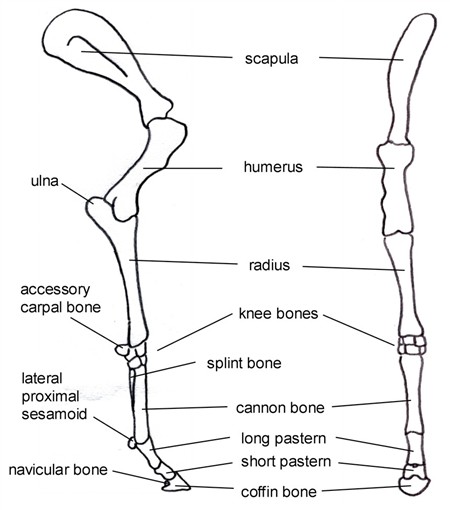
 Anatomy Of The Horse Osteology
 The Equine Hock What Horse Owners Should Know Thal Equine


Posting Komentar
Posting Komentar