Panoramic anatomythe following slides identify the anatomicalstructures found on panoramic radiographsin navigating through the slides you should clickon the left mouse button when you see themouse holding an x ray tubehead or you aredone reading a slide. It was also noted that the patient has very pronounced styloid processes bilaterally.
 Panoramic Anatomy Artifacts Dental Hygiene 309 With
This zone involves the dentition alveolar structures and condyles.

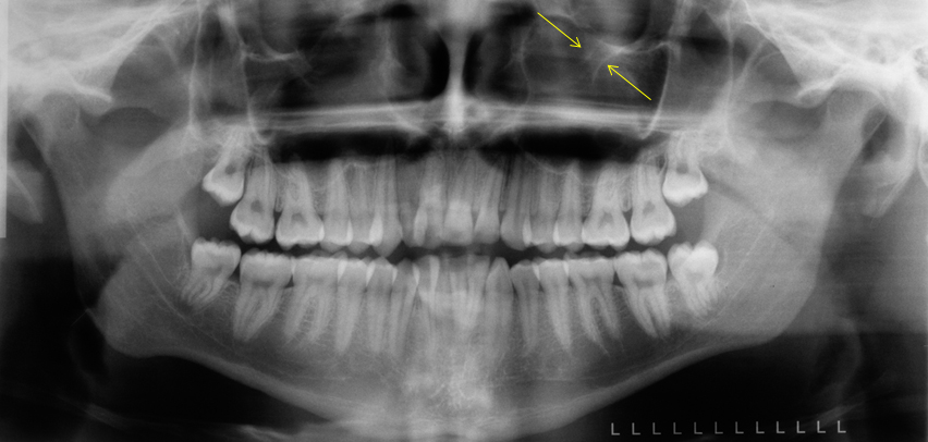
Panoramic radiograph anatomy. Anatomy of a panoramic radiograph. The maxillary sinus is located over the maxillary premolars and molars. Melanie helmke rdh msed discusses dental radiographic anatomy.
The panoramic image indicates a flattened condyle and significant wear of the glenoid fossa of the temporal bone due to constant force from bruxism and clenching. A zone of interest that is in focus with the panoramic film. Panoramic radiography is a form of focal plane tomography.
Simple panoramic radiograph landmarks part 3 condyle etc. Thus images of multiple planes are taken to make up the composite panoramic image where the maxilla and mandible are in the focal trough and the structures that are superficial and deep to the trough are blurred. The panoramic radiography is frequently used as initial diagnostic image of some alterations and based on it the professional will verify the need of other more detailed and more accurate examinations.
Interpreting panoramic radiographs examination is an orderly process. Hitting enter or pagedown will also work. It allows a general screening of the bony anatomy of the maxillofacial region in addition to identification of potential odontogenic sources of infection most commonly the maxillary or mandibular first or second molars.
The panoramic radiograph is the imaging study of choice for the evaluation of the odontogenic etiology of suspected vestibular and buccal space infections and is frequently the only imaging study that is necessary. The panoramic radiograph allows the dental professional to view a large area of the maxilla and mandible on a single film. Although it is obvious that a panoramic radiograph depicts the teeth and jaws in a single convenient view it may be less clear how the other structures of the head and neck become captured on the image.
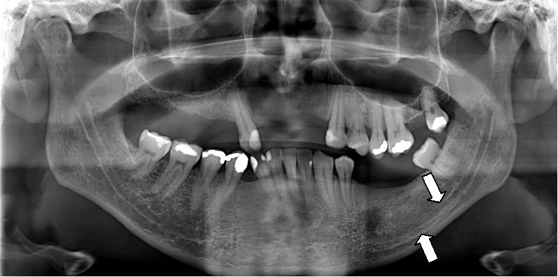
Mandible maxilla zygoma soft tissue air spaces teeth interpreting panoramic radiographs examine the borders of the bone first next examine the medullary bone check the internal structures such as canals foramina and sinuses. Provides overall survey of anatomy in the area being examined as well as an opportunity to discover findings otherwise missed by clinical or intra oral radiographic examination. Studentrdh dental hygiene online board review.
The maxillary sinus appears on the panoramic image as a radiopaque structure.
 Panoramic Radiograph Wikipedia
 Panoramic Radiograph Anatomy At University Of Minnesota
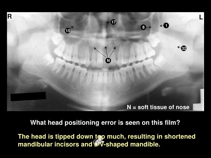
 Reading A Panoramic Radiograph
 Reading A Panoramic Radiograph
Automatic Synthesis Of Panoramic Radiographs From Dental
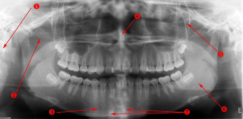
 Dentaltown Anatomical Landmarks On A Panoramic Radiograph
Art 3 Georgescu Qxp

 Self Study Pan Anatomy
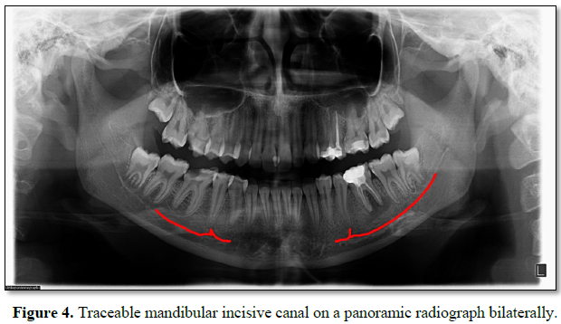 Scitech Detection Of Mandibular Incisive Canal By
 Panoramic Radiography Diagnosis Of Relevant Structures

 Pdf Visibility Of Mandibular Anatomical Landmarks In
 Panoramic X Ray Landmarks Purposegames
Planmeca Promax 2d S3 Panoramic X Ray Unit
 Use Of Panoramic Imaging For Patient Education Panoramic

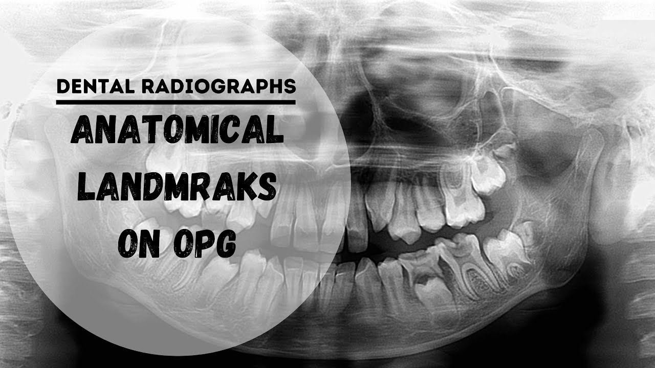 Dental Panoramic Radiographs Opg Dpt Anatomical Landmarks
 Ppt Panoramic Radiography Powerpoint Presentation Free
Panoramic Normal Anatomical Landmarks Flashcards Quizlet
 Panoramic Radiography Diagnosis Of Relevant Structures
 Panoramic Radiograph Pdf


Posting Komentar
Posting Komentar