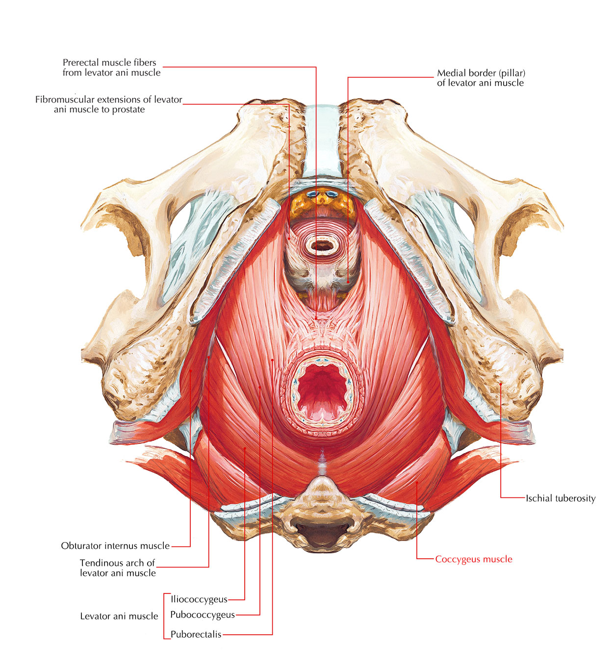
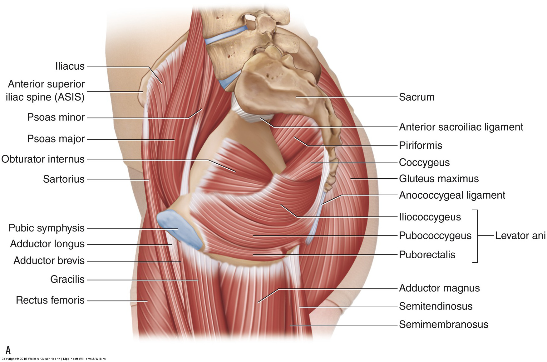
Posteriorly ischial spines of the pevlic bones. This muscle makes up most of the levator ani muscles.
 Easy Notes On Muscles Of The Pelvis Learn In Just 6
The right and left hip bones plus the sacrum and the coccyx together form the pelvis.

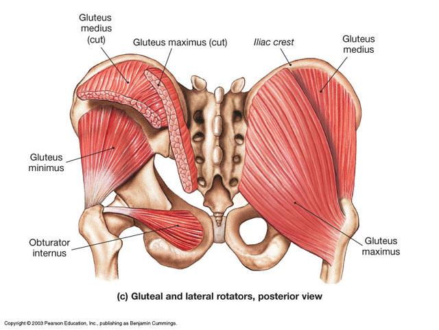
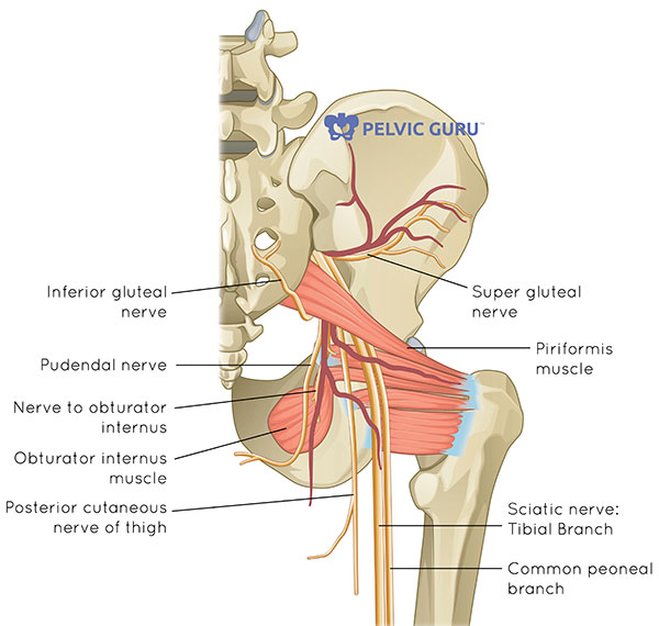
Pelvic anatomy muscles. Lying exposed between the protective bones of the superiorly located ribs and the inferiorly located pelvic girdle the muscles of this region play a critical role in protecting the delicate vital organs within the abdominal cavity. Laterally thickened fascia of the obturator internus muscle known as the tendinous arch. Innervated by the pudendal nerve.
The skin tissues and organs in the pelvis are supplied by the vasculature of the pelvis and innervated by many nerves of the pelvis including the pudendal nerve. The muscles of the pelvis form its floor. They form a large sheet of skeletal muscle that is thicker in some areas than in others.
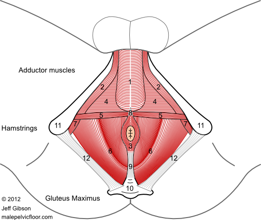
Female pelvis muscles puborectalis. There are many organs that sit in the pelvis including much of the urinary system and lots of the male or female reproductive systems. The muscles of the abdomen lower back and pelvis are separated from those of the chest by the muscular wall of the diaphragm the critical breathing muscle.
These muscles have attachments to the pelvis as follows. Innervated by pudendal nerve. This muscle is responsible for holding in urine and feces.
It originates from the ilium and ischium. This muscle forms most of the lateral wall of the pelvis and traverses through the lesser sciatic foramen attaching to the greater trochanter of the femur. The iliococcygeus has thinner fibers and serves to lift the pelvic floor as well as.
The floor of the pelvis is made up of the muscles of the pelvis which support its contents and maintain urinary and faecal continence. The pelvic girdle is formed by a single hip bone. Anterior pubic bodies of the pelvic bones.
Deep urogenital diaphragm layer. They form a large sheet of skeletal muscle that is thicker in some areas than in others. They support the pelvic organs especially during increases in intra abdominal pressure and also aid in urinary and faecal continence.
The hip bone attaches the lower limb to the axial skeleton through its articulation with the sacrum. Innervated by sacral nerve roots s3 s5. There are many muscles that form the pelvic floor including puborectalis pubococcygeus iliococcygeus and coccygeus.
The pelvic floor consists of three muscle layers. It is enervated by the nerve to the obturator internus. The muscles of the pelvic floor are collectively referred to as the levator ani and coccygeus muscles.
 Muscles Of The Pelvic Floor Preview Human Anatomy Kenhub
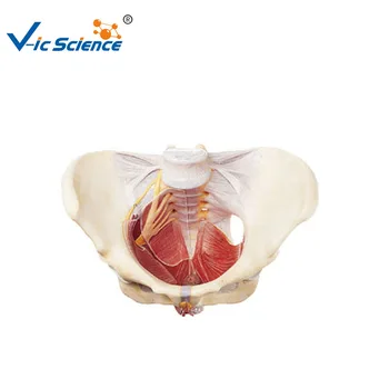 Female Pelvis And Pelvic Floor Muscles Anatomy Model Buy Female Pelvis Anatomy Model Pelvic Floor Muscles Model Pelvic Floor Muscles Anatomy Model
Hip Strains Orthoinfo Aaos
![]() Muscles Of The Pelvic Floor Anatomy And Function Kenhub
Muscles Of The Pelvic Floor Anatomy And Function Kenhub
 Anatomy Of The Pudendal Nerve Health Organization For
Pelvic Floor Dysfunction Cleveland Clinic
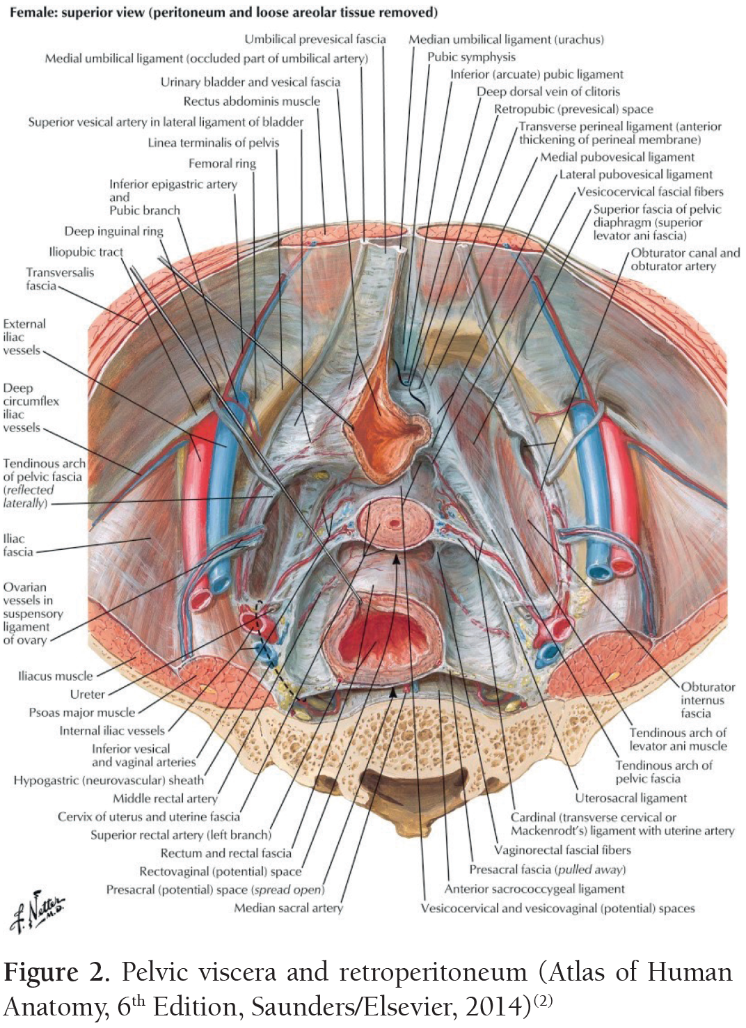
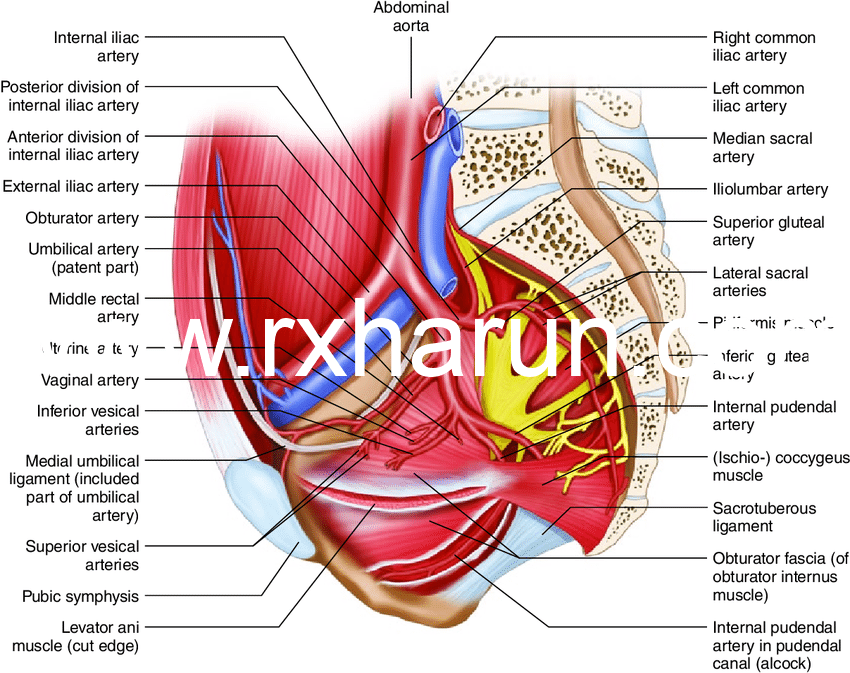
 Female Pelvic Anatomy Abdominal Key
 Pelvic Floor Muscles Mayo Clinic
 Pin By R On Massage Career Muscle Anatomy Yoga Anatomy
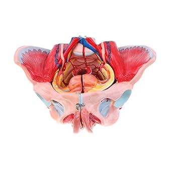 Magideal 1 1 Female Pelvis Vessels Muscles Nerves With
![]() Diagram Pictures Muscles Of The Pelvic Floor Anatomy
Diagram Pictures Muscles Of The Pelvic Floor Anatomy
 Female Pelvic Floor 1 Anatomy And Pathophysiology Nursing
 Pelvic Floor Wikipedia
:max_bytes(150000):strip_icc()/Depositphotos_19871399_original-56a05f523df78cafdaa14cd1.jpg) Hamstring Muscles
Hamstring Muscles
 Coccygeus Muscle An Overview Sciencedirect Topics
 Pelvic Floor Anatomy Pelvic Diaphragm Part 1
![]() Hip And Thigh Bones Joints Muscles Kenhub
Hip And Thigh Bones Joints Muscles Kenhub
 Pelvic Floor Muscles The Base For All Movement Anatomy
 Pregnancy Related Pelvic Girdle Pain Rayner Smale
 What Is The Pelvic Floor Your Pace Yoga
 Understand Hip Anatomy Muscles For Yoga Jason Crandell Yoga
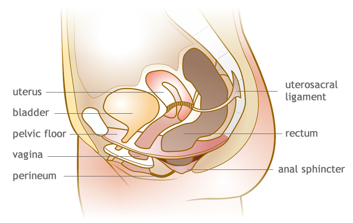
 Functions Of Pelvic Floor Muscle Anatomy Exercise Rx Harun
 Max The Muscle Skeleton On Pelvic Mounted Roller Stand 3b Smart Anatomy
 Pelvis Femur Muscles Pelvis Anatomy Muscle Anatomy Yoga
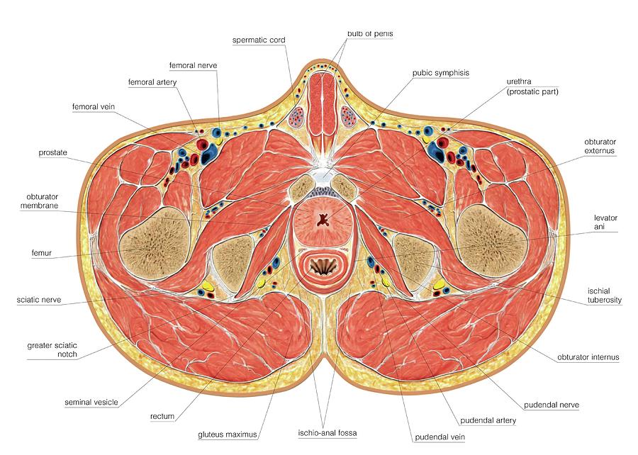 Muscles Of Pelvis Floor Cross Section
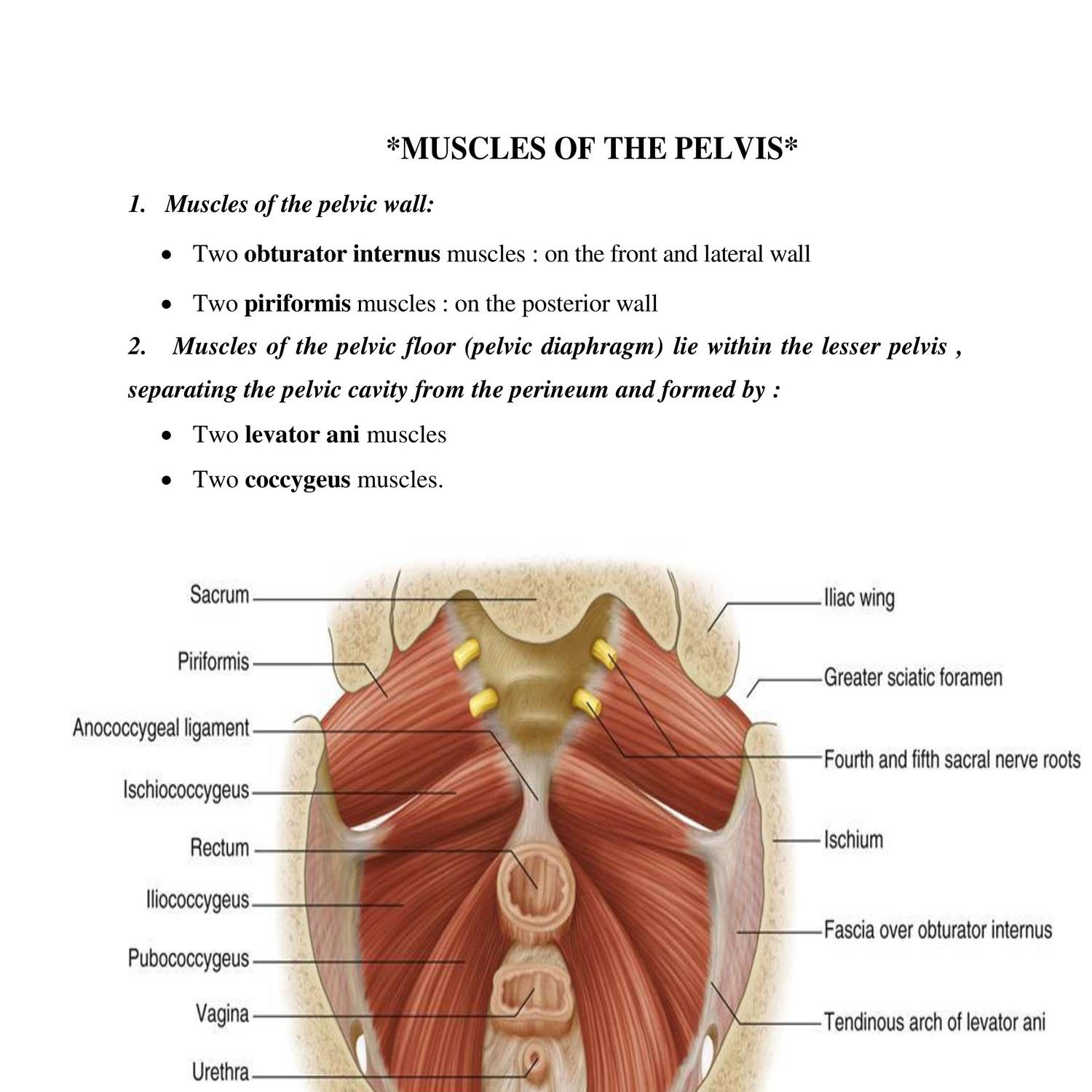 Anatomy Muscles Of Pelvis Doc Docdroid


Posting Komentar
Posting Komentar