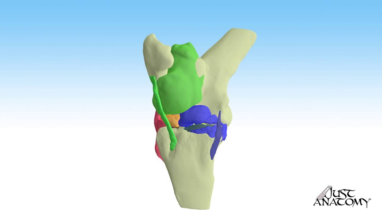
The most complex joint in the horse is the stifle joint. Wilson dvm ms diplomate acvs.
 Importance Of Proper Hind Leg Conformation Equimed Horse
How to take and interpret radiographs of the young performance horse.
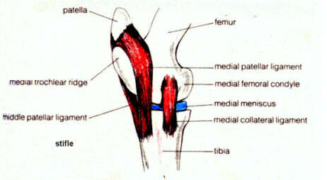
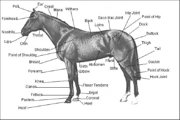
Stifle anatomy horse. Stifle joint the stifle is the equivalent of the human knee and it is the largest most complex joint in the horse. The joint includes the kneecap and its ligaments which give structural stability. The horses stifle is akin to a human knee and it usually bends forward.
A and b typical poor positioning for lateromedial view. As the leg moves the patella rides up and down the trochlear ridges of the femur in the femoropatellar joint. A horse with a locked stifle will likely hold its hind leg stiff and straight unable to unlock the joint.
Instead there is an intertubercular bursa. The stifle is the area where the tibia the bone that forms the gaskin meets the femur the bone that extends upward to the hip. The stifle lifts the leg upward and forward making it critical to moving and athletic pursuits.
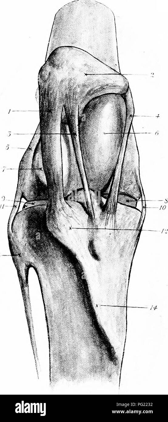
This bursa lies between the humeral tubercles cushioning the bicipital tendon but does not communicate with the cavity of the shoulder joint. In the horse there is no sheath surrounding the bicipital tendon. Stifle anatomy radiograph patella femur the equine stifle corresponds to the human knee.
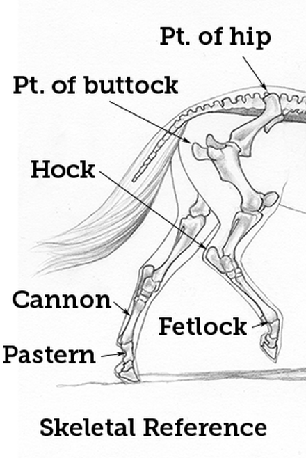
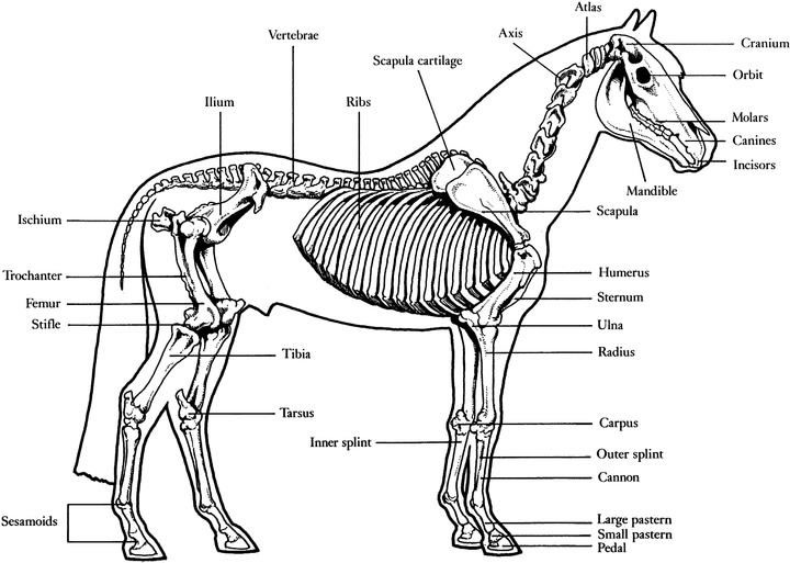
Encircling the whole stifle joint is a thin capsule that has a special fluid that assists with shock absorption and lubrication. Points of a horse equine anatomy refers to the gross and microscopic anatomy of horses and other equids including donkeys and zebras. Hind limb anatomy of most horses.
As a rule of thumb align the beam parallel to the heel bulbs of. The radiograph at left credit vetwerx is a lateral view of the stifle showing the knee cap or patella and the femur. The stifle is analogous to the human knee.
The reciprocal apparatus of the hind limb aids in preventing fatigue when the horse is standing and insures that there will be reciprocal flexing of the hock joint when the stifle joint is flexed or that the hock will extend when the stifle extends thereby preventing injuries. The bones that make up the stifle are the femur thigh tibia shin and patella kneecap. Observe your horse to see if it holds its leg taut and if it drags the toes of its hoof on the ground behind it.
When you pick up a horses hind leg the joint bends forward just as your knee does as you climb a staircase. How to take and interpret radiographs of the equine stifle david a. Similar to the human knee the stifle is located on the horses hind limbs.
 Stifle Joint Stock Photos Stifle Joint Stock Images Alamy
 How To Perform Arthrocentesis Of The Compartments Of The
 How To Ultrasound The Equine Stifle Joint
 Anatomy Of The Horse Osteology
 File The Anatomy Of The Horse A Dissection Guide 1922
 The Locations Of The Fetlock Carpal And Stifle Joints In
 Pdf Magnetic Resonance Imaging Of The Equine Stifle
 Behind The Bit The Stifle The Mother Of All Joints
 Horse Anatomy Mobility Health
 Horse Vertebrate Anatomy Diagram Of A Horse
 Hindlimb Anatomy Physiology Wikivet English
 Horse Anatomy Mobility Health
 The Stifle Is Comparable To The Human Knee What My Mare
 Stifle Helpful Horse Hints
 Locking Stifles Henderson Equine Clinic
 External Equine Anatomy All Things Equine
 Cranial Cruciate Ligament Disease In Dogs New England
 Equine Stifle Joint
 Joint Problems David Ramey Dvmdavid Ramey Dvm
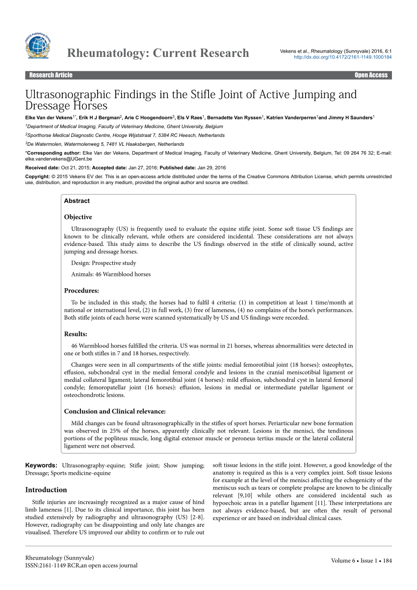 Pdf Ultrasonographic Findings In The Stifle Joint Of Active
 Horse Hock Massage For Better Performance Expert How To
 Equine Stifle Joint Anatomy Locked Versus Unlocked Position
:max_bytes(150000):strip_icc()/hind-leg-problems-in-horses-1886457_FINAL-5bf466b446e0fb0051770a3e.png) Hind Leg Problems In Horses Causes And Treatment
Hind Leg Problems In Horses Causes And Treatment
 Aec Client Education Upward Patellar Fixation
 Equine Stifle In Parker Berthoud Boulder Co Vetwerx Equine
 Equine Stifle In Parker Berthoud Boulder Co Vetwerx Equine
 The Stifle Joint Of The Horse Mackinnon
 3 Steps To Stronger Stifles Expert How To For English Riders
 Locking Stifles Henderson Equine Clinic


Posting Komentar
Posting Komentar