Muscles and tendons the motion of the ball and socket is controlled by several very powerful muscles which attach to the bones. In vertebrate anatomy hip or coxa in medical terminology refers to either an anatomical region or a joint.
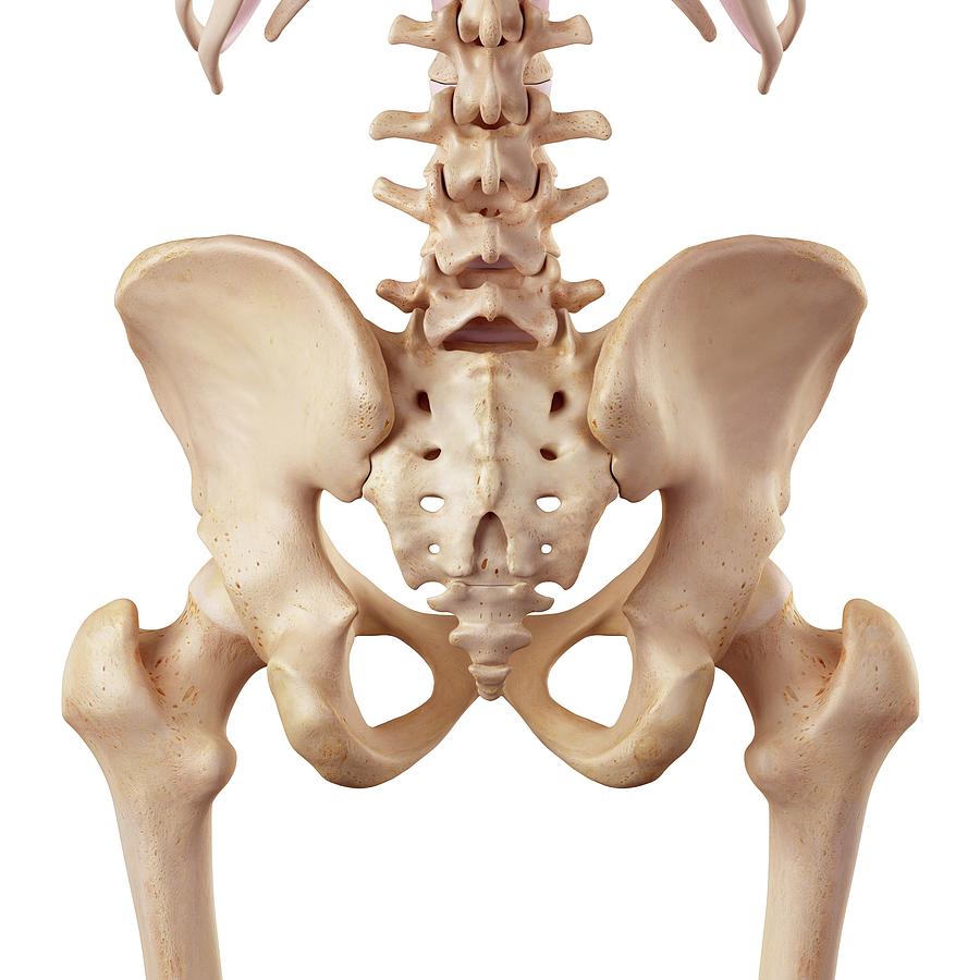
 The Pelvic Girdle Of Human Hip Bone Anatomy Vector
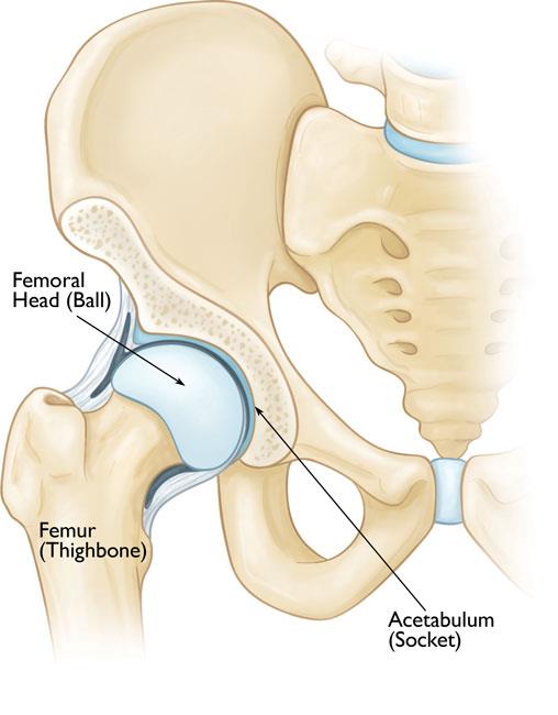
One of the bodys largest weight bearing joints the hip is where the thigh bone meets the pelvis to form a ball and socket joint.

Anatomy of the human hip. A round cup shaped structure on the os coxa known as the acetabulum forms the socket for the hip joint. Anatomy of the hip joint capsule of the hip like the shoulder the hip is a ball and socket joint but is much more stable. The thigh bone or femur and the pelvis join to form the hip joint.
The hip helps the body maintain balance and assists in ambulation. It is also referred to as a ball and socket joint and is surrounded by muscles ligaments and tendons. The hip region is located lateral and anterior to the gluteal region inferior to the iliac crest and overlying the greater trochanter of the femur or thigh bone.
The hip joint is a ball and socket synovial joint formed between the os coxa hip bone and the femur. The hip is a joint responsible for supporting the bodys weight during both movement and rest periods. The hip joint is a ball and socket joint.
The muscles you probably know the best are your glutes. The hip joint consists of two main parts. Femoral head a ball shaped piece of bone located at the top of your thigh bone or femur acetabulum a socket in your pelvis into which the femoral head fits.
Amphibians and reptiles have relatively weak pelvic girdles and the femur extends horizontally. The bones of the hip include the femur the ilium the ischium and the pubis. The round head of the femur rests in a cavity the acetabulum that allows free rotation of the limb.
The pubis ischium and ilium together constitute the pelvis while the thigh bone is the femur. The stability in the hip begins with a deep socketthe acetabulum. Hip anatomy the hip joint is the largest weight bearing joint in the human body.
 Vector Illustration Anatomy Of A Healthy Human Hip Joint Isolated

 Anatomy 101 Learn To Balance Mobility Stability In Your
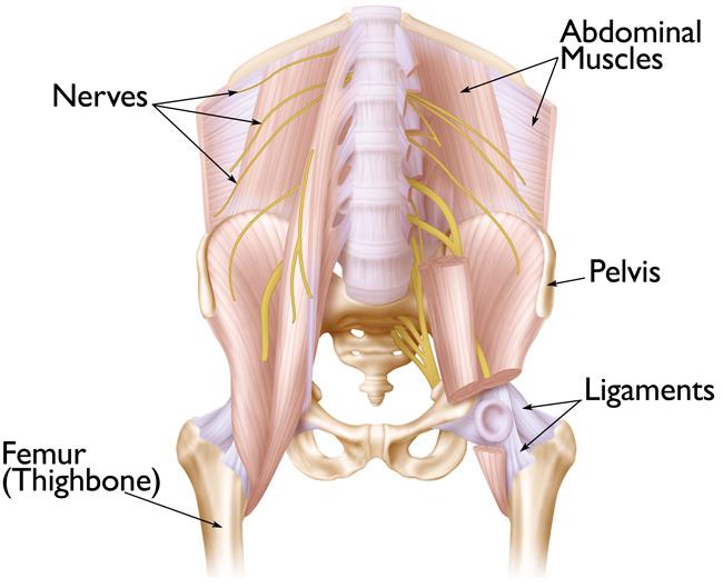
 Pelvis Nerve Hip Human Anatomy Human Body Nervous System
 List Of Human Anatomical Regions Wikipedia
 Anatomy Of Human Hip Joint Stock Image K17236400 Fotosearch
 Hip Muscles Lateral View Hip Muscles Hip Muscles
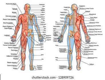
 Pin By Annie Matze On Anatomy Human Body Muscles Human
 Pelvis Human Anatomy Human Body Hip Bone Others Free Png
 Hip Bone Pelvis Human Skeleton Anatomy Png Clipart Abdomen
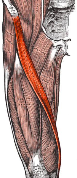
 Leg Muscles Anatomy Leg Anatomy Human Anatomy Hip Muscles
 Issues Around The Hip From Tendonitis To Bursitis Beacon
 Muscles Of The Hips And Thighs Human Anatomy And
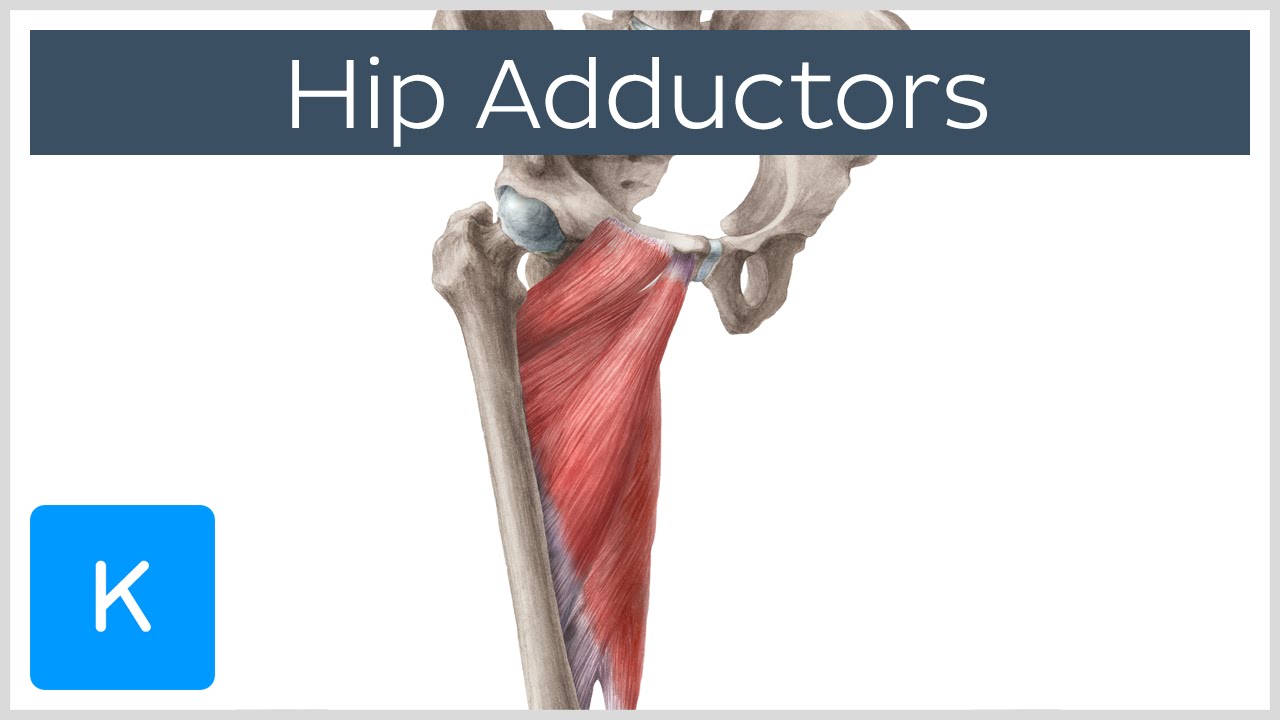 Anatomy Of The Hip Adductor Muscles Human Anatomy Kenhub
 Anatomy Hip Stock Illustrations 6 500 Anatomy Hip Stock
 Perthes Disease Legg Calve Perthes Orthoinfo Aaos
 Pelvis Hip Anatomy
 The Pelvic Girdle Of Human Hip Bone Anatomy Vector Illustration
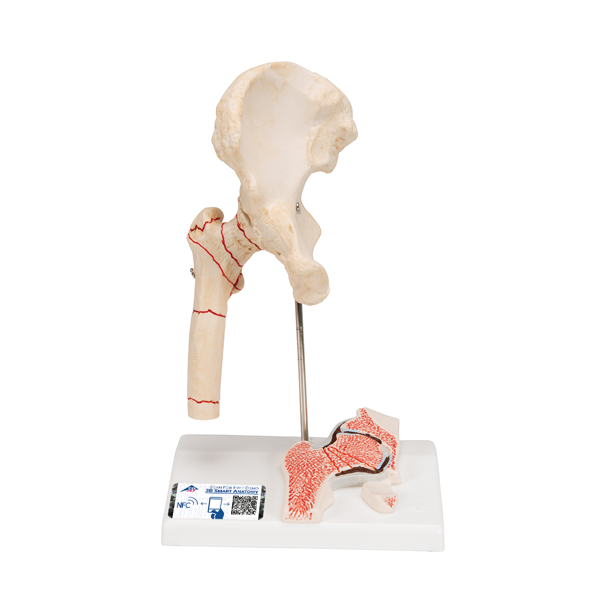
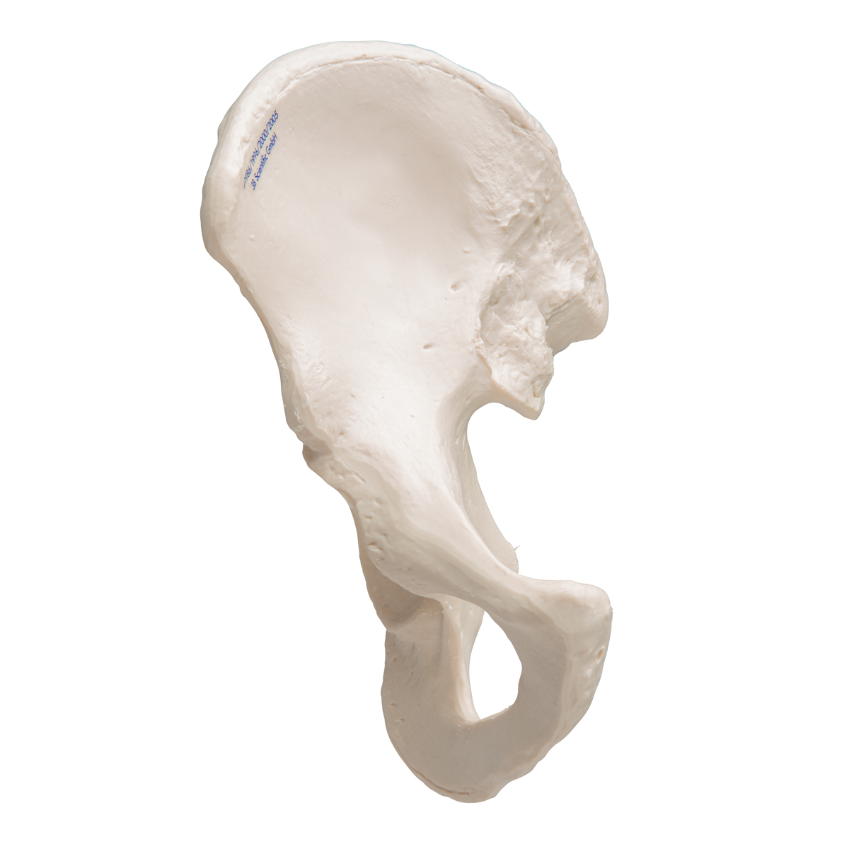 Human Hip Bone Model 3b Smart Anatomy 1019365 3b
 Hip Anatomy Pictures Function Problems Treatment
 Hip Human Anatomy Inflammation Articular Joint Pain Stock
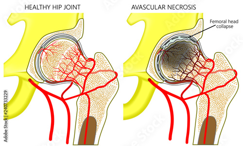 Vector Illustration Anatomy Of A Healthy Human Hip Joint And
 Anatomical Teaching Models Plastic Human Joint Models
 Hip Joint Anatomy Pictures And Information
 Human Hip Anatomy 1
 Human Hip Images Stock Photos Vectors Shutterstock
 Complications To Hip Fracture Fixation Medical
 Acetabular Fractures Orthoinfo Aaos
Hip Thigh Atlas Of Anatomy
 Leg Anatomy Britannica

 Videos Matching Introduction To Anatomy Movement Revolvy


Posting Komentar
Posting Komentar