The former is a thickening of the circular layer of the muscularis whereas the latter is a distinct muscle. Mri of peri anal fistulas mukesh g harisinghani md overview anatomy classification technique examples anal canal.
Anatomy Of The Rectum And Anus
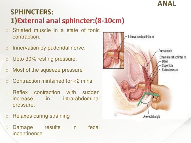
The muscle is in variable tonic contraction during waking hours and it can be contracted voluntarily.
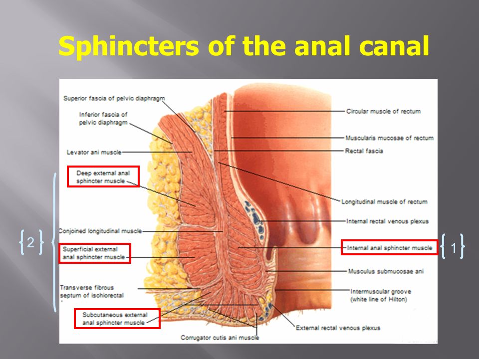
Anal sphincter anatomy. It consists of two strata superficial and deep. Circular muscles called the external sphincter ani form the wall of the anus and hold it closed. A second sphincter the external anal sphincter is composed of striated muscle and is divided into three parts known as the subcutaneous superficial and deep external sphincters.
An internal anal sphincter of smooth muscle and an external anal sphincter of skeletal muscle. In human digestive system. External anal sphincter voluntary muscle that surrounds the lower 23 of the.
Anal sphincter anatomy knowing the muscles that control your anal sphincter and how they work makes understanding the exercises much more clear. Glands release fluid into the anus to keep its surface moist. The internal anal sphincter the internal anal sphincter is an involuntary muscle which means you cannot consciously control it.
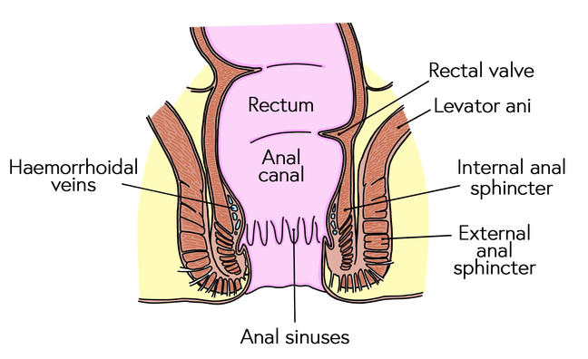
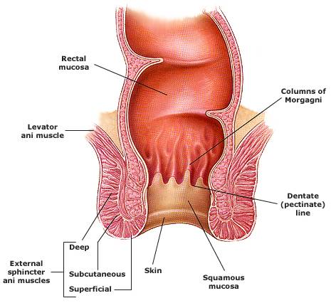
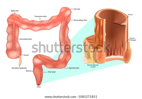
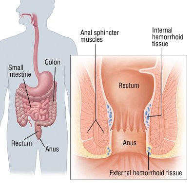
The external longitudinal muscle layer continues as the. The columns are vascular and enlargement of their venous plexus results in internal hemorrhoids. The involuntary autonomous internal anal sphincter is the lowermost continuation of the inner circular smooth muscle layer of the rectum.
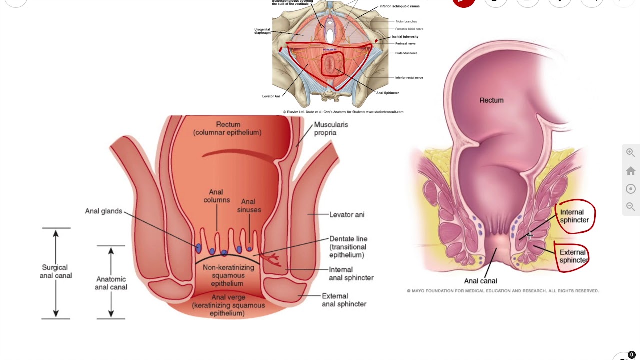
Internal anal sphincter surrounds the upper 23 of the anal canal. The external anal sphincter measures about 8 to 10 cm in length from its anterior to its posterior extremity and is about 25 cm opposite the anus when defecation occurs the sphincter muscle retracts. The wall of the anal canal contains two sphincter muscles.
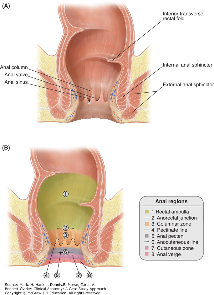
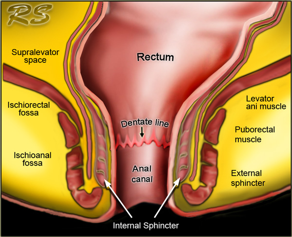
Anatomy dentate line perineal skin anorectal ring sphincter mechanism external sphincter internal sphincter intersphincteric spaceintersphincteric space puborectalis sling levator ani muscle. It is formed from a thickening of the involuntary circular smooth muscle in the bowel wall. Several vertical mucosal folds the anal formerly called rectal columns are usually visible in the upper half of the canal fig.
The sphincter ani internus is the thick lower end of the inner circular layer of the gut. To describe the various patterns of normal sphincter anatomy as seen at endoanal magnetic resonance mr imaging and to assess sex and age related variations in the dimensions of the anal sphincter to refine the diagnosis of sphincter disorders.
 Figure 2 From Perianal Fistulous Tract Perianal Sinus Tracts
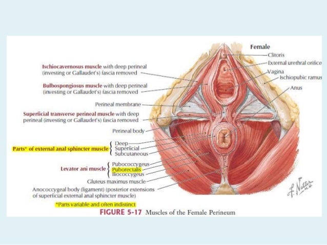
 Perineum Clinical Anatomy A Case Study Approach
 Anal Sphincter Iphone 7 Cases Fine Art America

 Anatomy Of Anal Sphincter And Perineal Body
 Rectum Anal Canal Ppt Video Online Download
 The Radiology Assistant Rectum Perianal Fistulas
 Surgical Anatomy Anal Canal
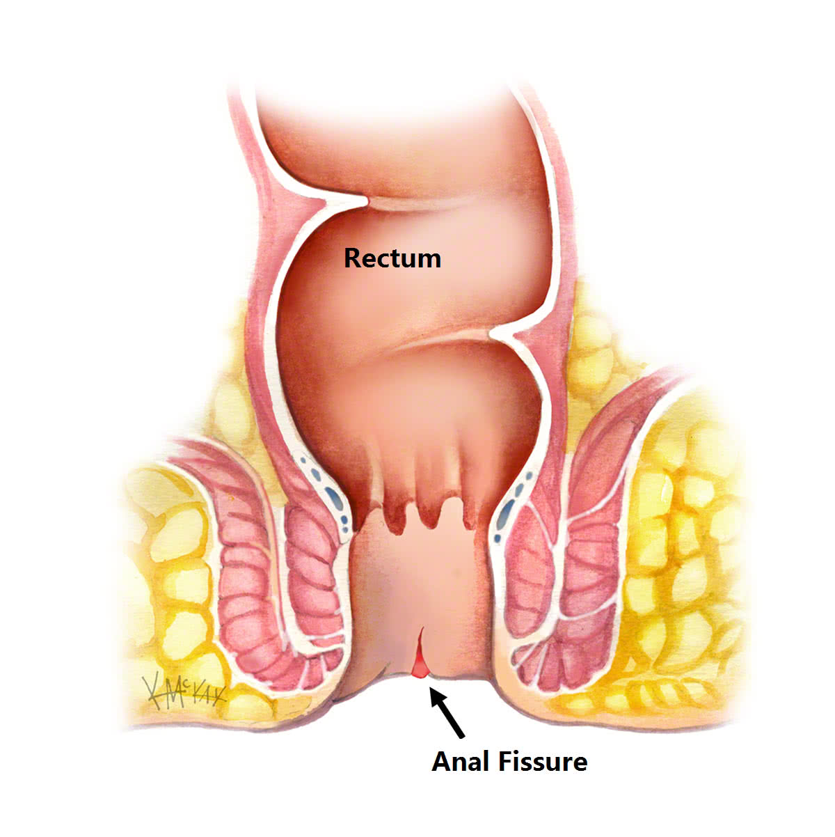 Anal Fissures Colorectal Surgeons Sydney

 Internal Anal Sphincter Rectum Anal Canal Stock Vector
 Anal Canal Sphincters Introduction Anatomy
 Why Does Anal Sphincter Muscle Damage Cause Faecal
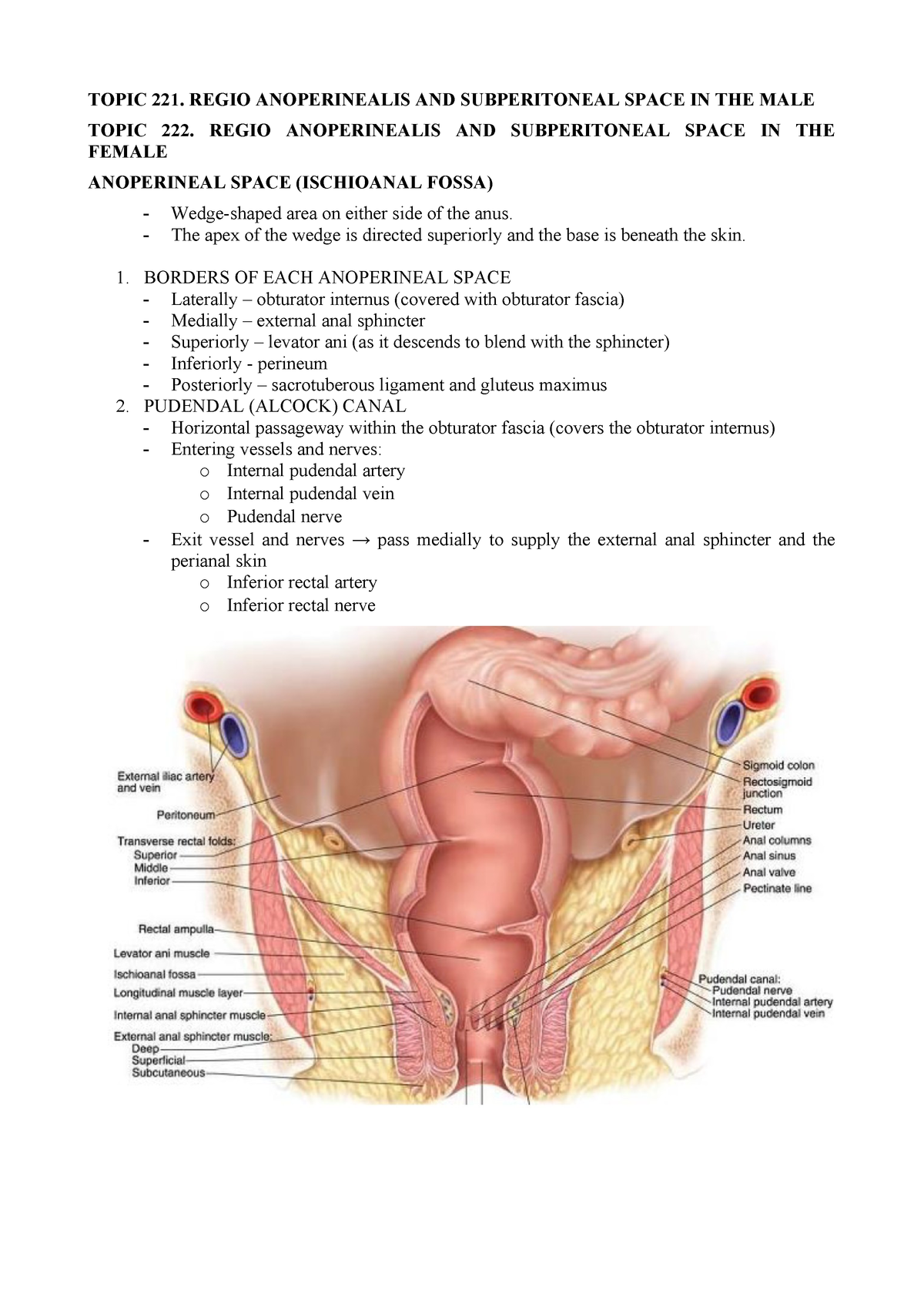 Topic 221 222 Regio Anoperinealis And Subperitoneal Space
 Common Anorectal Conditions Part I Symptoms And Complaints
 Anal Disorders Guide Causes Symptoms And Treatment Options
 Anal Canal Anatomy Qa

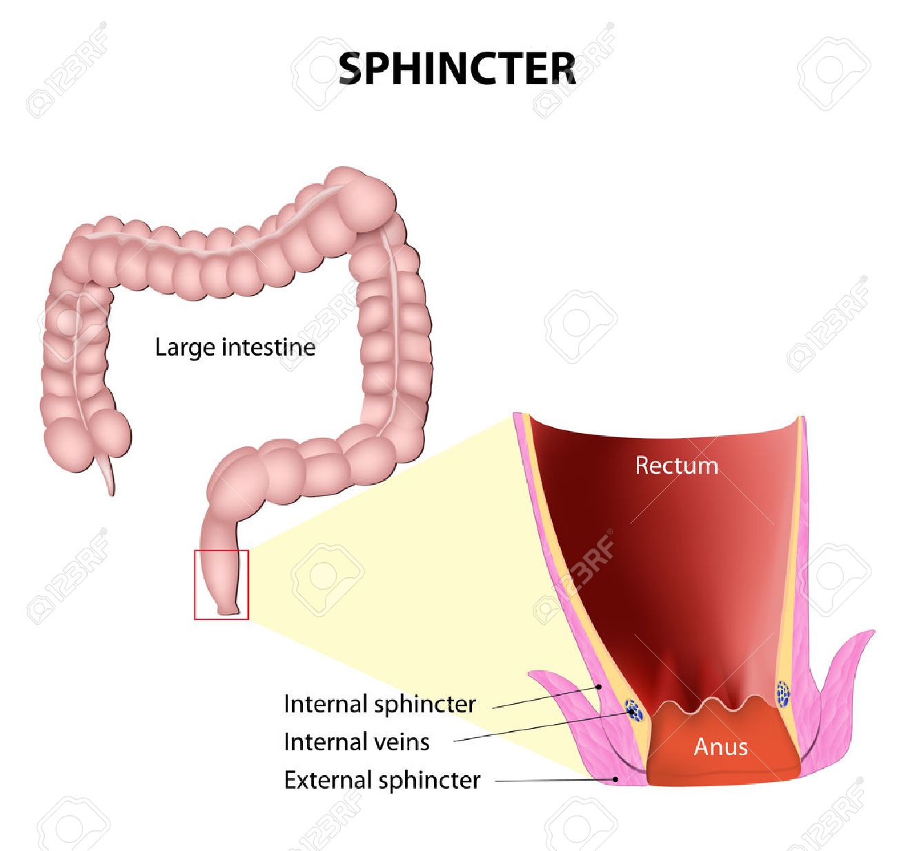 Sphincters Anatomy Of The Lower Rectum And Anus Showing The
 Obstetric Pelvic Floor And Anal Sphincter Injuries Lone
 Pediagenosis
 The Rectum Anatomy Of The Rectum Physiology Of The
The Structures Involved In Maintaining Continence And
 Anal Sphincter Complex Preservation Springerlink
 Anal Sphincter Imaging Sydney Pelvic Floor Health

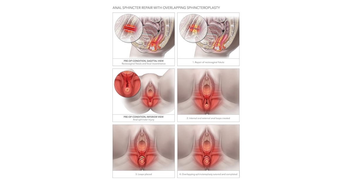 Anal Sphincter Repair With Overlapping Sphincteroplasty


Posting Komentar
Posting Komentar