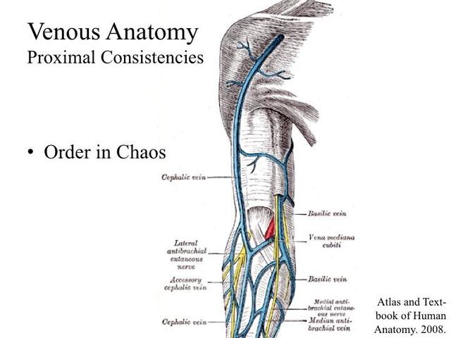
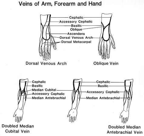
The major superficial veins of the hand forearm and upper arm exist as single structures and infrequently have accessory veins. Superficial veins of the upper limb in the cadaver.
 Arteriovenous Access Initial Evaluation And Follow Up
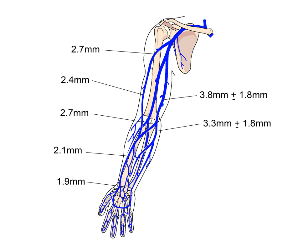
Arm like in the forearm the arm is drained by the brachial veins deep veins that accompany the brachial artery and all its branches.
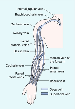
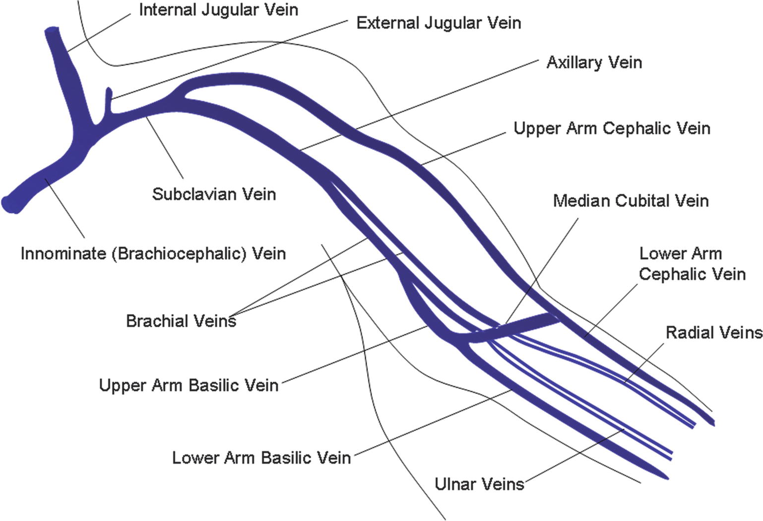
Arm venous anatomy. The primary venous return from the arm is through the axillary vein which continues centrally as the subclavian and brachiocephalic innominate veins before emptying into the superior vena cava. In common usage the arm extends to the hand. It can anatomically be divided into the superficial veins and the deep veins.
The venous system of the upper limb drains deoxygenated blood from the arm forearm and hand. Deep veins and superficial veins. In human anatomy the arm is the part of the upper limb between the glenohumeral joint shoulder joint and the elbow joint.
Each individual hands on training case is accompanied by image window specific expert instruction and probe positioning guidance. The vessels of the arms are part of the circulatory system which provides nutrients to the tissues. In common usage the arm extends to the hand.
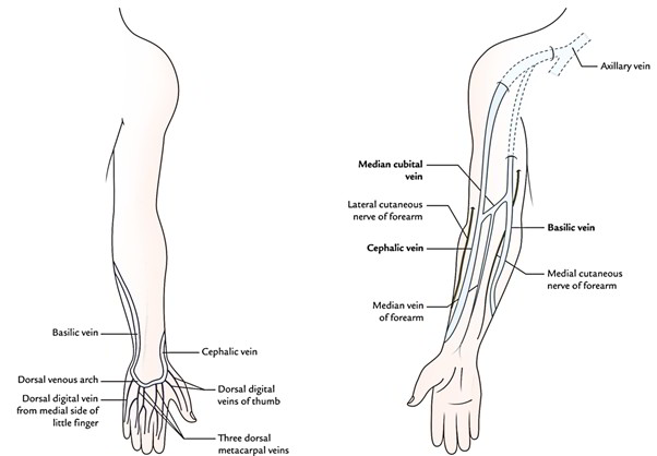
Anatomy physiology module provides a broad spectrum of adult male and female normal anatomy cases with varying body morphologies to maximize training efficacy. Starting around the radial area of what is known as the dorsal venous network the cephalic vein continues towards the upper part of the body in a circular fashion throughout the forearm interacting with tributaries along the way. As you reach the proximal arm the axillary vein will divide into the basilic and brachial veins.
Usually single but may be duplicated. In the hand forearm and upper arm the superficial system functions as the principal means for venous drainage. Continue from the axillary vein checking in transverse that the basilic and brachial veins of the upper arm are compressible.
The veins of the arm may be divided into two groups. Upper arm veins brachial basilic the basilic vein is the larger and is more superficial. As a result the caliber of the superficial veins is generally larger than the deep veins.
The arteries deliver freshly oxygenated blood to muscles and bone.
 Venipuncture Module 1 Anatomy Of The Arm And Vein Location
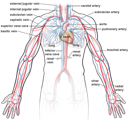
 Dentistry And Medicine Blood Supply Venous Drainage
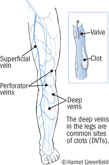 Deep Vein Thrombosis Blood Clots In Your Veins Harvard Health
 Median Cubital Antebrachial Veins Locations Functions
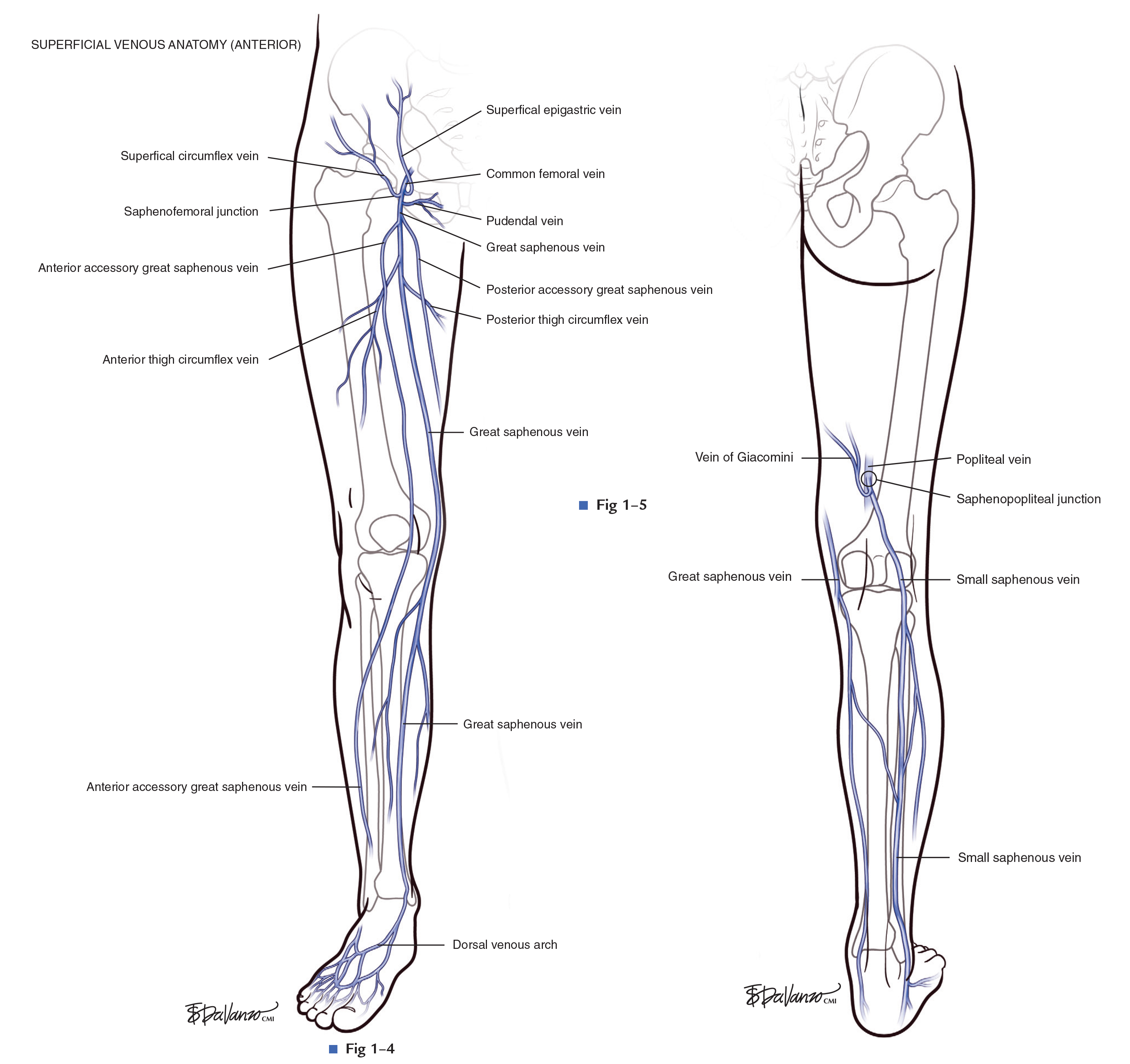
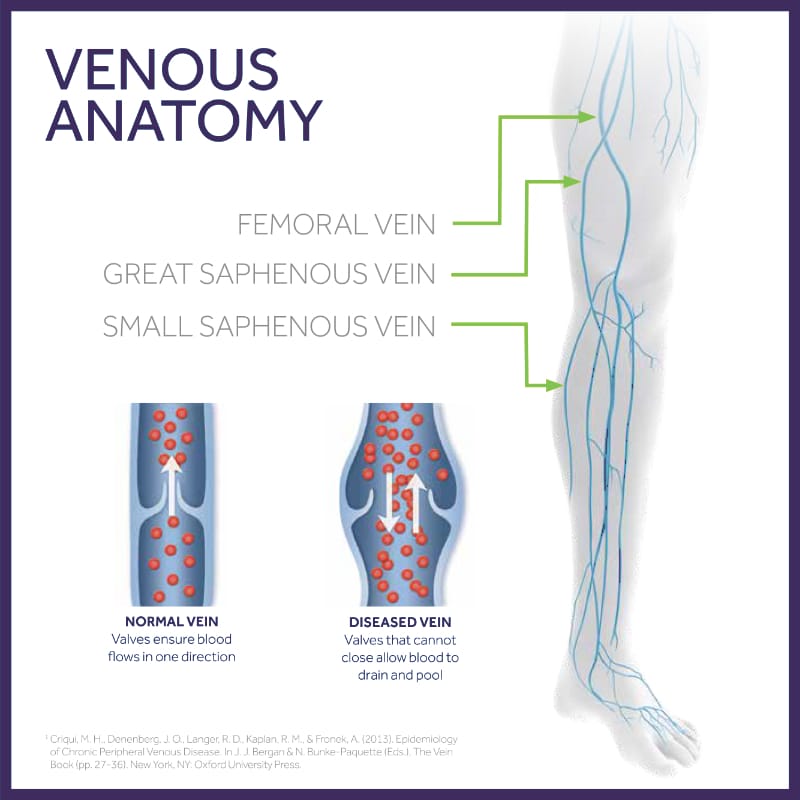
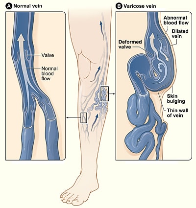 Varicose Veins Clinical Features Management

 Azygos Venous System Radiology Reference Article
 Easy Notes On Venous Drainage Of The Upper Limb
 The Peripheral Veins Radiology Key
 Picc Line Vein Anatomy Upper Limb Anatomy Anatomy Images
 Anatomy Of Veins Advanced Vein Care
 Forearm Artery And Venous System
 Fig Forearm And Hand Arterial And Venous Anatomy With The
![]() Brachial Vein Anatomy Course Tributaries Kenhub
Brachial Vein Anatomy Course Tributaries Kenhub
 The Peripheral Veins Radiology Key
/vascular-system-veins-56c87fa03df78cfb378b3e7c.jpg) What Is A Vein Definition Types And Illustration
What Is A Vein Definition Types And Illustration
 Vein Wikipedia
![]() Veins Of The Upper Limb Anatomy Kenhub
Veins Of The Upper Limb Anatomy Kenhub
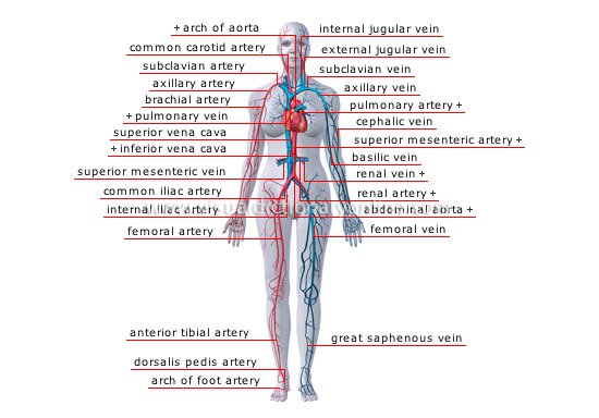
 Anatomy Atlases Illustrated Encyclopedia Of Human Anatomic
 Upper Extremity Venous Thrombosis Thoracic Key


Posting Komentar
Posting Komentar