Anchoring fibers type vii collagen penetrate into anterior stroma and attach with anchoring plaques type iv collagen to the stroma and to reticular fibers type iii collagen deep to the basement membrane. Corneal anatomy the cornea is the transparent front part of the eye that covers the iris pupil and anterior chamber.
 Anatomy And Physiology Of The Cornea Sciencedirect
The front part what you see in the mirror includes.

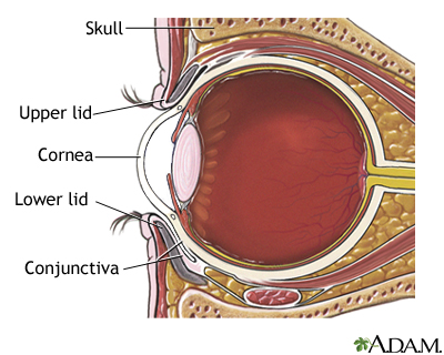
Cornea anatomy. This feature is not available right now. The area where the edge of the cornea meets the conjunctiva and sclera. The cornea lacks the neurobiological sophistication of the retina and the dynamic movement of the lens.
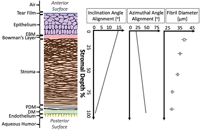
Please try again later. The stroma or supporting structure. The complexity of structure and function necessary to maintain such elegant simplicity is the wonder.
The anatomy and structure of the adult human cornea. Is made up of the cornea and the sclera. Together with the lens the cornea refracts light accounting for approximately two thirds of the eyes total optical power.
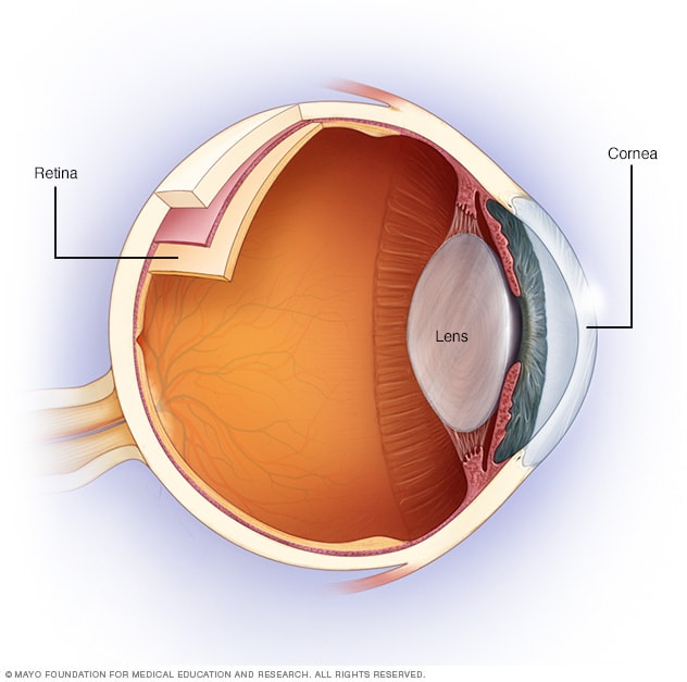
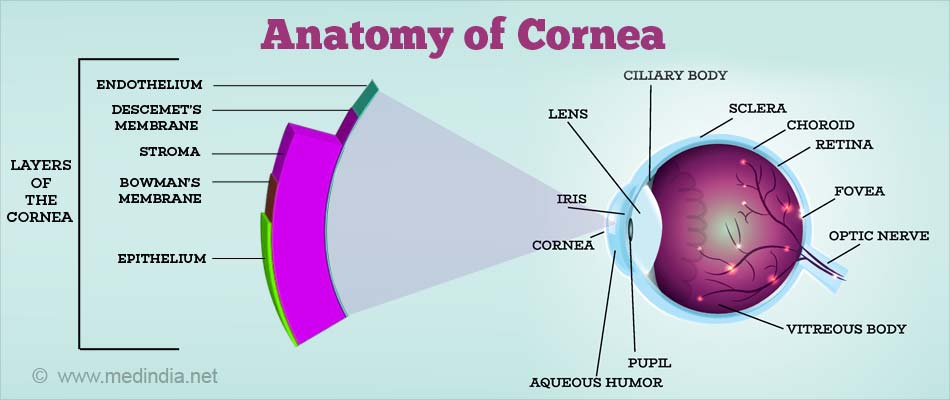
This magnified image of a section of the eye demonstrates the structure of the cornea and the limbus. With corneal edema the thickness of the cornea can substantially increase in the area of edema. The cornea is a transparent structure that together with the lens provides the refractive power of the eye.
The corneas main function is to refract or bend light. It contains five distinguishable layers. Yet without its clarity the eye would not be able to perform its necessary functions.
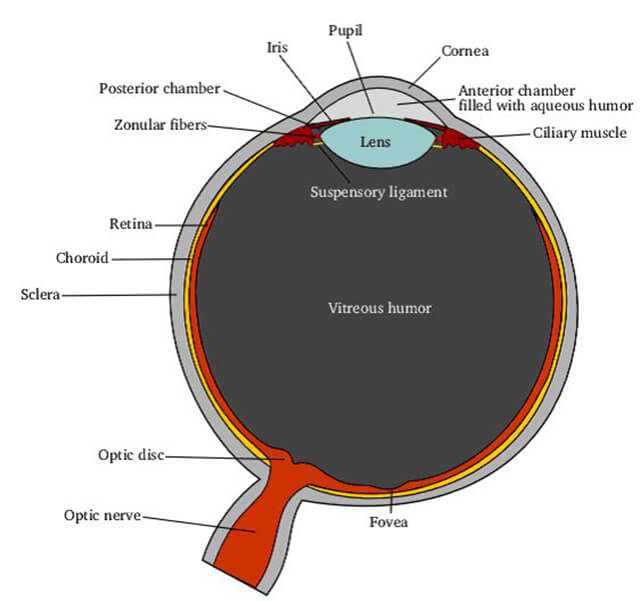
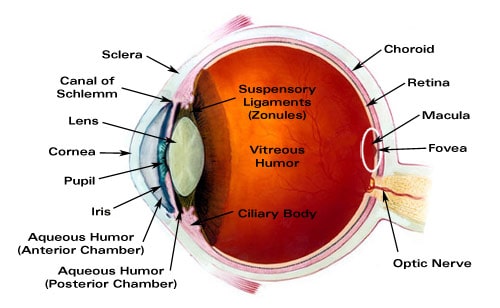
The epithelium or outer covering. The cornea is the transparent window of the eye. It covers the pupil the opening at the center of the eye iris the colored part of the eye and anterior chamber the fluid filled inside of the eye.
The cornea is the transparent front part of the eye that covers the iris pupil and anterior chamber. The black circular opening in the iris that lets light in. The cornea is the transparent part of the eye that covers the front portion of the eye.
The white of your eye. The cornea composes the outermost layer of the eye. When blood vessels invade the cornea they begin from the limbus.
A clear dome over the iris. The cornea with the anterior chamber and lens refracts light with the cornea accounting for approximately two thirds of the eyes total optical power. Part of the undergraduates course of ophthalmology.
A thin layer of tissue that covers the entire front of your eye except. The cornea is 0506 mm thick in the dog and cat and about 1 mm in horses. Anatomy and physiology of the cornea.
And the endothelium or inner lining.
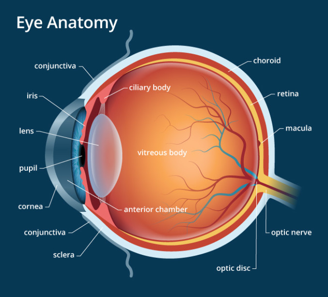
 Eye Anatomy
 Anatomy Of The Cornea
 Frontiers A Review Of Structural And Biomechanical Changes
 Vision And The Eye S Anatomy Healthengine Blog
Cornea Macular Degeneration Causes Diabetic Eye Problems
 The Cornea And Its Highlights Part 2 Anatomy Of The
 Simplified Anatomy Of The Eye Mayo Clinic


 Anatomy Of The Eye Vision Direct Uk
 Eye Anatomy A Closer Look At The Parts Of The Eye
 Evaluation And Management Of Corneal Abrasions American
 The Eyes Human Anatomy Diagram Optic Nerve Iris Cornea
 Corneal Transplantation
 Anatomy Of The Eye Moorfields Eye Hospital

 Corneal Anatomy At University Of Waterloo Studyblue

 The Anatomy And Physiology Of Cornea Download Scientific
 Cornea Definition And Detailed Illustration
 Eye Anatomy Medlineplus Medical Encyclopedia Image


Posting Komentar
Posting Komentar