Authors have used a variety of anatomic terms and descriptions that unfortunately have created ambiguity and confusion regarding this area of the knee. The medial anatomy of the knee consists of several layers of structures that work together to provide stability and function.
 Medial Collateral Ligament Of The Knee Physiopedia
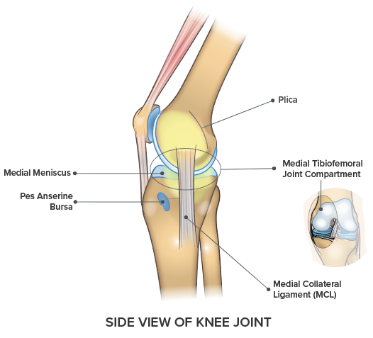
There are a number of structures on the medial side of the knee and problems in any one or more of these can cause pain.
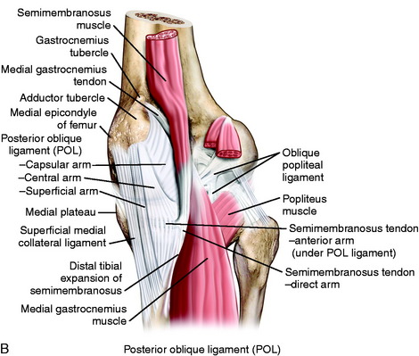
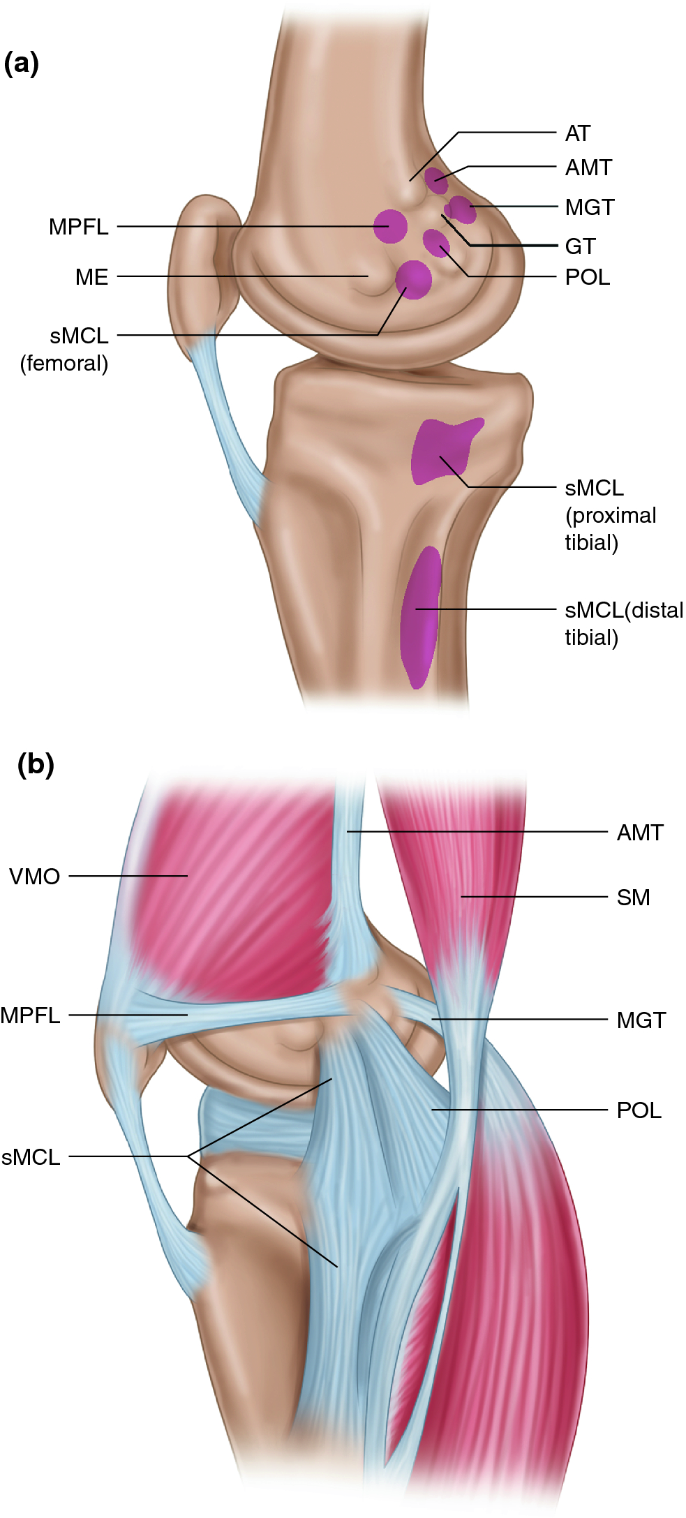
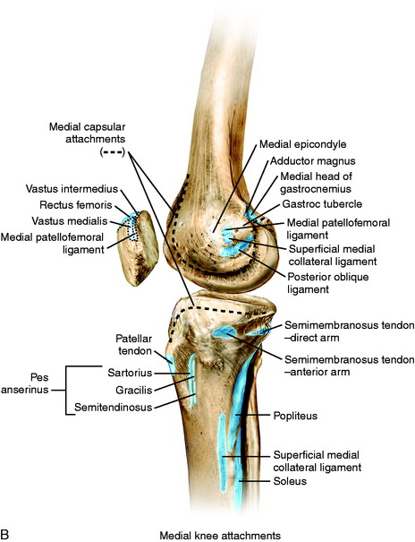
Knee anatomy medial. Medial anatomy of the knee. Medial the side of the knee that is closest to the other knee if you put your knees together the medial sides of each knee would touch lateral the side of the knee that is farthest from the other knee opposite of the medial side. The posterior oblique ligament femoral attachment was 77 mm distal and 64 mm posterior to the adductor tubercle and 14 mm distal and 29 mm anterior to the gastrocnemius tubercle.
The anatomy of the medial part of the knee plc studies combined posterior cruciate ligament and superficial medial collateral ligament knee reconstruction. The medial anatomy of the knee consists of several layers of structures that work together to provide stability and function. In knee joint anatomy knee ligaments are the main stabilising structures of the knee preventing excessive movements and instability.
The medial collateral ligament is recognised as being a primary static stabiliser of the knee and assists in passively stabilising the joint. Medial knee injuries are those to the medial side the inside of the knee are the most common. Medial knee pain is pain that occurs on the inner side of the knee and can be due to a number of problems.
Authors have used a variety of anatomic terms and descriptions that unfortunately have created ambiguity and confusion regarding this area of the knee. When stress is applied this ligament aids control in transferring the joint through a normal range of movement. The knee joins the thigh bone femur to the shin bone tibia.
Symptoms may come on gradually over time or may develop suddenly after a knee injury. The anatomy of the medial part of the knee. Medial anatomy of the knee.
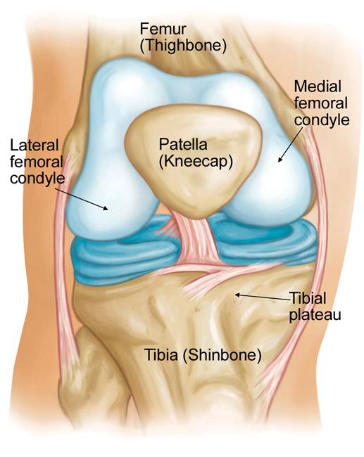
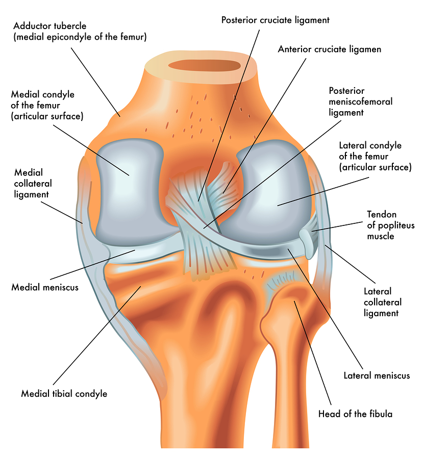
The mcl also prevents an anterior movement of the tibia and hyperextension. The deep medial collateral ligament consisted of meniscofemoral and meniscotibial portions. The knee is one of the largest and most complex joints in the body.
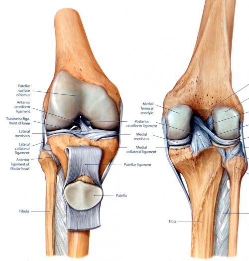
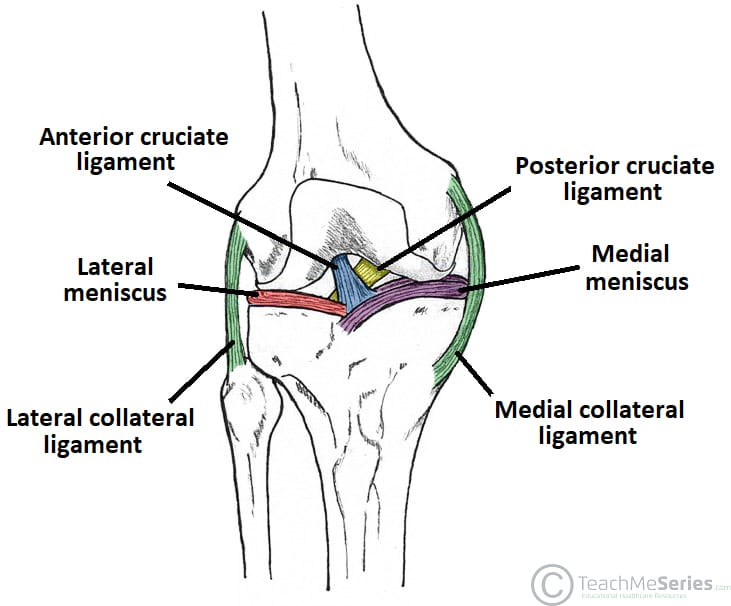
Ligaments are tough fibrous connective tissues which link bone to bone made of collagen. The medial ligament complex of the knee is composed of the superficial medial collateral ligament smcl deep medial collateral ligament dmcl and the posterior oblique ligament pol.
 Ligaments Of The Knee Knee Sports Orthobullets

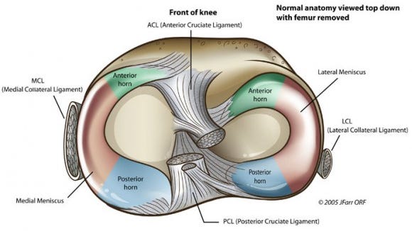 The Injury Zone Basic Anatomy And Function Of The Meniscus
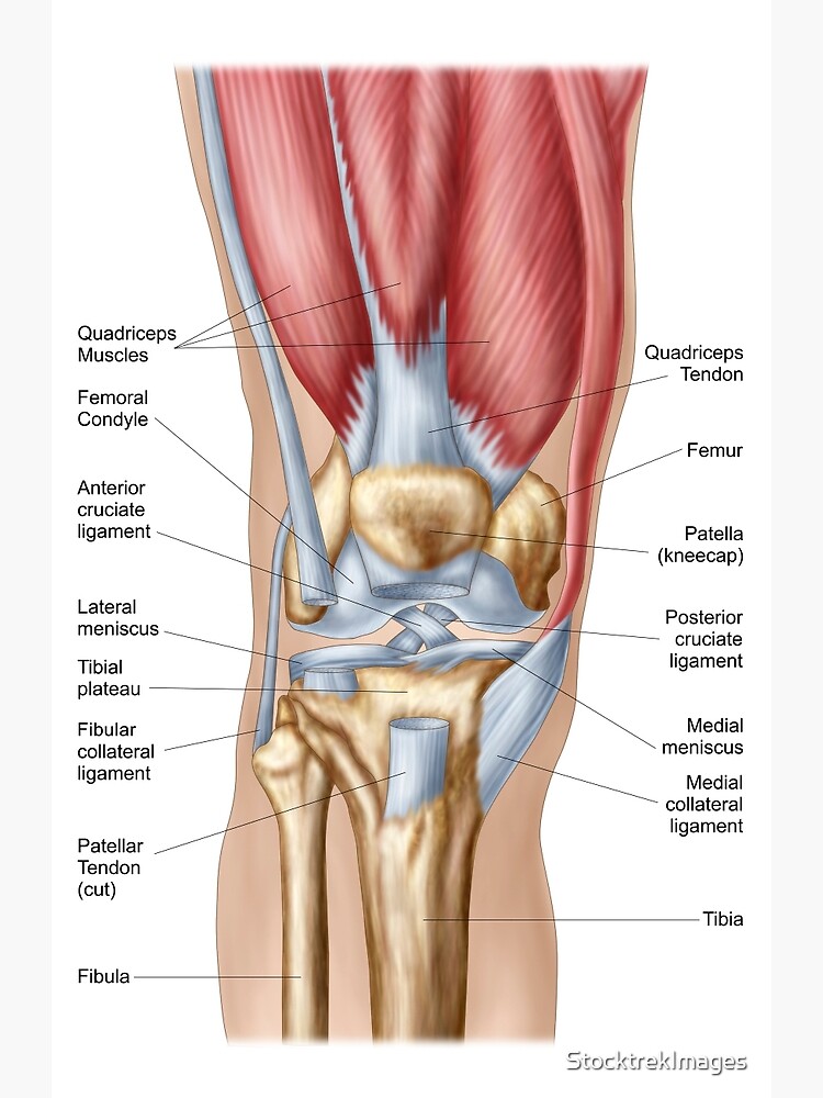
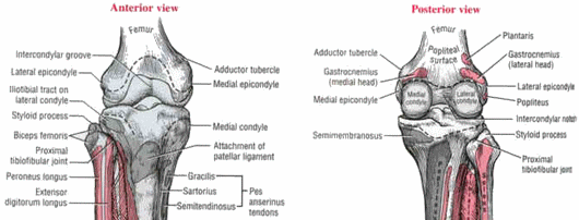
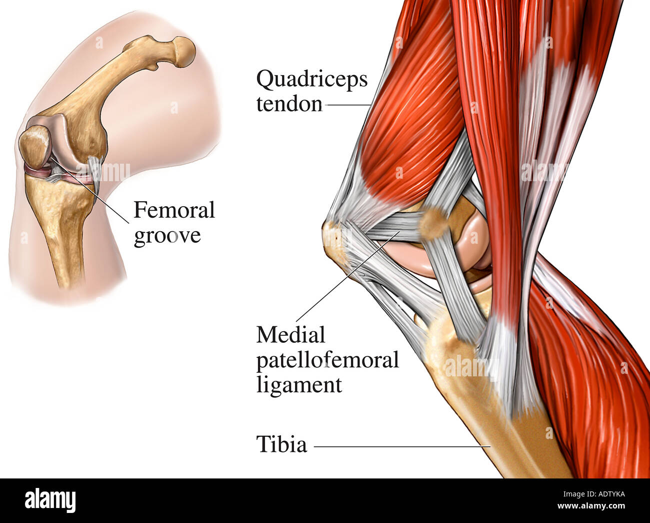
 Medial And Anterior Knee Anatomy Musculoskeletal Key
 Knee Physiopedia
 Knee Pain On The Inside Of Your Joint Causes Solutions
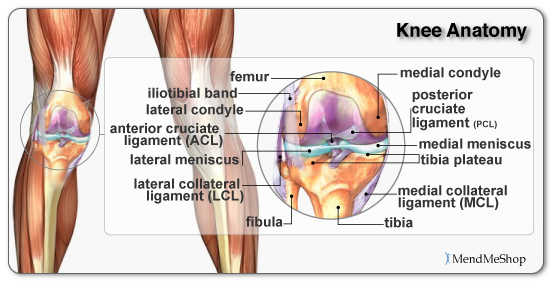
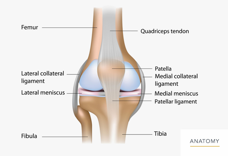
 Knee Anatomy
 Osteonecrosis Of The Knee Orthoinfo Aaos
 Applied Anatomy Of Knee Joint Epomedicine
 Ultrasound Guided Saphenous Adductor Canal Block Nysora
 Surgical Treatment Of Combined Acl And Medial Sided Knee
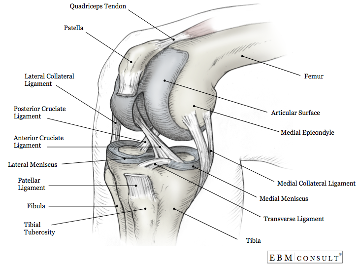
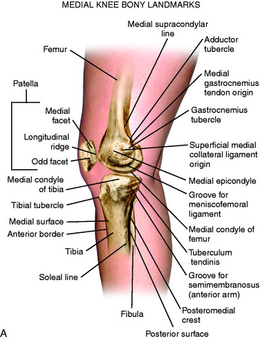
 Medial View Of The Knee
 The Knee Anatomy Carolineohare13
 The Unhappy Triad Anatomy Snippets Complete Anatomy

 Knee Calf Orthopedic Specialist Of Northern California
 Acl Vs Mcl Pcl Absolute Life Wellness Center
 Anatomy Of The Knee How The Knee Works Knee Anatomy
 Patella Approach Mid Axial Longitudinal Approach
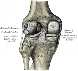
Anatomy Of Human Knee Joint Greeting Card
 Knee Joint Picture Image On Medicinenet Com
 Knee Joint Anatomy Bones Ligaments Muscles Tendons Function
 Acl Solutions Acl Knee Anatomy And Diagram Images
 4 Common Causes Of Knee Pain
 Medial And Anterior Knee Anatomy Musculoskeletal Key
 Patellofemoral Joint Physiopedia
 The Knee Joint Articulations Movements Injuries
 Knee Human Anatomy Function Parts Conditions Treatments
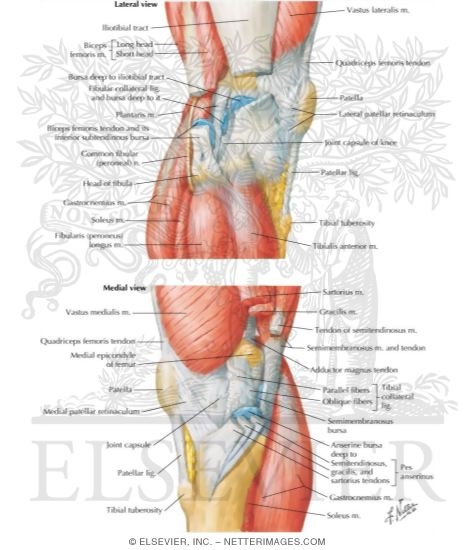 Knee Lateral And Medial Views
 Medial And Anterior Knee Anatomy Musculoskeletal Key
 Muscles Of The Knee Joint Medial View Stock Photo 7710841


Posting Komentar
Posting Komentar