In part ii we will discuss shoulder instability. Mri anatomy of shoulder dr.
 Shoulder Human Anatomy Image Function Parts And More
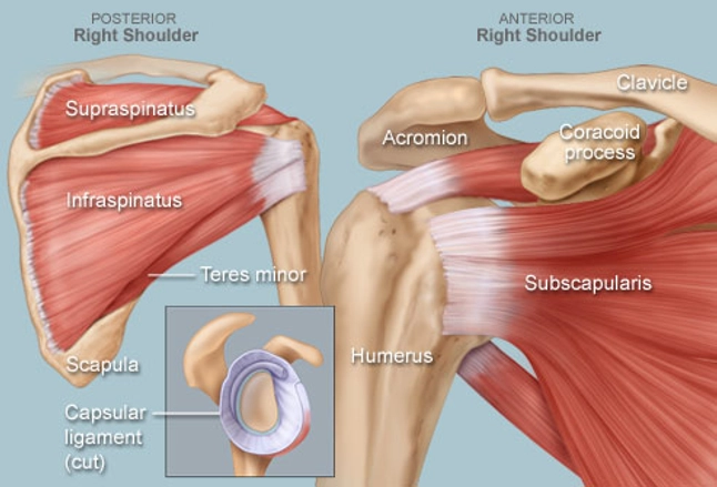
Simply put the shoulder or shoulder joint is the connection of the upper arm and the thoraxcomprising of numerous ligamentous and muscular structures the only actual bony articulations are the glenohumeral joint and the acromioclavicular joint acjthe shoulder allows for a large range of motion but is also more prone to dislocation and other injuries.

Shoulder anatomy mri. Confirmation of pathology in different planes and sequences increases diagnostic accuracy. For more information on shoulder anatomy please contact the office of dr. Mr is the best imaging modality to examen patients with shoulder pain and instability.
Knee shoulder shoulder arthrogram ankle elbow wrist hip. The joint is used very often so shoulder injuries are common in patients. Click on a link to get t1 axial view t2 fatsat axial view t1 coronal view t2 fatsat coronal view t2 fatsat sagittal view.
Use the mouse scroll wheel to move the images up and down alternatively use the tiny arrows on both side of the image to move the images on both side of the image to move the images. Atlas of shoulder mri anatomy. Mri of shoulder anatomy 1.
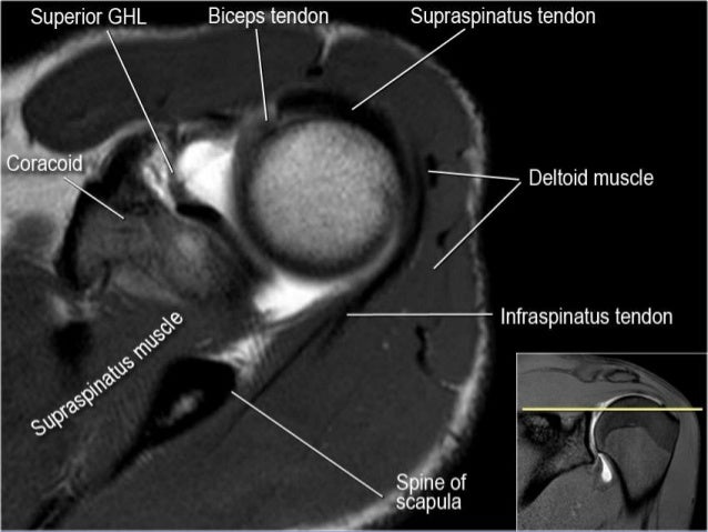
An mri of the shoulder of a healthy subject was performed in the 3 planes of space coronal axial sagittal commonly used in osteoarticular imagery with two weightings most commonly used to explore the musculo skeletal pathology of the shoulder. In part iii we will focus on impingement and rotator cuff tears. Nikhil verma in chicago illinois.
Use the mouse to scroll or the arrows. Spin echo t1 and proton density with fat saturation sequences. The anatomy of the shoulder is complex but the shoulder joint allows a wide range of movement.
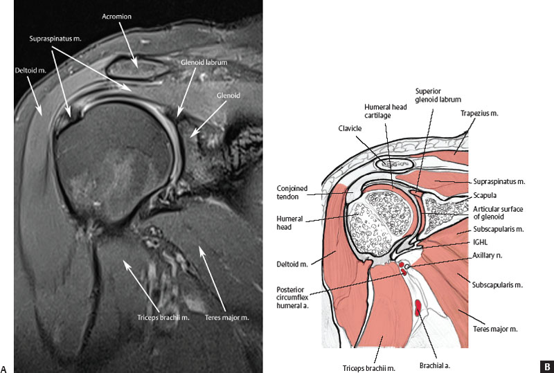
The shoulder in a conventional mri exam is acquired in partial external rotation in the axial coronal and sagittal planes. Magnetic resonance imaging. An mri scanner uses a high powered magnet and a computer to create high resolution images of the shoulder and surrounding structures.
This webpage presents the anatomical structures found on shoulder mri. In shoulder mr part i we will focus on the normal anatomy and the many anatomical variants that may simulate pathology. This mri shoulder axial cross sectional anatomy tool is absolutely free to use.
Muhammad bin zulfiqar pgr iii fcps new radiology department services hospital services institute of medical sciences special thanx to radiology assistant 2.
 Shoulder Anatomy And Normal Variants
 Mri Of Shoulder Anatomy
 Normal Anatomy Variants And Pitfalls On Shoulder Mri
 Normal Mri Anatomy Of The Musculoskeletal System Radiology Key
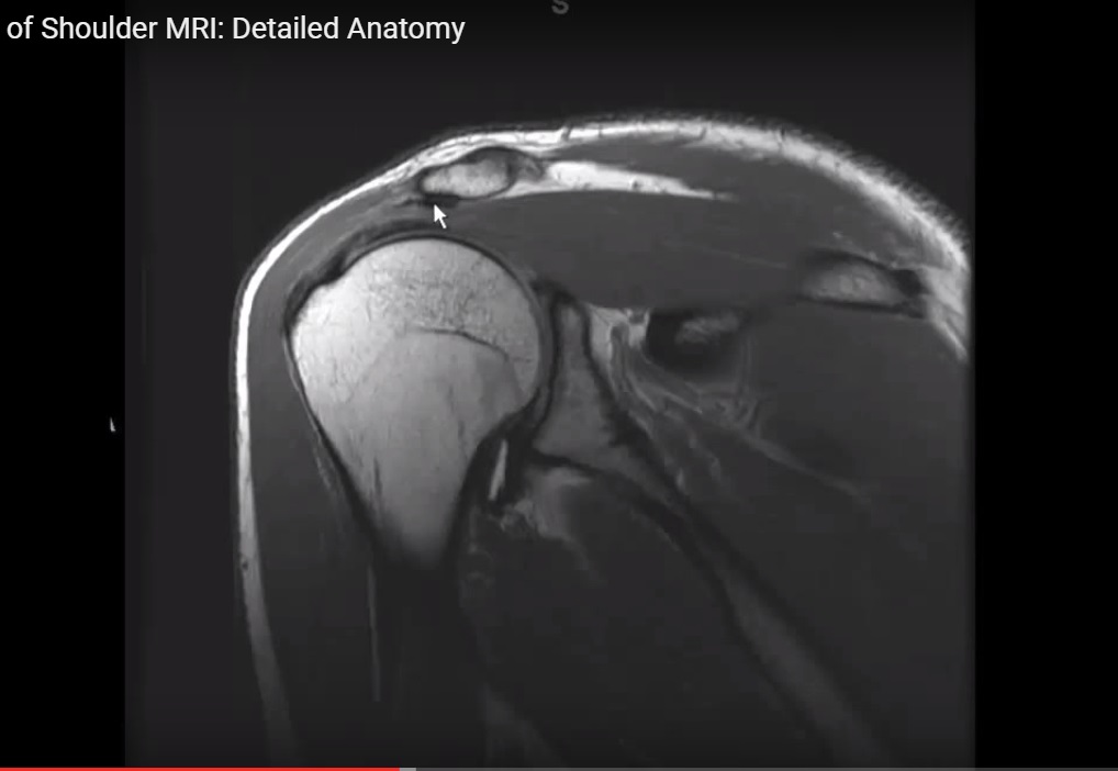
 Figure 4 From Normal And Variant Anatomy Of The Shoulder On

 Shoulder Mri For Rotator Cuff Tears
Mri Findings For Frozen Shoulder Evaluation Is The
Stanford Msk Mri Atlas C 2019
 Teaching Files University Of North Dakota
 Shoulder Radiology Key

 Mri Anatomy Of The Shoulder Ppt Video Online Download
 Radiology Anatomy Images Mri Shoulder Anatomy
 Mri Shoulder Anatomy Shoulder Coronal Anatomy Free Cross
Shoulder Anatomy Mri Shoulder Axial Anatomy Free Cross
 The Radiology Assistant Shoulder Mr Anatomy
 Shoulder Mri Basic Axial Anatomy

 Shoulder Mri Radiographical And Illustrated Anatomical Atlas
 Mri Of Shoulder Anatomy
 Mri Shoulder Anatomy Shoulder Coronal Anatomy Free Cross

 Shoulder Mri
 Supraspinatus Muscle Radiology Reference Article
 Shoulder Mri Radiographical And Illustrated Anatomical Atlas

 Msk Clinical Cases Traumatology Ppt Video Online Download


Posting Komentar
Posting Komentar