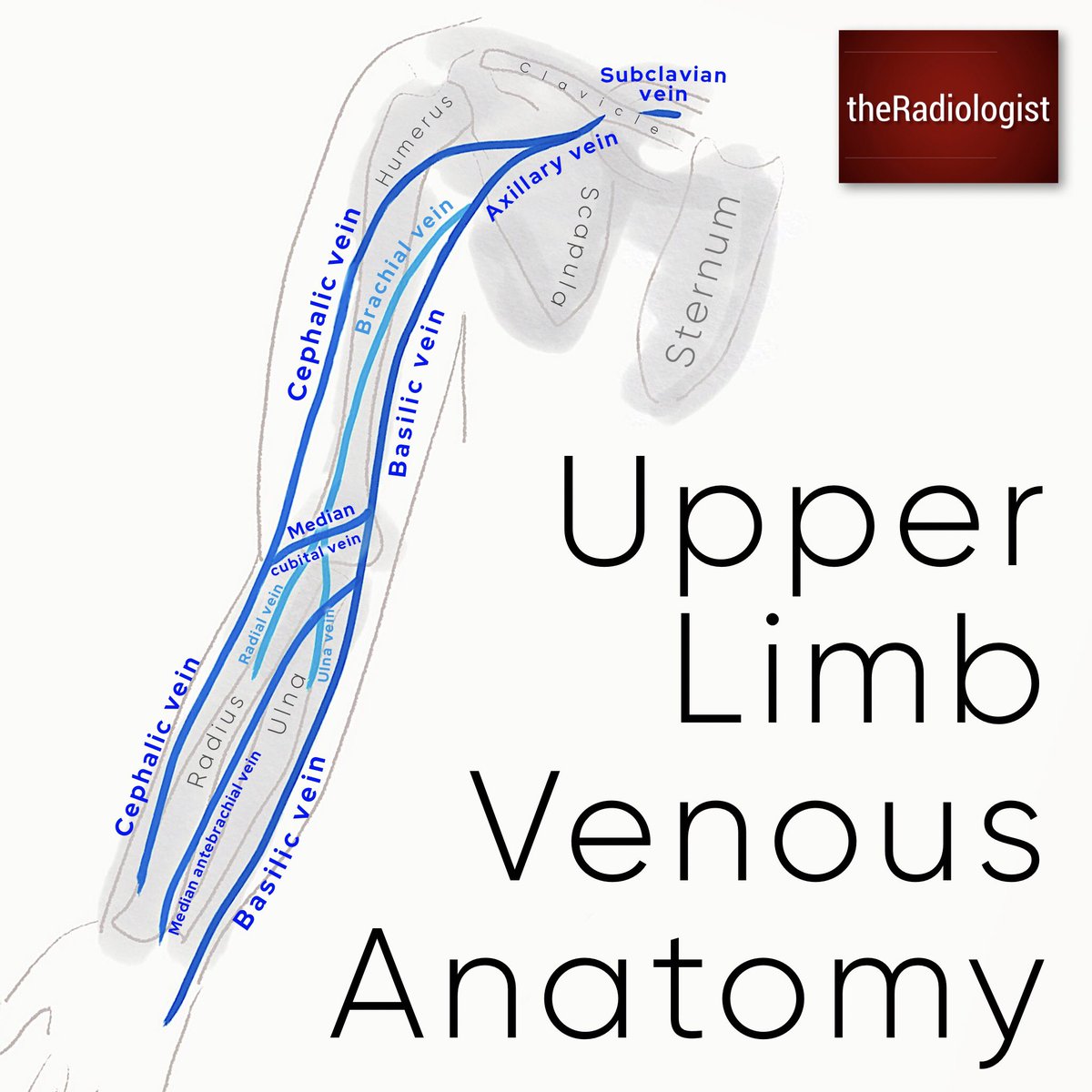
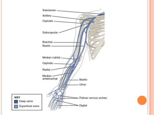
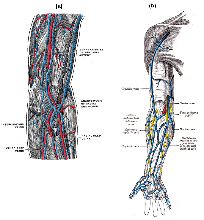
The veins of the upper extremity are divided into two sets superficial and deep. The deep veins accompany the arteries and constitute the venæ comitantes of those vessels.
 The Veins Of The Upper Extremity And Thorax Human Anatomy
Well start by looking at the superficial venous system and well start distally.
Upper extremity venous anatomy. The deep veins follow the same course as the arteries of the upper limb. In the upper extremity the deep veins share the name of the artery they accompany. Chronic venous disease may affect the upper extremity after an acute thrombotic event of any cause or in any patient with longterm catheterization.
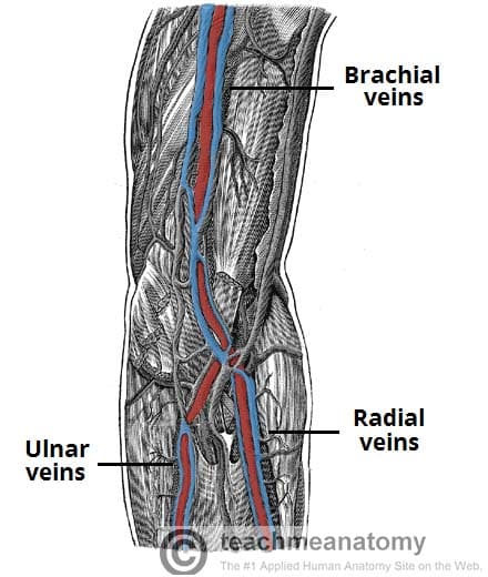
Vena comitantes and superficial veins. Upper limb dvt ultrasound normal anatomy basic deep venous anatomy of the arm. The brachial veins are the largest in size and are situated either side of the brachial artery.
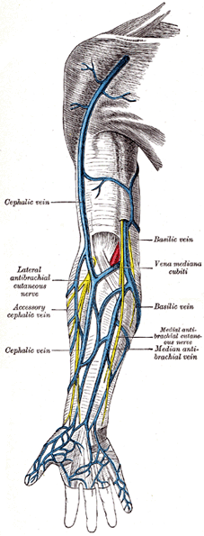
This vein as well as the deep veins act as counterparts to the arteries supplying the arm by bringing deoxygenated blood back to the heart. The diagnosis of chronic venous disease is considerably more challenging than acute venous disease because enlarged thrombusfilled veins are not present. Dorsal digital veins dorsal metacarpal veins palmar digital veins intercapitular veins dorsal venous network palmar venous network cephalic vein basilic vein median antebrachial vein.
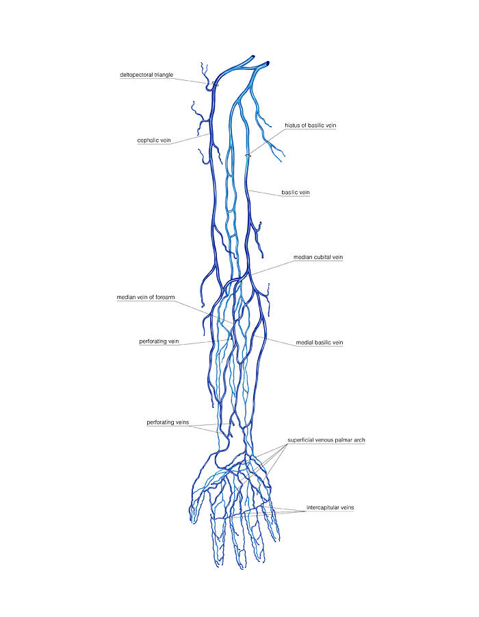
The superficial veins are placed immediately beneath the integument between the two layers of superficial fascia. Chronic upper extremity venous disease. The deep venous system of the upper limb is situated underneath the deep fascia.
There is more of a variation in the anatomy of the upper extremity. In the hand forearm and upper arm the superficial system functions as the principal means for venous drainage. True most of this variation involves the median cubital veins and their connection with the basilic and cephalic veins.
The two sets anastomose frequently with each other. Veins for the upper extremity direct blood flow from the hand wrist forearm upper arm and shoulder to the ipsilateral central thorax veins and ultimately the superior vena cava. It is formed by paired veins which accompany and lie either side of an artery.
Veins of the upper extremities are grouped into deep veins which are accompanying veins of arteries from which they derive their names latin. Basic superficial venous anatomy of the arm. The venous drainage of the upper limb consists of a superficial system which drains to a higher pressure deep venous system.
The hand is a very mobile part of the upper limb and we perform very specialised tasks with it every day key adaptations can be seen in the specialised structures of the hand.
 Theradiologist On Twitter Diagram Simplified Diagram Of
 Ultrasonography For Deep Venous Thrombosis Radiology Key
Venous System Of Upper Limb
 Ultrasound Guided Peripheral Intravenous Access Pocket
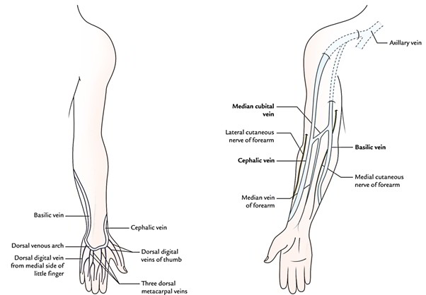
 Venous Drainage Of The Upper Limb Basilic Cephalic
 Venous Lymphatic Drainage Of Upper Limb
![]() Veins Of The Upper Limb Anatomy Kenhub
Veins Of The Upper Limb Anatomy Kenhub
 Upper Limb Anatomy
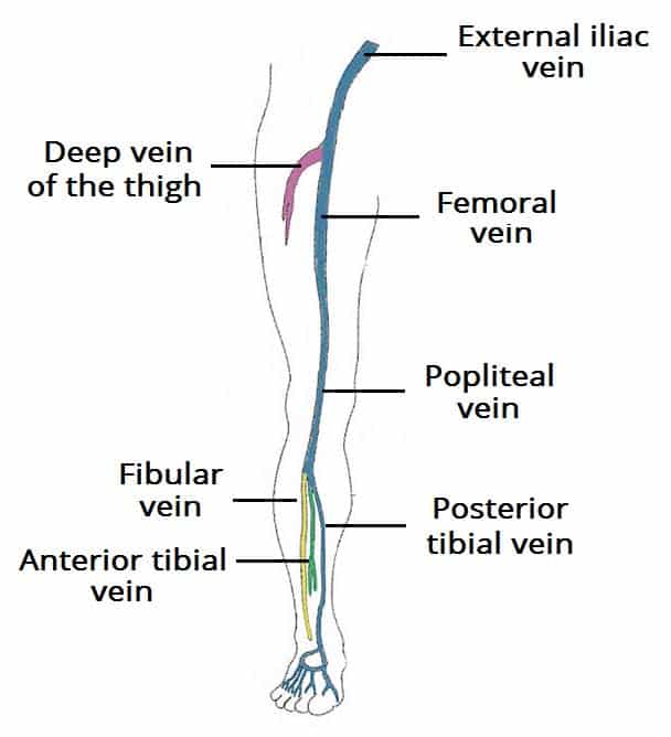 Venous Drainage Of The Lower Limb Teachmeanatomy
 Easy Notes On Venous Drainage Of The Upper Limb
 Emergency Ultrasound
Superficial Venous Palmar Arch
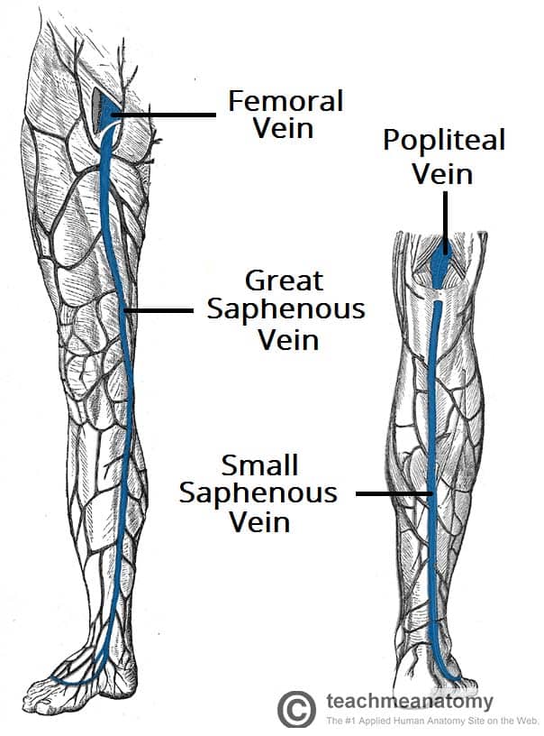 Venous Drainage Of The Lower Limb Teachmeanatomy
 Ultrasound Evaluation Of The Peripheral Vascular System
 Upper Extremity Venous Exam Technique And Interpretation
 Head And Neck Venous Anatomy Vascular And Non Vascular
 Arm Dvt Normal Ultrasoundpaedia
 Image Result For Ultrasound Upper Extremity Venous Anatomy
 Online Cme Upper Extremity Venous Evaluation
 Upper Extremity Venous Doppler Sonographic Tendencies
 Upper Extremity Venous Thrombosis
 Venous Sonography Of The Upper Extremities And Thoracic
 Leg Dvt Normal Ultrasoundpaedia
 Ecr 2014 C 1039 Normal Vascular Variants Of The Upper
 Venous Anatomy And Upper Extremity
 Dentistry And Medicine Blood Supply Venous Drainage
 Arm Dvt Normal Ultrasoundpaedia
 Venous System Of The Upper Limb Artwork Stock Image
 Upper Extremity Venous Imaging Alexander Street A
 Upper Limb Veins Illustrations Radiology Case
 Arm Venous Anatomy Physiology Module Sonosim
 Cephalic Vein Wikipedia
 Circulatory Routes Boundless Anatomy And Physiology


Posting Komentar
Posting Komentar