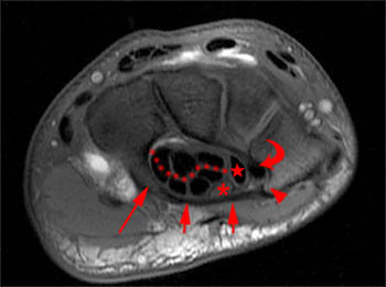
3 extensor carpi ulnaris t. Wrist ligaments are best assessed with dedicated wrist mri.
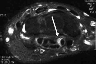 Carpal Tunnel Syndrome Imaging Practice Essentials
Hobby jl dixon ak bearcroft pw et al.

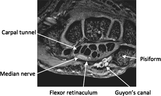
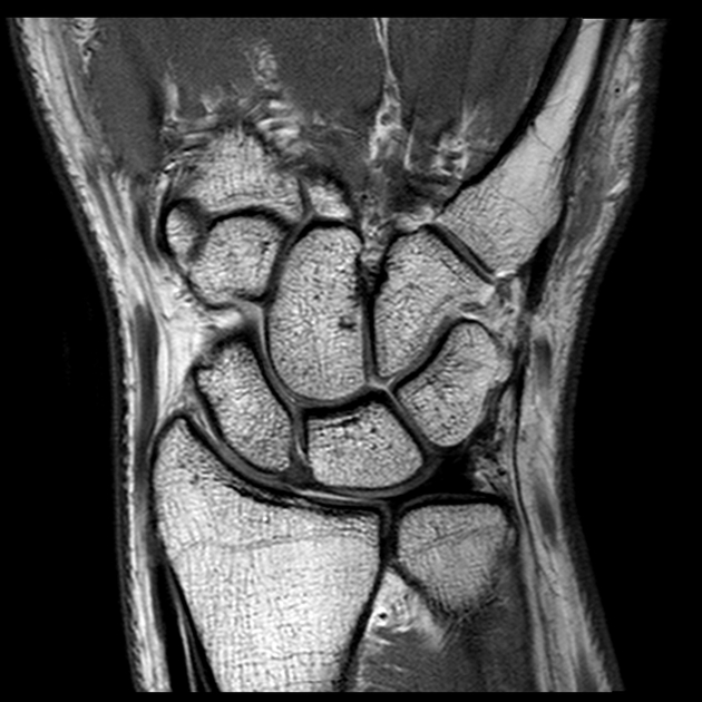
Wrist anatomy mri. In recent years magnetic resonance imaging mri has become a very important modality for diagnosing wrist and hand diseases including osteoarthritis rheumatoid arthritis ra occult fracture avascular necrosis avn ligamentoustendinous injuries impaction syndrome and nerve entrapment syndrome. 4 extensor digiti minimi t. Mr imaging of the wrist.
Use the mouse to scroll or the arrows. The intrinsic and extrinsic wrist ligaments play a vital role in the stability of the wrist joint. Mri of the wrist allows physicians to examine the wrist anatomy to rule out any structural abnormalities.
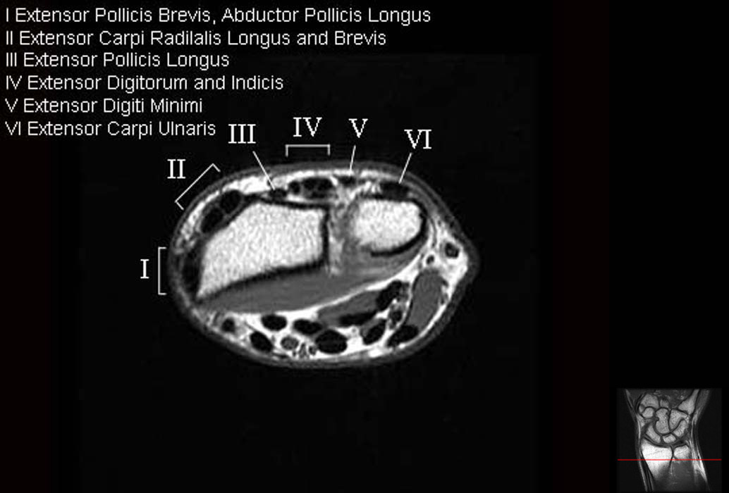
5 extensor digitorum indicis tt. There are numerous ligaments but included below are the most clinically significant. 8 extensor carpi radialis brevis t.
6 extensor pollicis longus tendon. Mri uses a magnetic field radio waves and a computer to create images soft tissues bones and internal body structures. Use the mouse scroll wheel to move the images up and down alternatively use the tiny arrows on both side of the image to move the images.
Understanding the complex anatomy of the wrist and more common disease of the ligamentous osseous and tendinous structures allows the radiologist to efficiently and accurately evaluate mri of the wrist with improved diagnostic capabilities. This mri wrist axial cross sectional anatomy tool is absolutely free to use. 1 2 mri is a noninvasive and nonirradiative imaging tool and can provide high soft tissue contrast resolution.
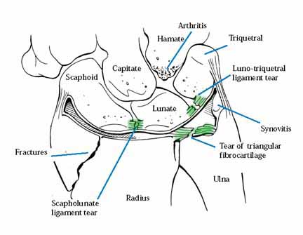
Although much attention is paid to the scapholunate ligament lunotriquetral ligament and the triangular fibrocartilage complex additional intrinsic and extrinsic ligaments in the wrist play an important part in carpal stability. Your doctor has ordered a mri magnetic resonance imaging of your wrist. Mri of the wrist.
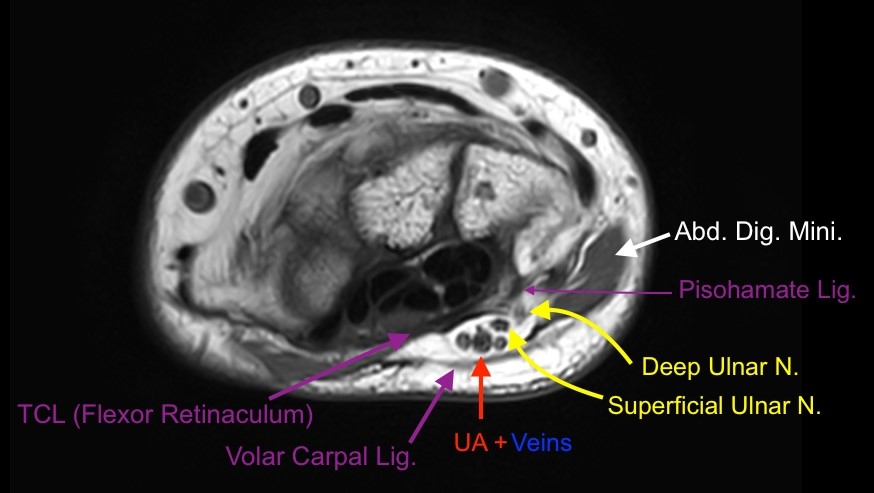
1 flexor carpi ulnaris m t. Effect on clinical diagnosis and patient care. With improved mri techniques the radiologist can increasingly visualize these ligaments.
To sum up mri of the wrist is a relevant tool for diagnosis and clinical management of wrist pain including the evaluation of traumatic injuries and chronic syndromes. This ultimately leads to more efficient treatment and better patient outcomes. All the ligaments of the wrist visible in mri are shown on this anatomical module including collateral ligaments the radiocarpal and ulnocarpal ligaments as well as the intercarpal ligaments.
9 extensor carpi radialis longus t.
 Wrist Mri

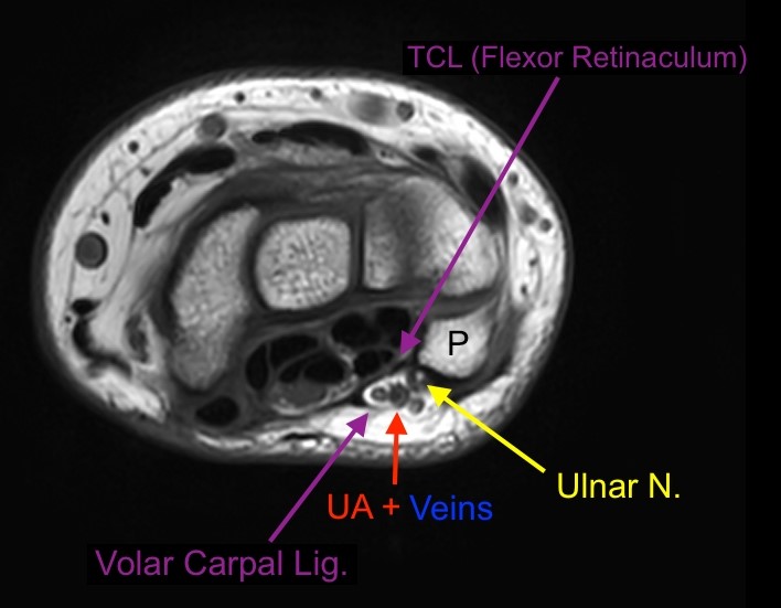
Wrist Anatomy Mri Wrist Axial Anatomy Free Cross

 The Radiology Assistant Wrist Carpal Instability
 Normal Proton Density Pd Weighted Sagittal Mri Of The
 Ulnar Sided Wrist Pain Part I Anatomy And Physical
 Normal Wrist Mri Radiology Case Radiopaedia Org

 Sports Related Extensor Carpi Ulnaris Pathology A Review Of

 Wrist Magnetic Resonance Imaging Anatomy T1 Weighted Axial
 Mri Wrist Anatomy
 The Hand Mr Medical Imaging Anatomy Atlas
 Mri Wrist Coronal Anatomy Wrist Tendon And Ligaments
 Mri Anatomy Of Tfcc
 Dorsal Wrist Impingement Raleigh Hand Surgery Joseph J

Mri Of The Extensor Tendons Of The Wrist
 Scaphoid Bone An Overview Sciencedirect Topics
Mri Of The Extensor Tendons Of The Wrist
 Ct Mri Wrist Anatomy Dr Ahmed Esawy
 Wrist Anatomy Mri Wrist Axial Anatomy Free Cross
 Wrist Anatomy Mri Wrist Axial Anatomy Free Cross
Magnetic Resonance Imaging Of The Hand And Wrist In A
![]() Mri Types Indications Contraindications Advantages Kenhub
Mri Types Indications Contraindications Advantages Kenhub
 Musculoskeletal Mri
Analysis Of Mr Imaging Of Wrists In Female Patients With


Posting Komentar
Posting Komentar