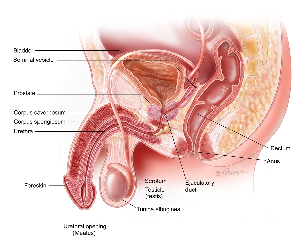
It is situated in the region called the bladder base. The greater part of this surface is directly continuous with the bladder wall.
 Prostatitis Infection Of The Prostate Symptoms Diagnosis
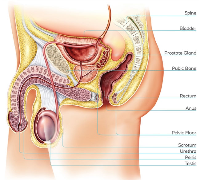
It is the size of a walnut and anatomically sits in front of the rectum and between the bladder and the penis.
Prostate bladder anatomy. The apex is directed downward and is in contact with the superior fascia of the. The prostate gland is part of the male reproductive system. The median lobe is found just posterior to the urethra along the midline of the prostate.
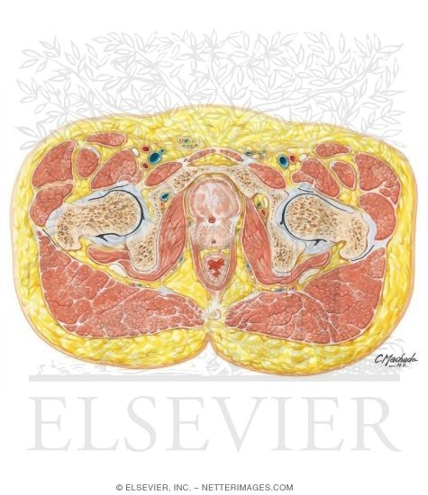
Cross sectional anatomy of the male pelvis on mr imaging prostate bladder genital organs rectum male pelvis. The volume of the prostate can be estimated by the formula 052 length width height. Within it sits the urethra coming from the bladder which is called the prostatic urethra and which merges with the two ejaculatory ducts.
Apex of the prostate. The median umbilical ligament is a fibrous cord anterior to the bladder and develops from the urachus. The prostate makes a protein called psa.
The prostate gland is an inverted pyramid that surrounds the proximal urethra which traverses the prostate close to its anterior surface. Tubes called vasa deferens are conduits for sperm that move from the testes to the seminal vesicles. Anatomy of the prostate base of the prostate.
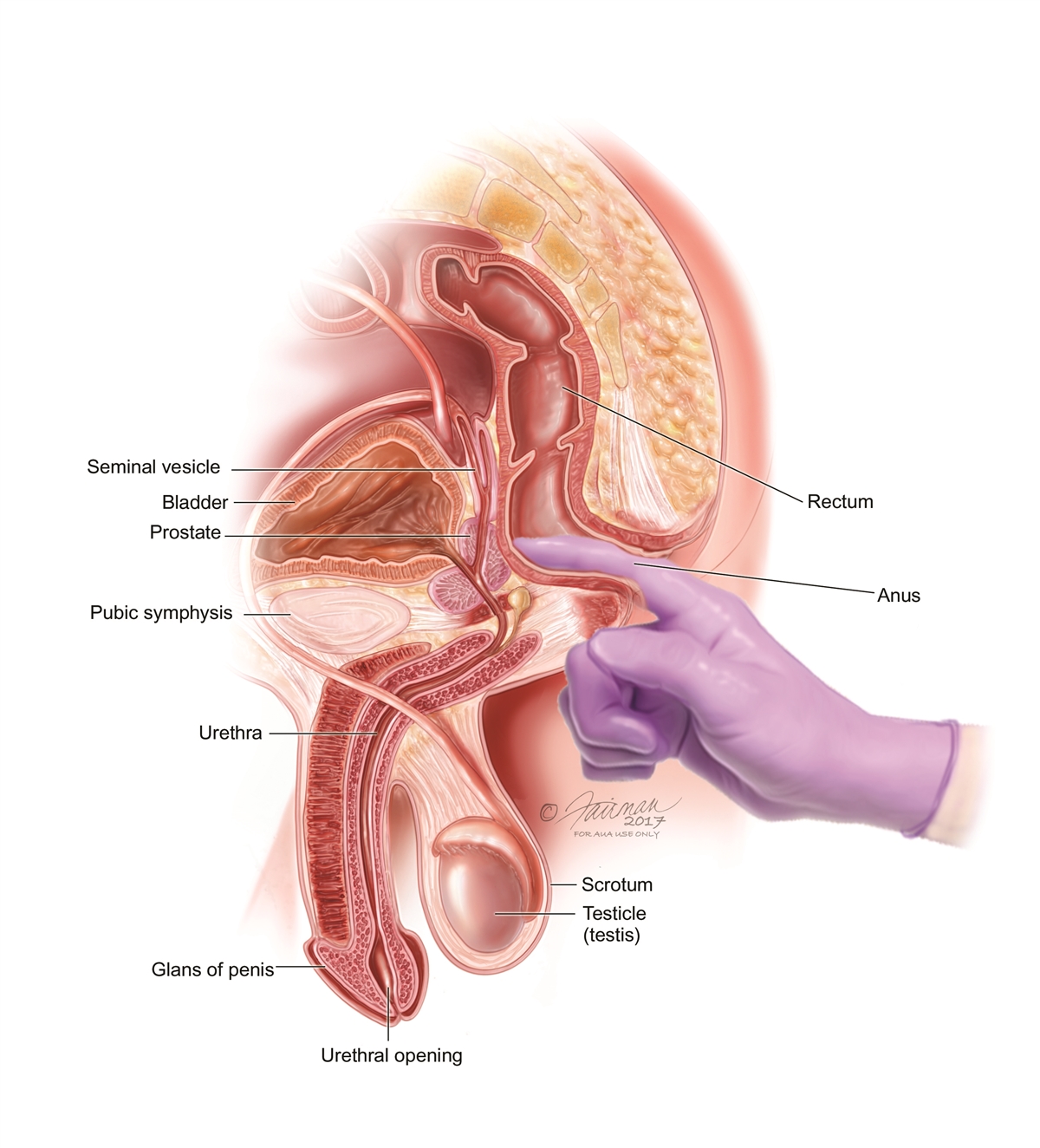
Most importantly posteriorly to the prostate lies the ampulla of the rectum this anatomical arrangement is utilised during digital rectal examinations dre allowing physicians to examine the gland. Prostate tests digital rectal examination dre. The mean weight of the normal prostate in adult males is about 11 grams usually ranging between 7 and 16 grams.
It has a superior base inferior apex and anterior inferolateral and posterior surfaces. The base is directed upward near the inferior surface of the bladder. Anatomy of the male pelvis prostate bladder genital organs perineum on mr imaging.
Anatomy of the bladder and prostate the urinary bladder. The base of the prostate is in continuity with the bladder. On the anterior end of the prostate are the two lateral lobes.
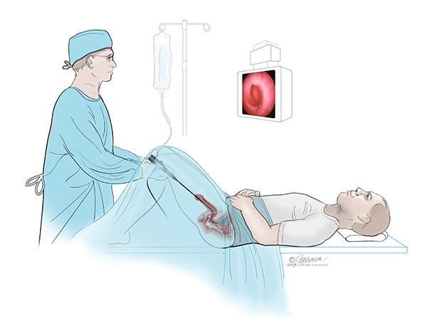
Prostate ultrasound transrectal ultrasound. Zonal anatomy of prostate mcneal mri. A needle is inserted.
Mri coronal section. Posterior and medial to the lateral lobes is the much smaller anterior lobe. The prostate gland is a mass of tissue just below the urinary bladder in males about the size of a walnut.
An ultrasound probe is inserted into the rectum. Sperm are generated in the testes. The prostate surrounds the urethra.
Several distinct lobes make up the structure of the prostate. The prostate is positioned inferiorly to the neck of the bladder and superiorly to the external urethral sphincter with the levator ani muscle lying inferolaterally to the gland. The bladder develops from the urogenital sinus but the base or bladder trigone is of mesonephric duct origin.
A doctor inserts a lubricated.
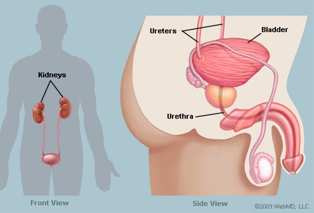
 Anatomy Of The Male Urinary Tract
Holmium Laser Enucleation Of The Prostate Holep
 Patient Education Benign Prostatic Hyperplasia Bph
 If You Have Prostate Cancer
 Prostate Cancer Signs Diagnosis Treatment Understanding
 What Is Prostatitis Symptoms Causes Treatment Antibiotics
 Bladder
 Prostate Wikipedia
 What Is Ultrasound Imaging Urology Care Foundation
 The Bladder Human Anatomy Function Picture Location
 The Prostatitis Foundation Urethral Strictures
 Male Pelvis Bladder Prostate Junction
Painful Urination Dysuria Cleveland Clinic
 Bladder Cancer Treatment Bladder Cancer Pictures Signs
 Amazon Com Antique Print Anatomy Urinary Bladder Prostate
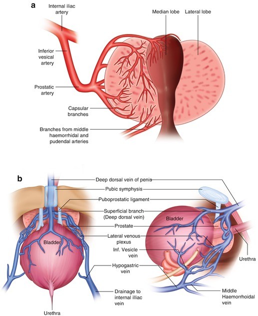 Anatomy Physiology And Pathology Of The Large Prostate
 Men How To Insert A Catheter
 Benign Prostatic Hyperplasia Wikipedia
Prostatic Urethral Lift Cleveland Clinic
 What Is Prostate Cancer Koelis
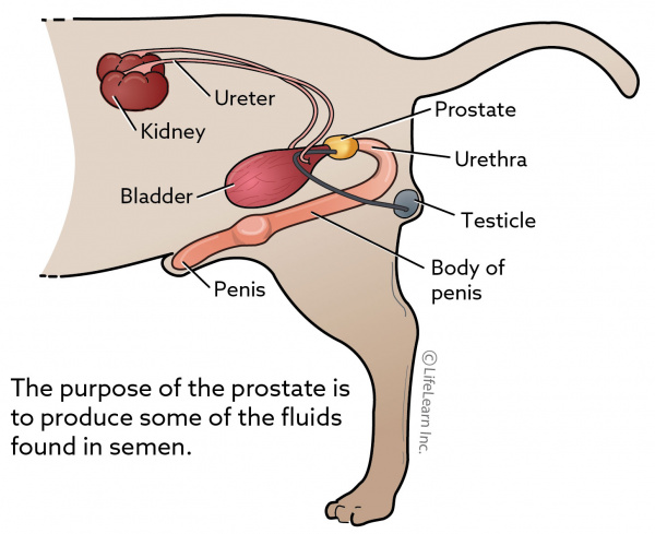 Prostatic Disease In Dogs Vca Animal Hospital

 Prostatitis Infection Of The Prostate Symptoms Diagnosis
 Benign Prostate Disorders Endotext Ncbi Bookshelf
Male Reproductive System Information Cleveland Clinic
 Prostate Anatomy Centro Gil Vernet De Urologia
 Figure Anatomy Of The Male Reproductive Pdq Cancer


Posting Komentar
Posting Komentar