Capitate trapezium and flexor retinaculum. Anatomy of the middle finger metacarpal.
Adult Thumb Metacarpal Fractures Midwest Bone And Joint
1st metacarpal radial border adductor pollicis origin.

Anatomy of metacarpals. This metacarpal has multiple articular facets on its base which is shaped somewhat like a styloid process with an articular facet on the radial dorsal surface for articulating with the distal carpal bone capitate. The metacarpus of the hand is composed of five bony structures known as the metacarpals. Theyre conventionally numbered 1 to 5 from lateral radial to medial ulnar side.
They consist of a proximal base a shaft and a distal head. The articulation with the hamate and the fourth and fifth metatarsal bases allows for much freer movement but the cmc and metacarpal ligaments are still very strong. Capitate base and bases of 2nd and 3rd metacarpal.
The insertion of the flexor carpi radialis at the base of the second metacarpal contributes as well. They can be divided into three categories. The second with the trapezium trapezoid capitate and third metacarpal.
Besides the metacarpophalangeal joints the metacarpal bones articulate by carpometacarpal joints as follows. The metacarpus is composed of 5 metacarpal bones. They consist of a proximal base a shaft and a distal head.
The first with the trapezium. Capitate base and bases of 2nd and 3rd metacarpal. The metacarpals together are referred to as the metacarpus the tops of the metacarpals form the knuckles where they join to the wrist.
Theyre conventionally numbered 1 to 5 from lateral radial to medial ulnar side. Each finger has three phalanges. The metacarpal bones form the skeleton of the palm articulating proximally with the carpus and distally with the digits.
The fourth with the. Carpal bones proximal a set of eight irregularly shaped bones. These are located in the wrist area.
Muscular attachments in the metacarpal area. On the palm side they are covered with connective tissue. Metacarpals there are five metacarpals each one related to a digit.
The third with the capitate and second and fourth metacarpals. Anatomy of the metacarpals parts of a metacarpal. They are five in number one for each digit and lie side by side and slightly divergent from each other being separated by intervals called interosseous spaces.
Scaphoid bone and trapezium flexor retinaculum. Phalanges distal the bones of the fingers. The metacarpals are long bones within the hand that are connected to the carpals or wrist bones and to the phalanges or finger bones.
 Handcare Org Anatomy Photo Gallery
Adult Thumb Metacarpal Fractures Midwest Bone And Joint
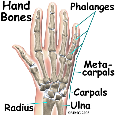 Physical Therapy In Lansing For Hand Anatomy
:background_color(FFFFFF):format(jpeg)/images/library/4687/5Pyr4Ltbc4LJ5MxszANmmw_First_metacarpal_bone_02.png) Metacarpal Bones Anatomy And Clinical Aspects Kenhub
Metacarpal Bones Anatomy And Clinical Aspects Kenhub
 Hand Bones And Wrist Bones Mnemonics Anatomy And Physiology
 Bones Of The Arm And Hand Interactive Anatomy Guide
 Metacarpus Human Skeleton 3d Model Stock Illustration
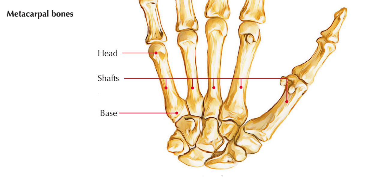 Easy Notes On Bones Of The Hand Learn In Just 3 Minutes
 Hand Bones Phalanges Metacarpals And More Preview Human Anatomy Kenhub
 Bison Metacarpals And Carpals Anatomy Veterinary Studies
 7 6c Carpals Metacarpals And Phalanges The Hand
 Hand And Finger Bones Kirkland Wa Evergreenhealth
 Paint Draw Paint Learn To Draw Anatomy Basics Metacarpals
 Anatomy Of Hand Wrist Bones Muscles Tendons Nerves
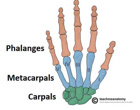 Bones Of The Hand Carpals Metacarpals Phalanges
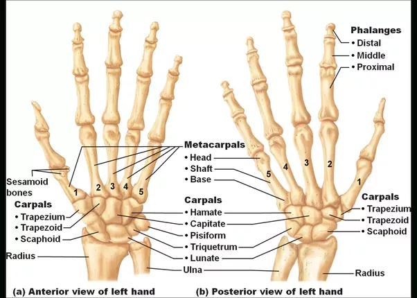 Hand Bones Carpals Metacarpals Phalanges Anatomy Info
First Metacarpal Musculoskeletal Skeletal Anatomyzone
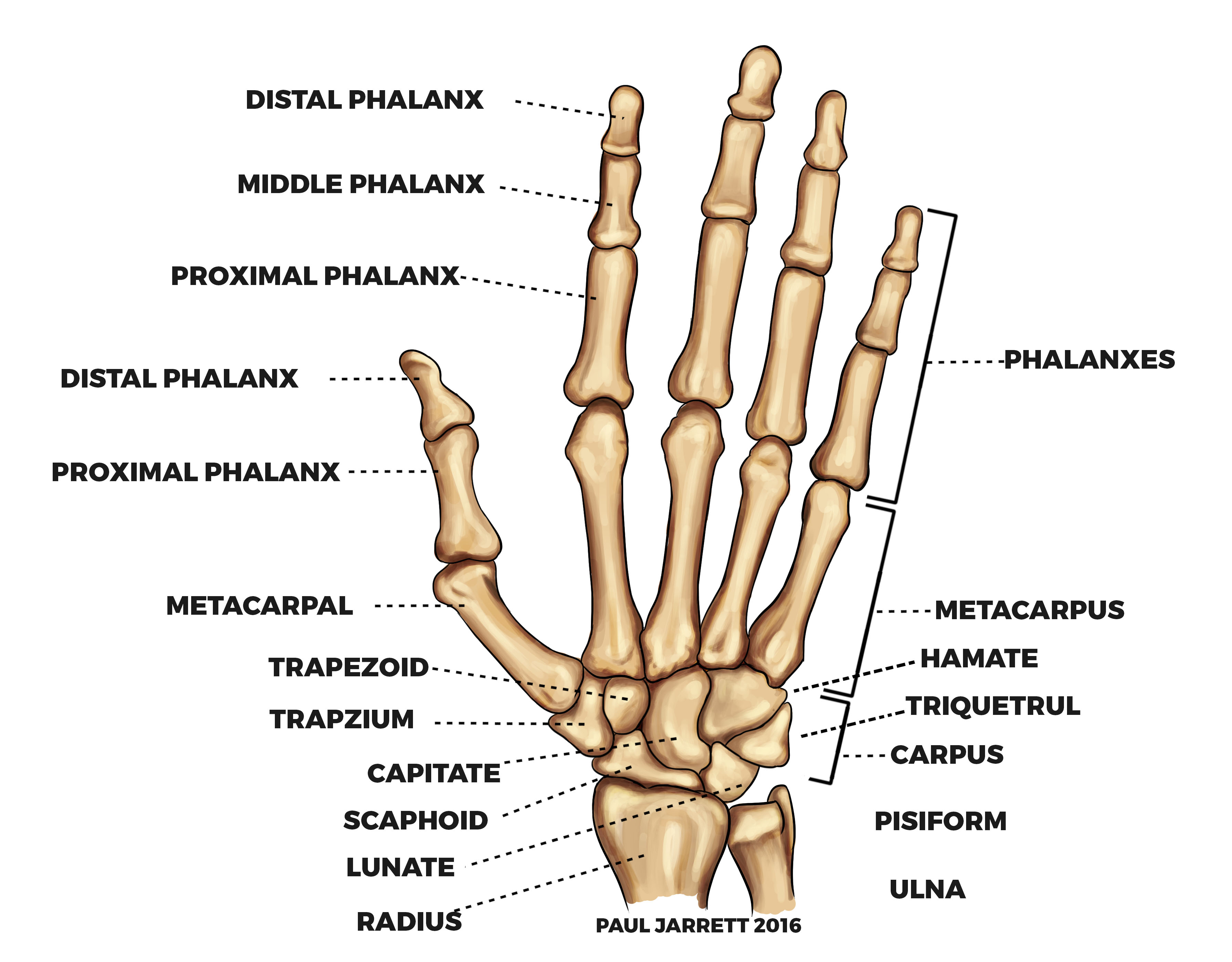 Hand And Wrist Anatomy Murdoch Orthopaedic Clinic
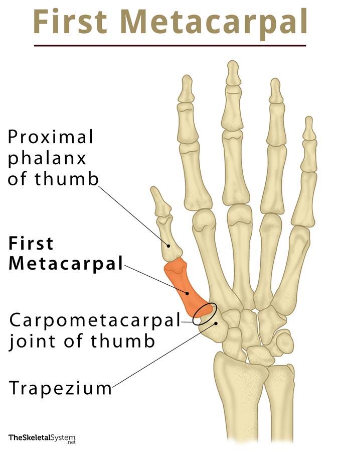 First Metacarpal Definition Location Anatomy Diagram
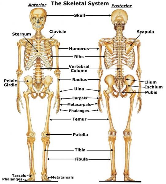 Scma Specialty Certified Medical Assistant
Anatomy 101 Finger Bones The Handcare Blog
 Anatomy Of The Hand Palmar View Doctor Stock

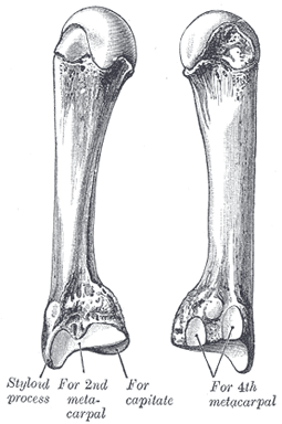
Posting Komentar
Posting Komentar