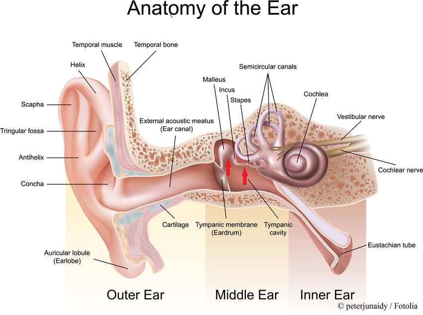
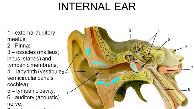
Sound travels through the auricle and the auditory canal a short tube that ends at the eardrum. The skin of the ear canal is very sensitive to pain and pressure.
 Anatomy And Physiology Of Ageing 6 The Eyes And Ears
Under the skin the outer one third of the canal is cartilage and inner two thirds is bone.

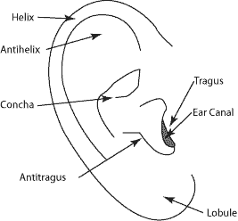
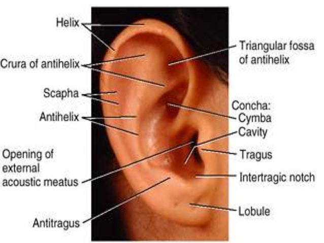
External ear anatomy. The asymmetric shape of the external auricle introduces delays in the path of sound that assist in sound localization. The auricle and external acoustic meatus or external auditory canal compose the external ear. Ear anatomy outer ear.
The external ear can be divided functionally and structurally into two parts. The outer ear external ear or auris externa is the external portion of the ear which consists of the auricle also pinna and the ear canal. In human hearing sound waves enter the outer ear and travel through the external auditory canal.
The canal is approximately an inch in length. This article will focus on the anatomy of the external ear its structure neurovasculature and its clinical correlations. The auricle or pinna and the external acoustic meatus which ends at the tympanic membrane.
The motion of the stapes against the oval window sets up waves in the fluids of the cochlea. It gathers sound energy and focuses it on the eardrum tympanic membrane. The ear has outer middle and inner parts.
Ear canal the ear canal starts at the outer ear and ends at the ear drum. The outer ear consists of three parts. The first is an outer funnel like structure called the auricle or pinna.
The external ear functions to collect and amplify sound which then gets transmitted to the middle ear. The outer ear includes. The ear also functions in the sense of equilibrium.
Auricle cartilage covered by skin placed on opposite sides of the head auditory canal also called the ear canal eardrum outer layer also called the tympanic membrane the outer part of the ear collects sound. When the waves reach the tympanic membrane they cause the membrane and the attached chain of auditory ossicles to vibrate.
 Ear Picture Image On Medicinenet Com
 Ear Anatomy Tragus Human Anatomy For The Artist The External
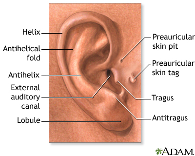 Medical Findings Based On Ear Anatomy Medlineplus Medical
 Ear Anatomy Pediatric Ear Disorder Treatment Ent For Children
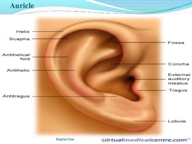 Anatomy And Physiology Of Ear By Maghan Das

 Human Ear Structure Function Parts Britannica
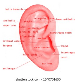 Outer Ear Images Stock Photos Vectors Shutterstock
![]() Basic Human Ear Anatomy And Physiology Outer Middle And
Basic Human Ear Anatomy And Physiology Outer Middle And
 Chapter 47 Diseases Of The External Ear Current Diagnosis
Medicine Decoded External Ear Anatomy
 Types Of Hearing Impairment University Of Iowa Hospitals
Anatomy Of The Ear Audiologist In Littleton
 Anatomy Of External Ear D Alessandro 2012 Download
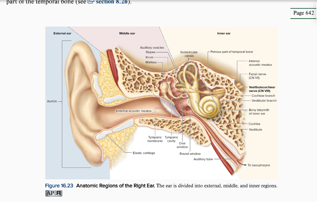 Solved 1 Study The Diagram Of The Anatomy Of The Ear Abo
 The External Ears Are Made Of Cartilage And Skin There Is A
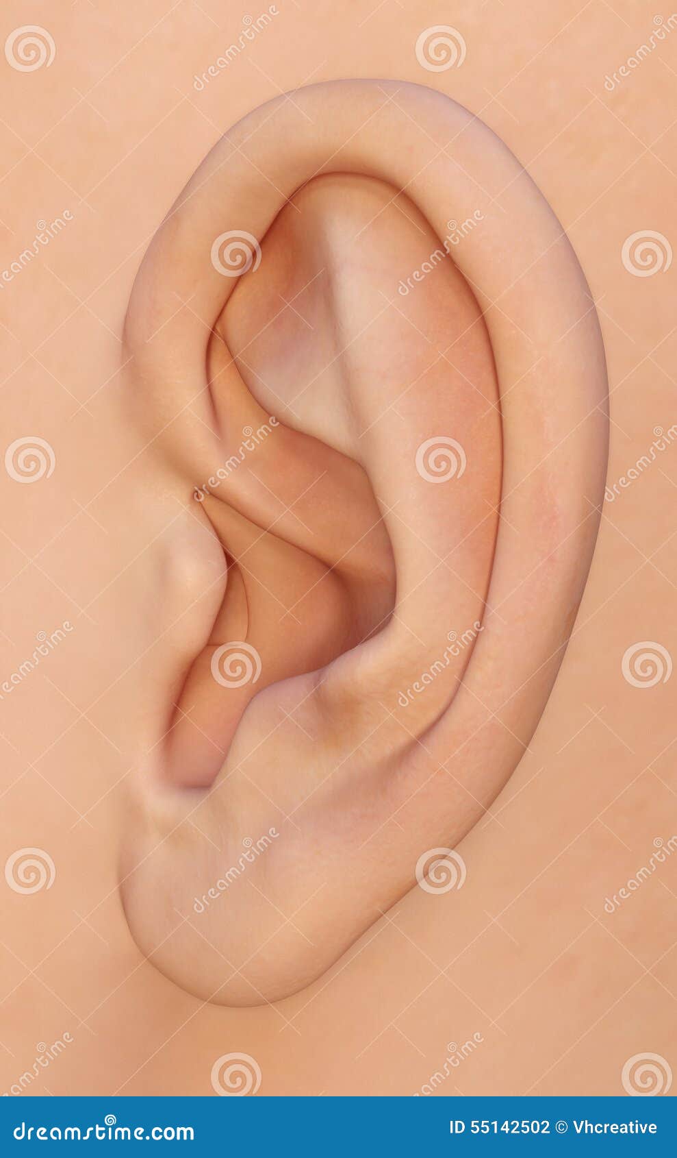 Ear Auricle The Human Outer Ear Anatomy Stock Photo
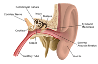 Anatomy And Physiology Of The Ear
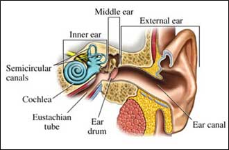 Do You Hear What I Hear Signs And Symptoms Of Tinnitus
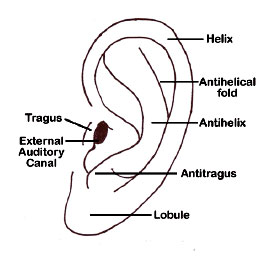 Hearing Outer Ear Development Embryology
 Human Ear Structure And Anatomy Online Biology Notes
 Anatomy And Physiology Of The Auditory System Ento Key
 The Anatomy Of The Outer Ear Health Life Media
 The Outer Ear Functions Parts Of The Outer Ear Pinna
 Ear Anatomy Outer Ear Mcgovern Medical School
 Human Ear Structure And Anatomy Online Biology Notes
 2012 Pearson Education Inc Figure The Anatomy Of The Ear
 Ent Lectures Anatomy Of Ear The External Ear


Posting Komentar
Posting Komentar