If you would like to learn all the parts of the foot structure you have come to the right place. The anatomy of the foot.
 Anatomy Lesson 59 Complete Feet Outlander Anatomy
Medial inside part of foot.

Foot anatomy diagram. Want to learn more about it. This consists of five long metatarsal bones and five shorter bones that form the toes phalanges. Medicinenet does not provide medical advice diagnosis or treatment.
Diagram of normal foot and ankle anatomy. Bones of the foot. The foot is a part of vertebrate anatomy which serves the purpose of supporting the animals weight and allowing for locomotion on land.
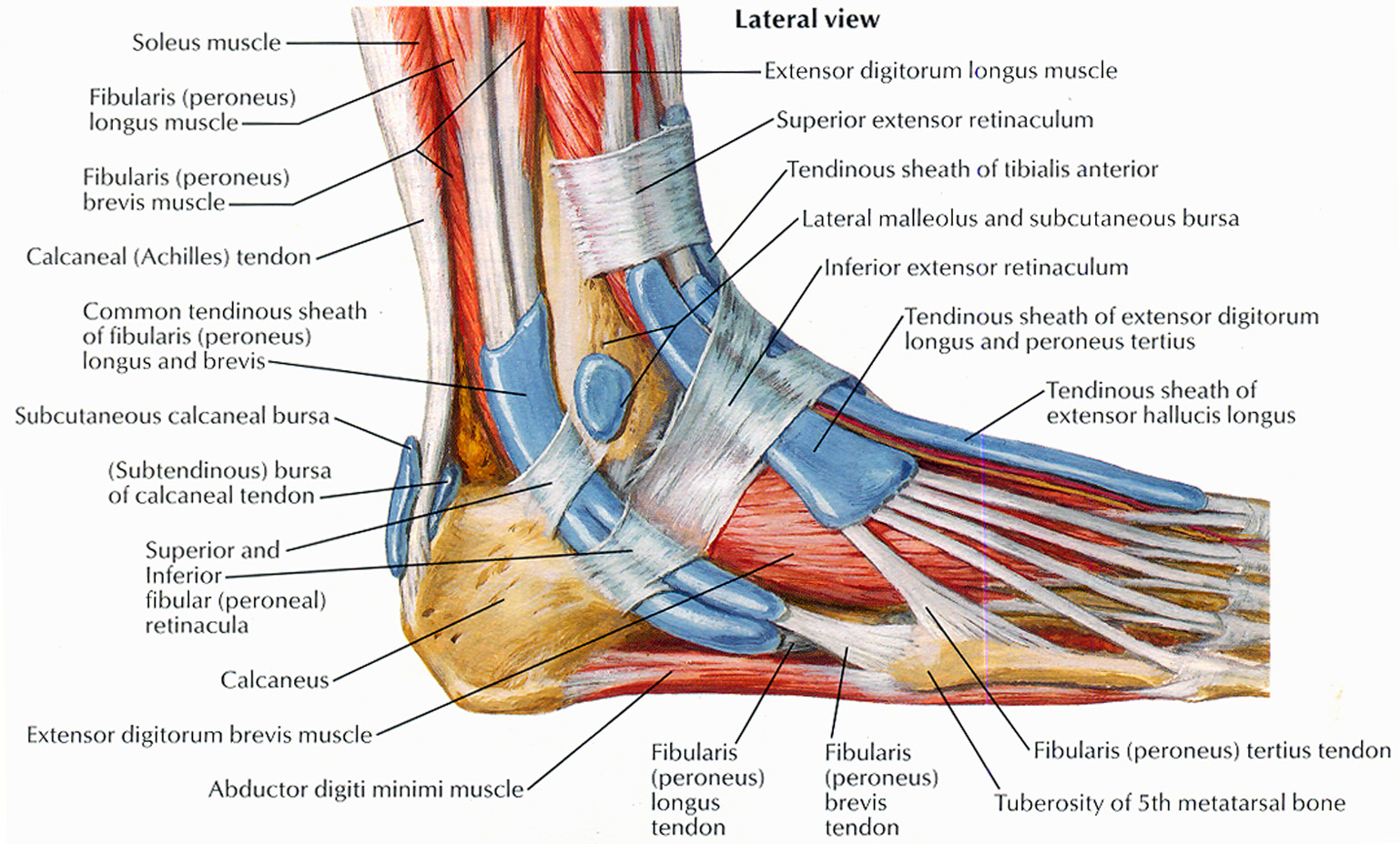
The foots shape along with the bodys natural balance keeping systems make humans capable of not only walking but also running climbing. In humans the foot is one of the most complex structures in the body. Lateral outside part of foot.
The foot is an extremely complex anatomic structure. The foot contains a lot of moving parts 26 bones 33 joints and over 100 ligaments. The foot is divided into three sections the forefoot the midfoot and the hindfoot.
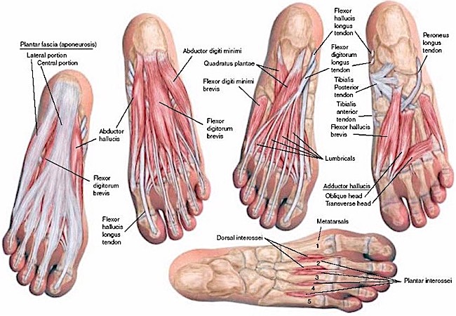
Understanding the structure of the foot is best done by looking at a foot diagram where the anatomy has been labeled. The 20 plus muscles in the foot help enable movement while also giving the foot its shape. The foot diagram has a complex structure made up of bones ligaments muscles and tendons.
Foot anatomy reference author. Picture of foot anatomy detail. Sign up for your free kenhub account today and join over 1234952 successful anatomy students.
The end of the leg on which a person normally stands and walks. Use it to pin point where you are having foot or ankle pain. Podiatric medical review board home foot anatomy.
Our engaging videos interactive quizzes in depth articles and hd atlas are here to get you top results faster. Home image collection a z list foot anatomy detail picture article medical illustrations. Like the fingers the toes have flexor and extensor muscles that power their movement and play a large.
It is made up of over 100 moving parts bones muscles tendons and ligaments designed to allow the foot to balance the bodys weight on just two legs and support. The foot is the lowermost point of the human leg. Marc mitnick dpm reviewed by.
Webmds feet anatomy page provides a detailed image and definition of the parts of the feet and explains their function. The contributors to this site are all board certified orthopaedic surgeons who specialize in treating patients with foot and ankle problems.
 Anatomy Of The Foot North Arkansas Podiatry
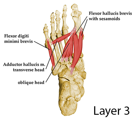 Layers Of The Plantar Foot Foot Ankle Orthobullets
 Anatomical Structure Of The Human Foot The Image Shows The
 Foot And Ankle Skeletal Diagram Anatomy Poster
Foot Human Leg Muscle Peroneus Longus Human Anatomy Tendon
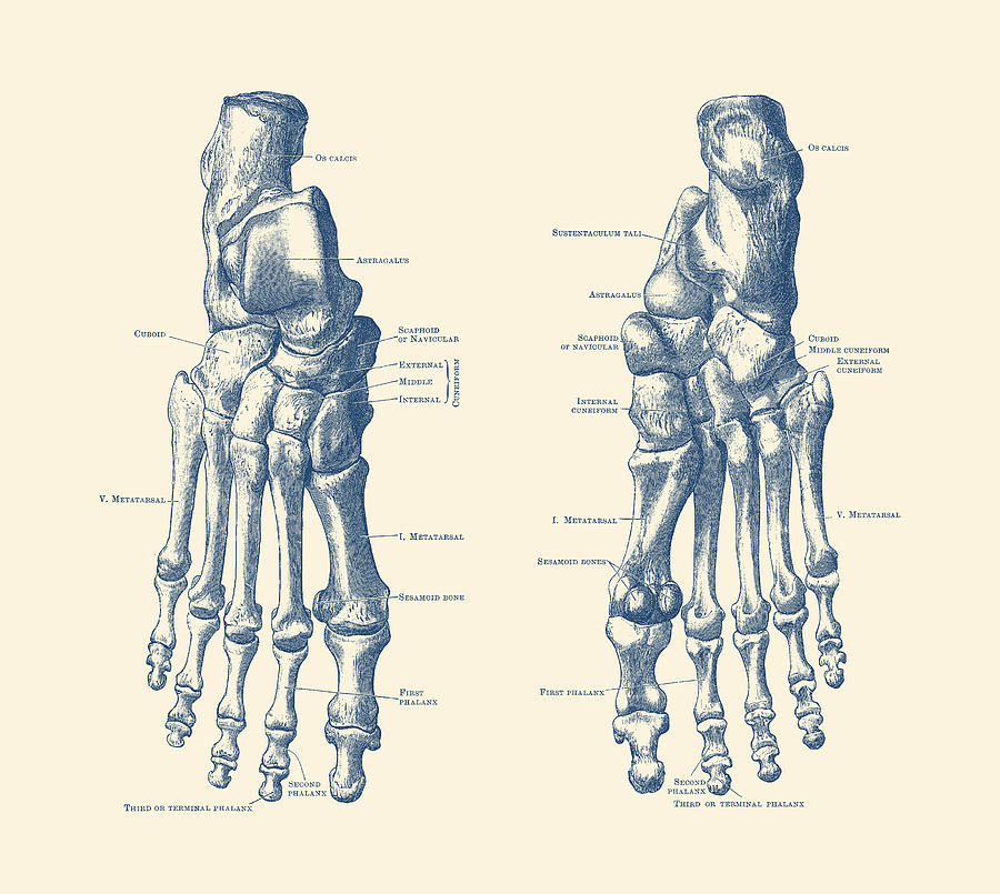
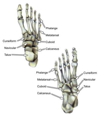
 Skeletal Foot Diagram Dual View Anatomy Print
 Foot Anatomy Detail Picture Image On Medicinenet Com
 The Anatomical And Physiological Overview Of The Human Foot
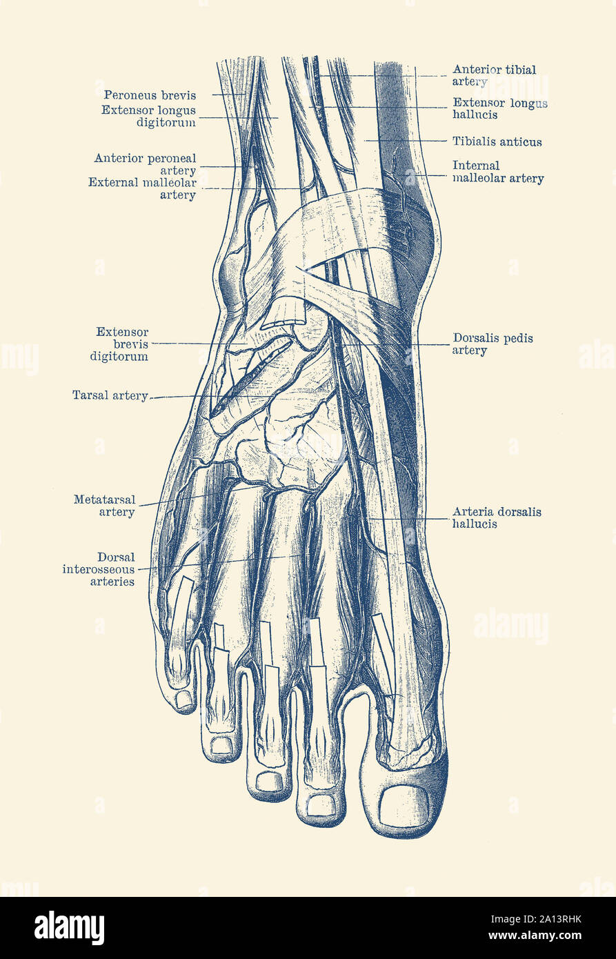
 Nerves And Arteries Of The Foot Preview Human Anatomy Kenhub

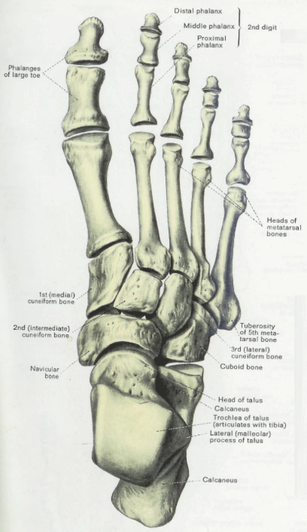
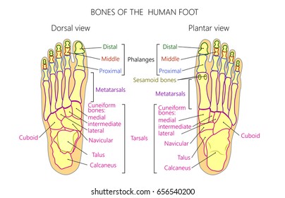
 Bones Of Foot Human Anatomy The Diagram Shows The Placement
 Bones The Of Foot Stock Vector Illustration Of Orthopedic
 Foot And Ankle Anatomy Allen Tx Foot Doctor

 Ankle Joint Anatomy Overview Lateral Ligament Anatomy And
 Knee Joint Picture Image On Medicinenet Com
 Achilles Tendon Human Anatomy Picture Definition
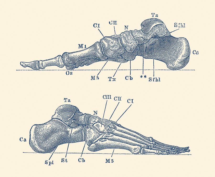
 Vintage Anatomy Print Of The Human Foot Showcasing The
 Ankle Foot Anatomy Diagram Quizlet
 Chapter 38 Foot The Big Picture Gross Anatomy
 Foot Bone Anatomy Overview Tarsal Bones Gross Anatomy
 Foot Anatomy Stock Vectors Images Vector Art Shutterstock


Posting Komentar
Posting Komentar