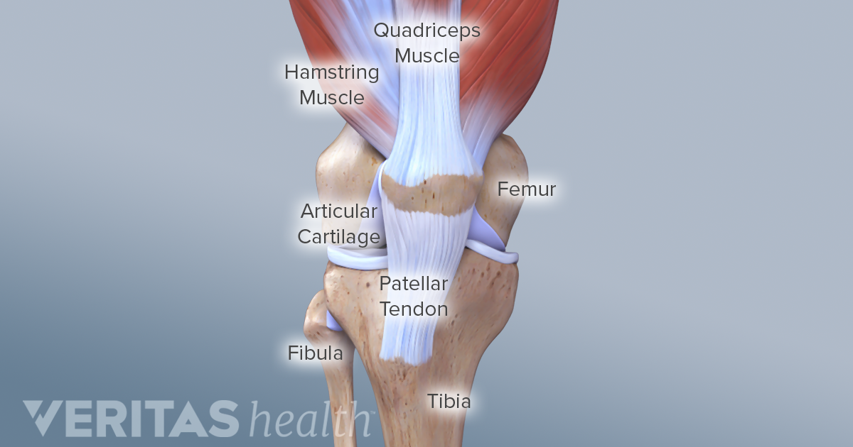
The knee is a hinge joint that is responsible for weight bearing and movement. Around the knee there are two types of tendons.
 Knee And Meniscus Medical Study Student Anatomy Model Showing
This lies on the front of the knee and connects the quadriceps muscles of the thigh to the tibia via the patella and patellar ligament or tendon.
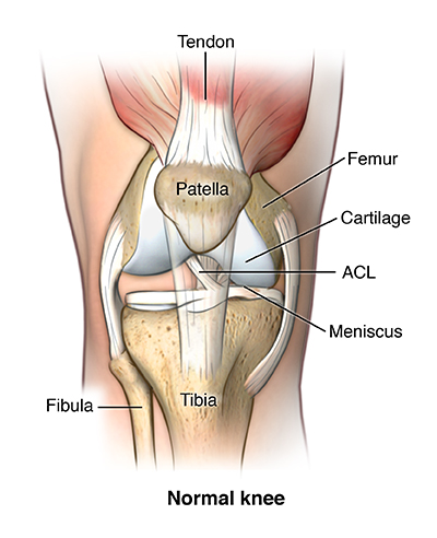
Knee anatomy tendons and ligaments. They cross each other to form an x with the anterior cruciate ligament in front and the posterior cruciate ligament in back. The anterior cruciate ligament prevents the femur from. The patella tendons surround the kneecap and the quadriceps tendons are toward the back of the knee and leg.
Forming an x on the inside of the knee are the anterior cruciate ligament acl as well as the posterior cruciate ligament pcl. The two prevent back and forth sliding of the knee during movement. Muscles are connected to bones by tendons.
The quadriceps tendon connects the muscles in the front of your thigh to your patella. The knee joint is a complex structure that involves bones tendons ligaments muscles and other structures for normal function. Like the knee ligaments the knee tendons can also break and tear.
When there is damage to one of the structures that surrounds the knee joint this can lead to discomfort and disability. These two prevent sideways sliding of the knee joint ad also brace it against unusual movement. Knee anatomy share on pinterest the knee is the most complex joint in the human body.
The muscles rest up against the bones will the tendons preserve this connection. On the sides of the knee are the medial collateral ligament mcl and the lateral collateral ligament lcl. One of the most important tendons is the quadriceps tendon.
These ligaments are frequently damaged by sudden twisting movements eg. There are numerous tendons around the knee that also help to stabilize the knee. Stretching from your patella to your shinbone is the patellar tendon.
Tendons connect the knee bones to the leg muscles that move the knee joint. Tendons are the connection between bones and muscles. The stability of the knee joint is maintained by four ligaments thick bands of tissue that stabilize the joint.
Ligaments join the knee bones and provide stability to the knee. The anterior cruciate ligament and posterior cruciate ligament provide front and back anterior and posterio. Changing direction quickly when running or a force through the knee eg.
The knee includes four important ligaments all of which connect the femur to the tibia. A fall or tackle. There are two pairs of ligaments in the knee the collateral ligaments at the side and the cruciate ligaments in the middle.
These are found inside your knee joint. They are associated with muscles discussed in the section above see above. It consists of bones meniscus.
The cruciate ligaments control the back and forth motion of your knee. The medial collateral ligament mcl and lateral collateral ligament lcl are on the sides of the knee and prevent the joint from sliding sideways.


 Collateral Ligaments Of The Knee Joint Patellar Tendon
 Knee Joint Picture Image On Medicinenet Com
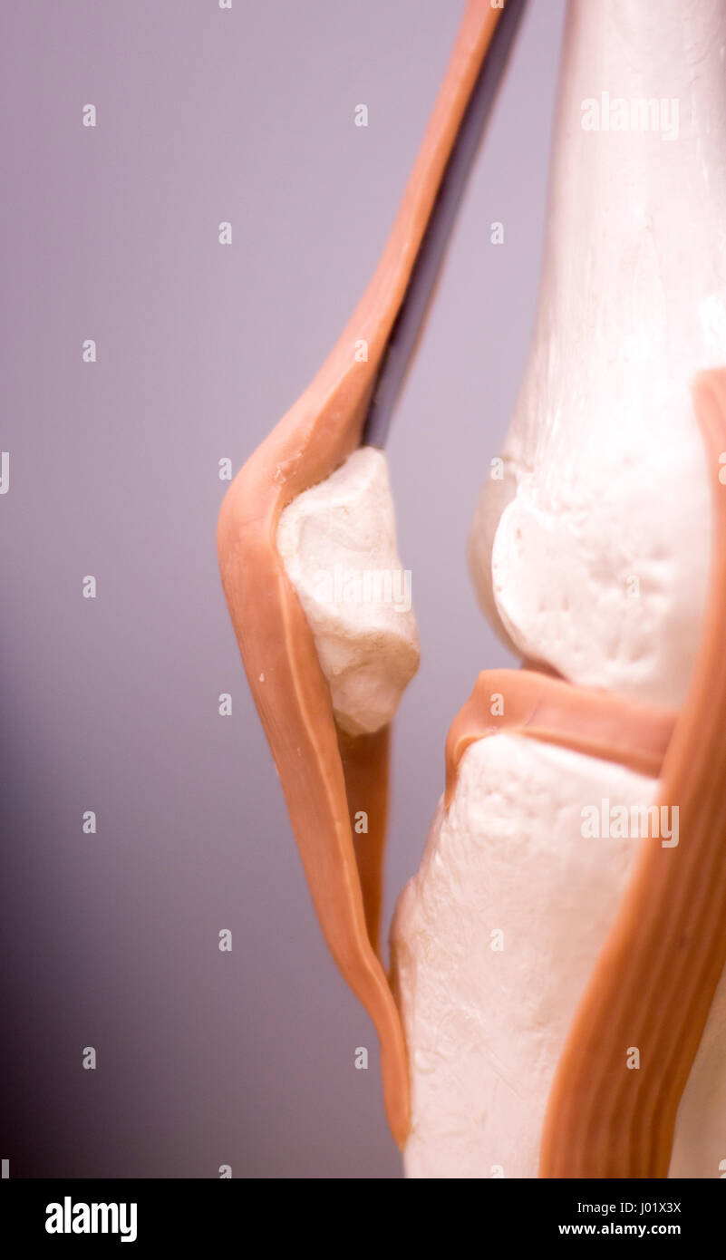 Knee And Meniscus Medical Study Student Anatomy Model
 Collateral Ligament Cl Injury Aftercare Medlineplus
 Quadriceps Tendonitis Of The Knee Richmond Va Sports Medicine
The Knee Anatomy Injuries Treatment And Rehabilitation
 Knee Anatomy
Common Knee Injuries Orthoinfo Aaos
 How To Deal With Knee Injury Symptoms Lcr Health

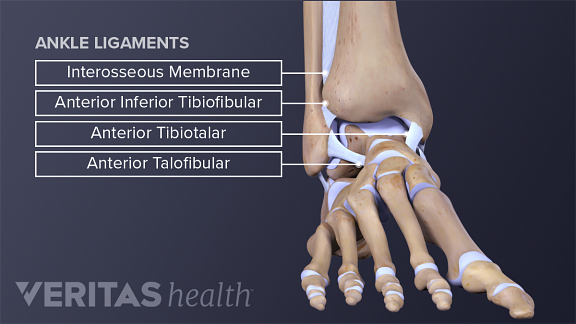 Ankle Anatomy Muscles And Ligaments
 Leg Knee Anatomy
 Anatomy Of The Knee Central Coast Orthopedic Medical Group
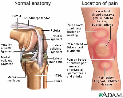
/188058334-crop-56aae7425f9b58b7d0091480.jpg) What Is Causing Your Knee Pain
What Is Causing Your Knee Pain
 Posterior Cruciate Ligament An Overview Sciencedirect Topics

 Knee Joint Picture Image On Medicinenet Com
 Knee Anatomy
 Cruciate Ligament Wikipedia
 Ligaments Of The Foot Muscles Tendons Ligaments Of The
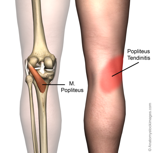 Popliteus Tendinopathy Physiopedia


Posting Komentar
Posting Komentar