Most commonly 33 the ascending and descending branches of the left colic artery communicate through the marginal vessels. Also called pedicel.
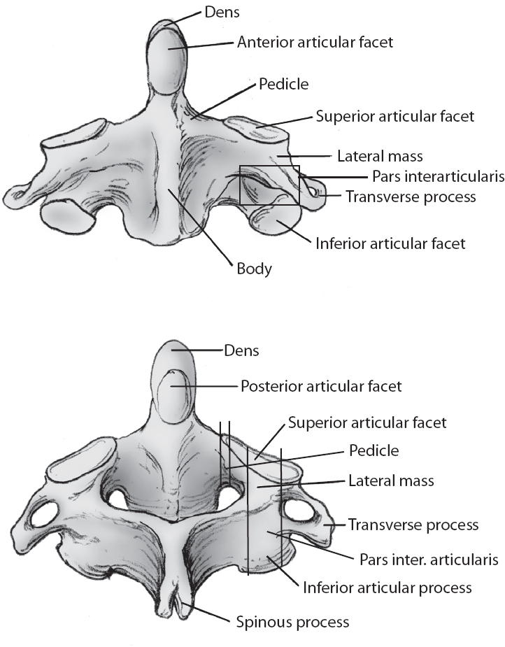 C1 C2 Techniques Neupsy Key
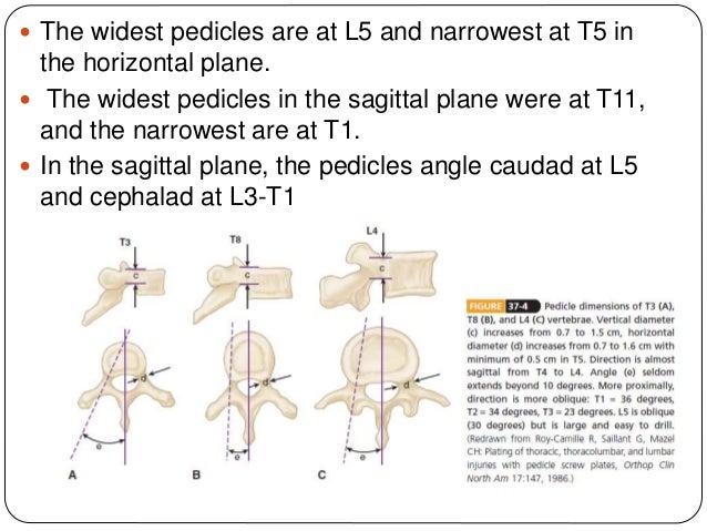
T12 usually has larger pedicle diameter than l1.
Pedicle anatomy. The interpedicular distance was measured at the midshaft of the pedicle. Pedicle cervidae the attachment point for antlers in cervids. Pedicel or petiole insect the stem formed by a restricted.
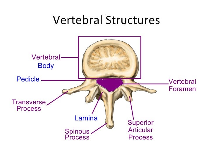
Glomerular filtration by slender cytoplasmic extensions called pedicels foot processes. The most narrow dimensions in both the transverse and sagittal planes were chosen as the transverse pedicle width and the sagittal pedicle width respectively. Two short stout processes extend from the sides of the vertebral body and joins with broad flat plates of bone laminae to form a hollow archway that protects the spinal cord.
Pedicel antenna the second segment of the antenna in the class insecta. Connections between the left and the middle colic arteries are common. In anatomy the pedicle is a short protrusion on the inner part of each vertebra in the spine of all humans and many animals.
In inarticulate larvae the pedicle a stalklike organ develops from a so called mantle fold along the valve margin. It is important to remember that the pedicle size and angulation varies throughout the spinal column. These processes are slightly expanded at their point of contact with the basement membrane and are separated from each other by slitlike spaces about 20 to 30 nanometres across.
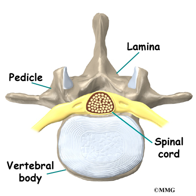
The pedicle is a stub of bone that connects the lamina to the vertebral body to form the vertebral arch. The vascular anatomy distal to the middle colic artery and near the splenic flexure is variable. Strong cylindrical anatomic bridge between the dorsal spinal elements and the vertebral body.
Consists of a strong shell of cortical bone and a core of cancellous bone. T4 has the narrowest pedicle diameter on average t7 can be irregular and have a narrow diameter on the concave side in ais. Pedicle diameter the pedicle wall is twice as thick medially as laterally.
From a distance the spine often looks like a solid column but in fact it is made of individual vertebrae which are ring shaped bones that together form something. In articulates it develops from the caudal or hind region. Biology a small stalk or stalklike structure especially one supporting or connecting an organ or other body part.
Animal anatomy pedicle in brachiopods a fleshy line used to attach and anchor brachiopods. Spine anatomy interactive video. The thoracic pedicle anatomy in all specimens was assessed by one researcher.
 Schematic Presentation Of The Postero Inferomedial Pedicle
:max_bytes(150000):strip_icc()/GettyImages-87293476-56e435703df78c5ba0571226.jpg) Spinal Anatomy Including Transverse Process And Lamina
Spinal Anatomy Including Transverse Process And Lamina

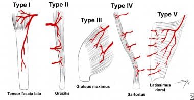 Tissue Flap Classification Classification And Principles Of

 Pedicle Identification Spine Surgery
 Baastrup Syndrome Physiopedia
 Pedicle Screws Fixation Of Thoraco Lumbar Spine


 Ao Surgery Reference
 Vertebra Wikipedia
Plos One Comparison Of Cervical Spine Anatomy In Calves


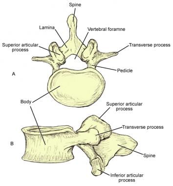
 Anatomy Of Spine

Lumbar Spine Anatomy Overview Gross Anatomy Natural Variants
 Vertebra Wikipedia

Biomechanical Study Of The Fixation Stability Of Broken
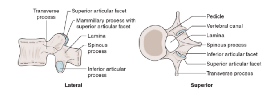
 Lumbar Spine Anatomy Eorthopod Com
 Vertebral Pedicle Anatomy In Relation To Pedicle Screw
 Thoracic Spine Anatomy Spine Orthobullets


Posting Komentar
Posting Komentar