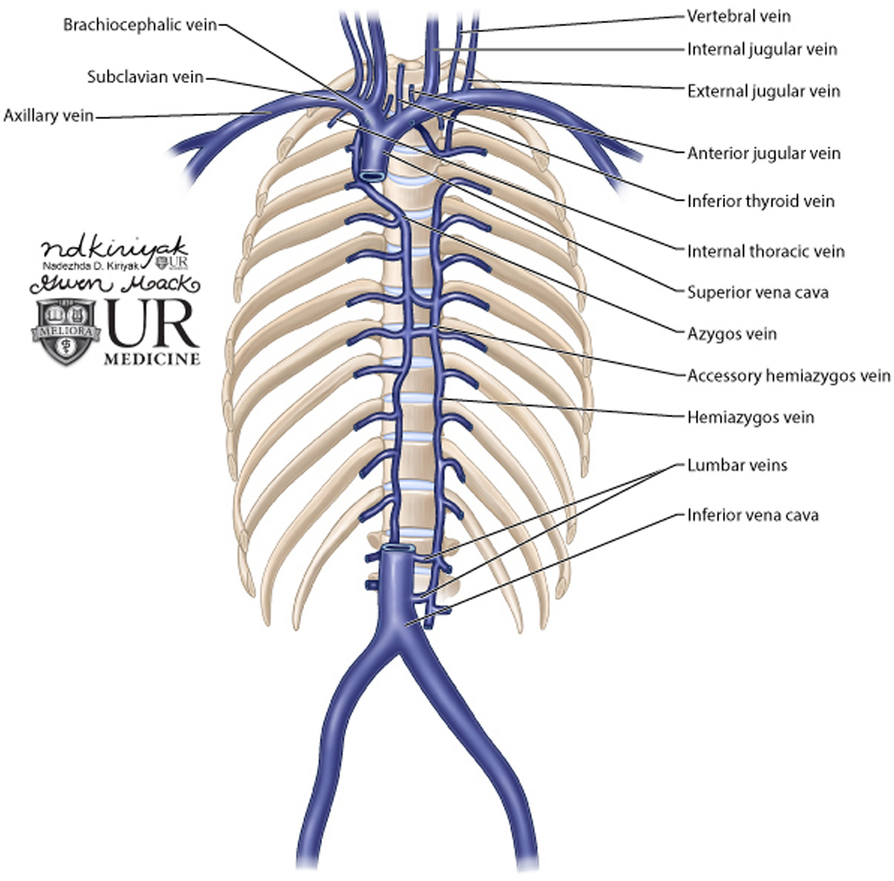
It is a large diameter 24 mm short length vein that receives venous return from the upper half of the body above the diaphragm. Its latin name is related to its large pipe appearance in cadavers cava meaning hollow.
Plos One Ct Of The Paraumbilical And Ensiform Veins In
Thin walls of tissue called fissures separate the different lobes.

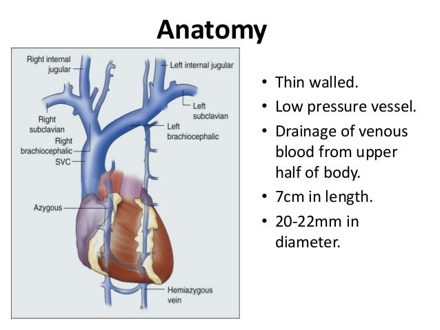
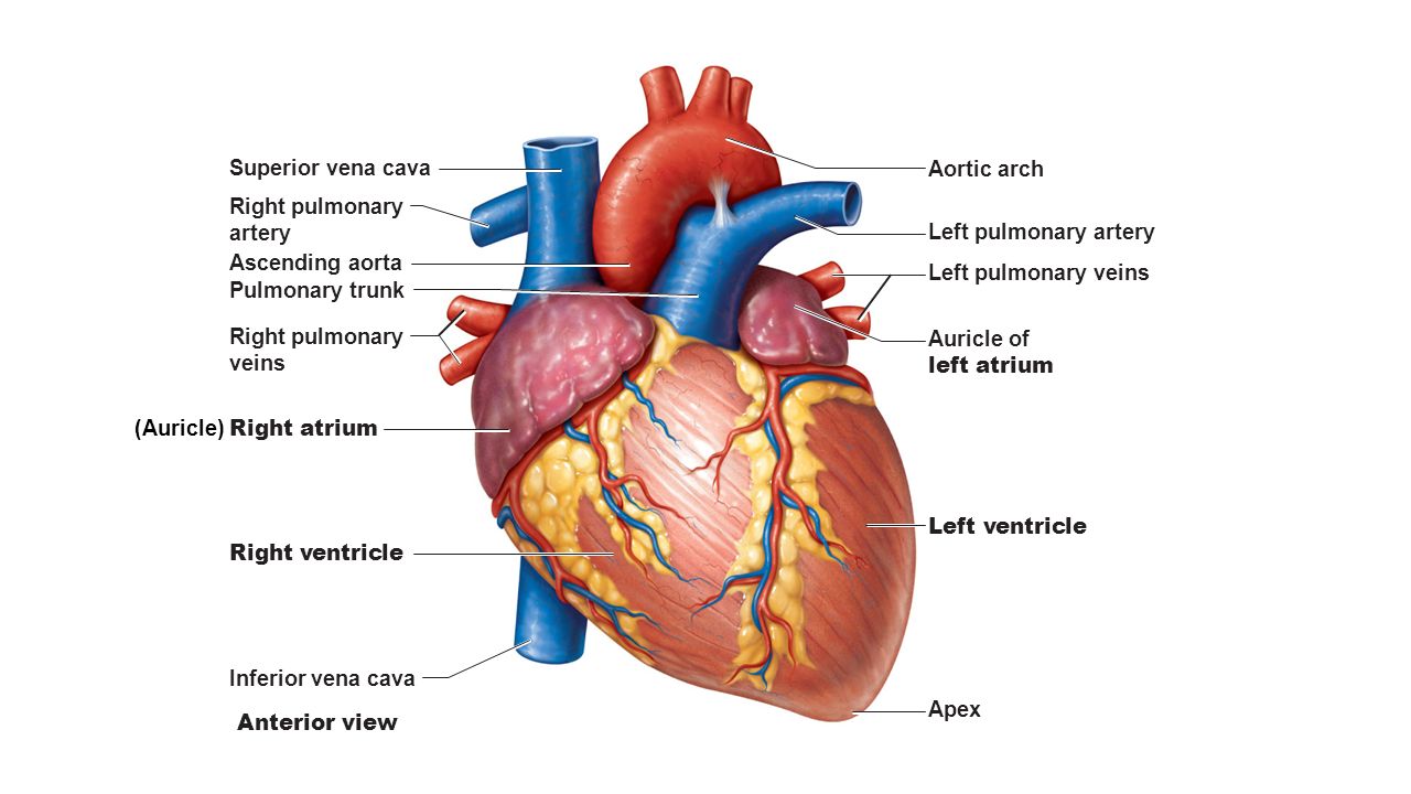
Superior vena cava anatomy. It is present within the superior and middle mediastinum. The lung consists of five lobes. The superior vena cava svc is a large valveless vein that conveys venous blood from the upper half of the body and returns it to the right atrium.
Although the vena cava is very large in diameter its walls are incredibly thin due to the low pressure exerted by venous blood. Gross anatomy the svc be. The superior vena cava svc also known as the cava or cva is a short but large diameter vein located in the anterior right superior mediastinum.
The superior lobes of each lung are the uppermost pieces also called the upper lobes. In this article we will look at the anatomy of the superior vena cava its position tributaries and clinical correlations. The left lung has a superior and inferior lobe while the right lung has superior middle and inferior lobes.
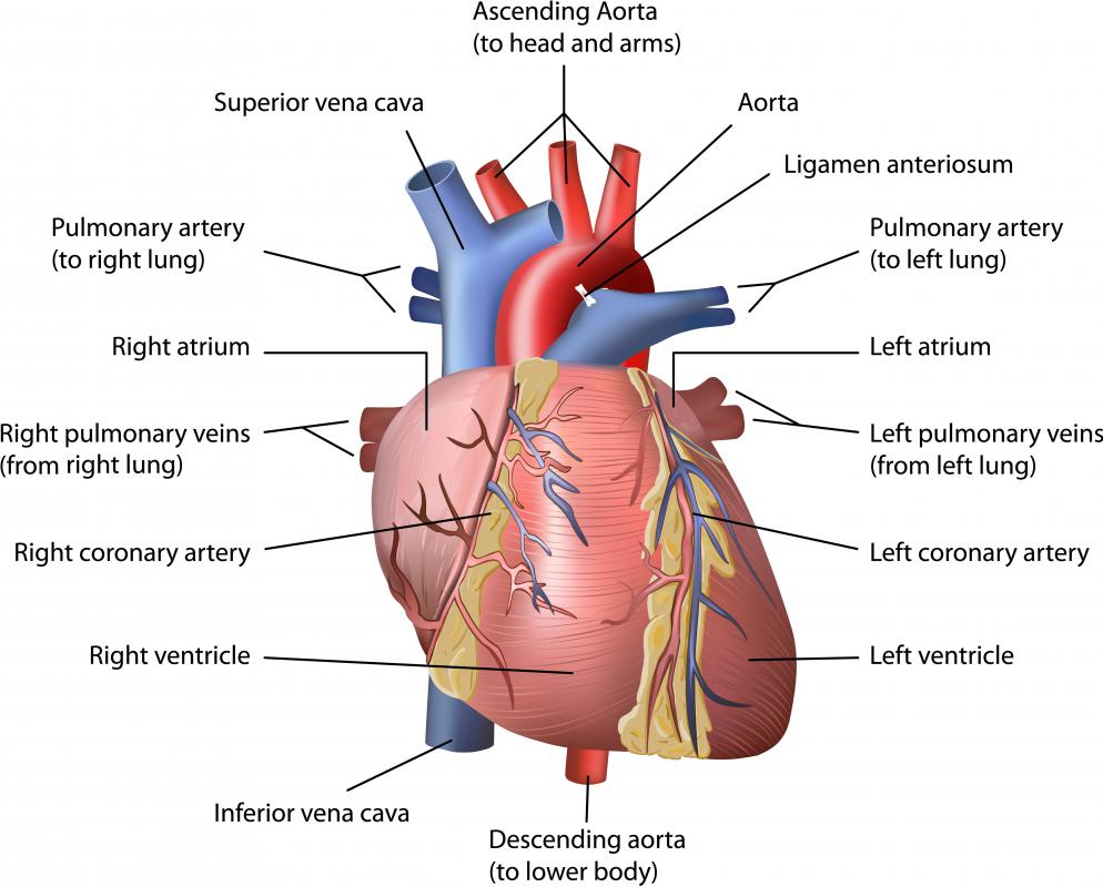
The superior vena cava svc is the superior of the two venae cavae the great venous trunks that return deoxygenated blood from the systemic circulation to the right atrium of the heart. The superior vena cava is a large significant vein responsible for returning deoxygenated blood collected from the body back into the heart. The superior vena cava handles the venous return of blood from structures located superior to the diaphragm.
The superior vena cava svc is a large valveless venous channel formed by the union of the brachiocephalic veins. Function of the venae cavae superior vena cava. In humans these veins are respectively called the superior and inferior venae cavae.
Whereas many mammals including humans have only one anterior vena cava other animals have two. This large vein brings de oxygenated blood from the head neck arm and chest regions of the body to the right atrium. This vein brings de oxygenated blood from the lower body regions legs back abdomen and pelvis to the right.
The inferior vena cava forms at the superior end of the pelvic cavity when the common iliac veins unite to form a larger vein. It receives blood from the upper half of the body except the heart and returns it to the right atrium. The superior vena cava svc also known as the cava or cva is a short but large diameter vein located in the anterior right superior mediastinum.
The anterior vena cava also known as the precava drains the head end of the body while the posterior vena cava or postcava drains the tail or rear end.
 Superior Vena Cava Radiology Reference Article
 Superior Vena Cava Syndrome Urine Or Urout

![]() Superior Vena Cava Anatomy Function Clinical Aspects
Superior Vena Cava Anatomy Function Clinical Aspects
 Persistent Left Superior Vena Cava Mcmahon Biovisuals
 Superior Vena Cava Central Venous Catheter Medical
 Common Variants Of Clinical Significance In The Central
 Radiologic Stages Of Vena Cava Obstruction The Stanford
 Solved Correctly Label The Following External Anatomy Of
 Superior Vena Cava Functions Structure
 Superior Vena Cava Syndrome
 Left Superior Vena Cava With Associated Venous Variations
 Blood Finds A Way Pictorial Review Of Thoracic Collateral
 Kenhub On Twitter Not Sure If You Re Pronouncing Superior
 What Is The Superior Vena Cava With Pictures

 Cv Physiology Cardiac Anatomy
 Figure 4 From 23 Superior Vena Cava Syndrome Semantic Scholar

 A The Trunk Of The Superior Vena Cava System It Shows That
 Superior Vena Cava Aorta Pulmonary Trunk Pericardium Cut

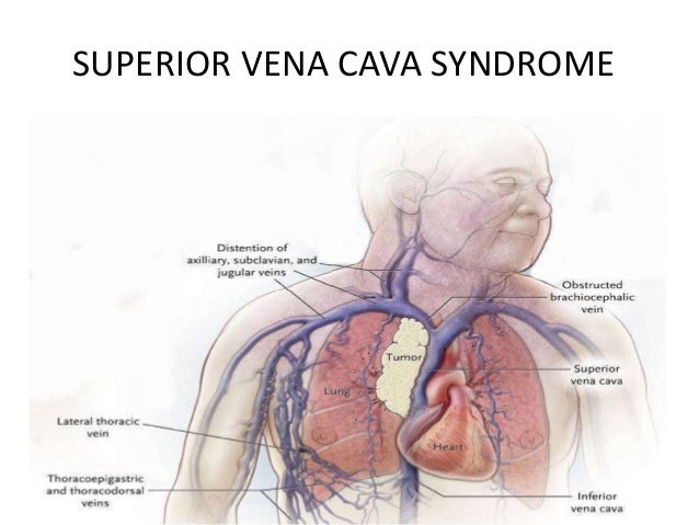 Superior Vena Cava Syndrome
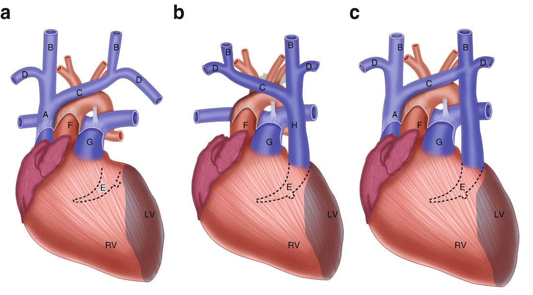 Persistent Left Superior Vena Cava Springerlink

Ultrasound Of The Week Svc Syndrome
:max_bytes(150000):strip_icc()/heart_and_major_vessels-5820b6ba3df78cc2e887becd.jpg) Superior And Inferior Venae Cavae
Superior And Inferior Venae Cavae
![]() Superior Vena Cava Anatomy Function Clinical Aspects
Superior Vena Cava Anatomy Function Clinical Aspects


Posting Komentar
Posting Komentar