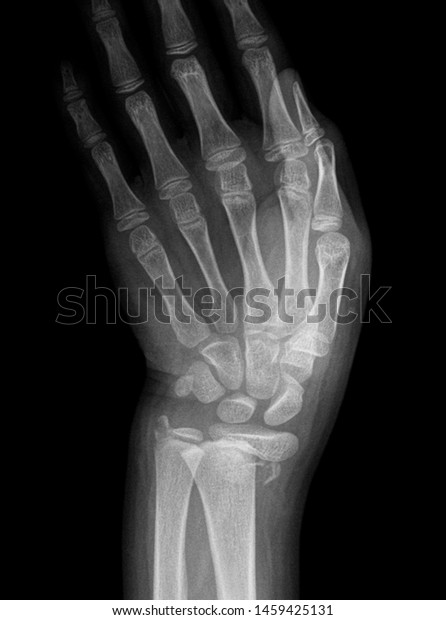
They are commonly performed in the paediatric and elderly populations after a fall on an outstretched hand as well as in patients after higher force trauma. Normal wrist x ray in an adult female for reference.
 Ecr 2015 C 2327 Commonly Missed Fractures In The
This webpage presents the anatomical structures found on wrist mri.

Wrist anatomy xray. Fall onto flexed wrist or direct blowback of wrist. There are numerous joints of the wrist named according to their. Copyright c 2005 2019 alex freitas md.
Atlas of wrist mri anatomy. Check for errors and try again. Knee shoulder shoulder arthrogram ankle elbow wrist hip contact.
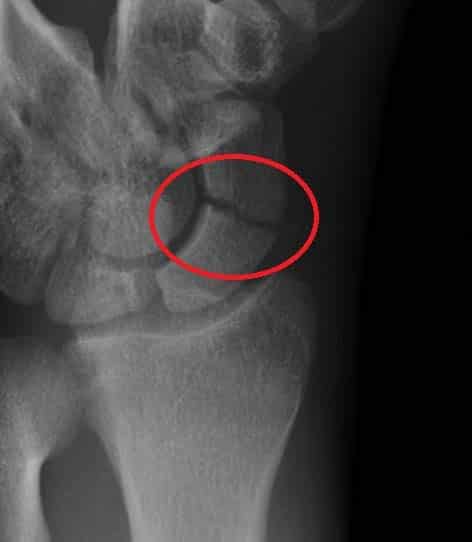
The scaphoid bone is the most commonly fractured wrist bone. Trauma x ray upper limb wrist x ray scaphoid fractures. Wrist radiographs are ubiquitous in the emergency departments.
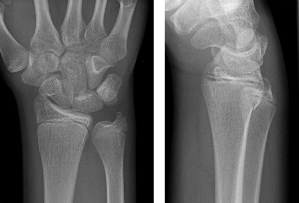
Classically an extra articular distal radius fracture with palmar angulation of distal fracture fragment. Moreover they may be performed as part of a skeletal survey looking for metabolic disease. Use the mouse to scroll or the arrows.
Fall on an outstretched hand. It has an elongated triangular shape with the apex pointing at the radius 5 and consists of the triangular fibrocartilage disc proper along with 12. The tfcc is located on the ulnar aspect of the wrist joint between the ulna and the lunate and triquetrum of the proximal carpal row.
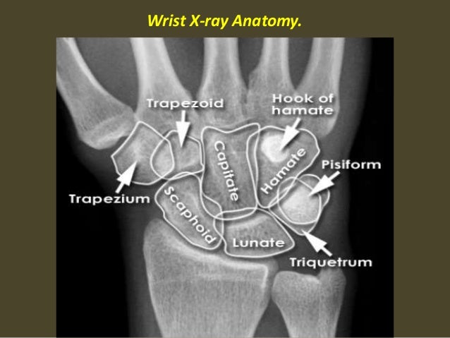
Stanford bone tumor bayesian network issssr msk lectures for residents ocad msk cases from around the world stanford msk mri atlas has served almost 800000 pages to users in over 100 countries. Normal radiographic anatomy of the wrist. 80 of all carpal bone fractures.
Click on a link to get t1 axial view t1 coronal view. The anatomy mri appearance and clinical significance of the scapholunate ligament lunotriquetral ligament triangular fibrocartilage complex carpal metacarpal ligaments and volar and dorsal extrinsic ligaments are reviewed. Also called the reverse colles.
If a carpal bone injury is suspected and not visible on the pa or lateral image. Unable to process the form.
 Carpal Bones Wikipedia
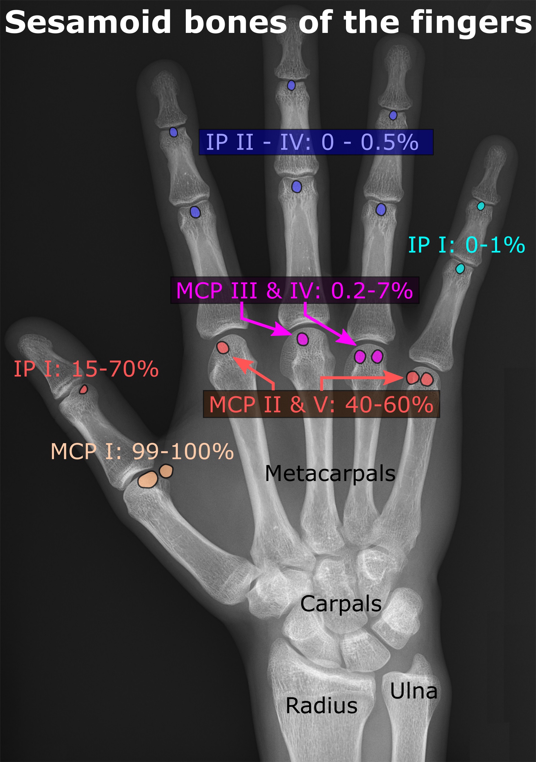 Sesamoid Bone Wikipedia
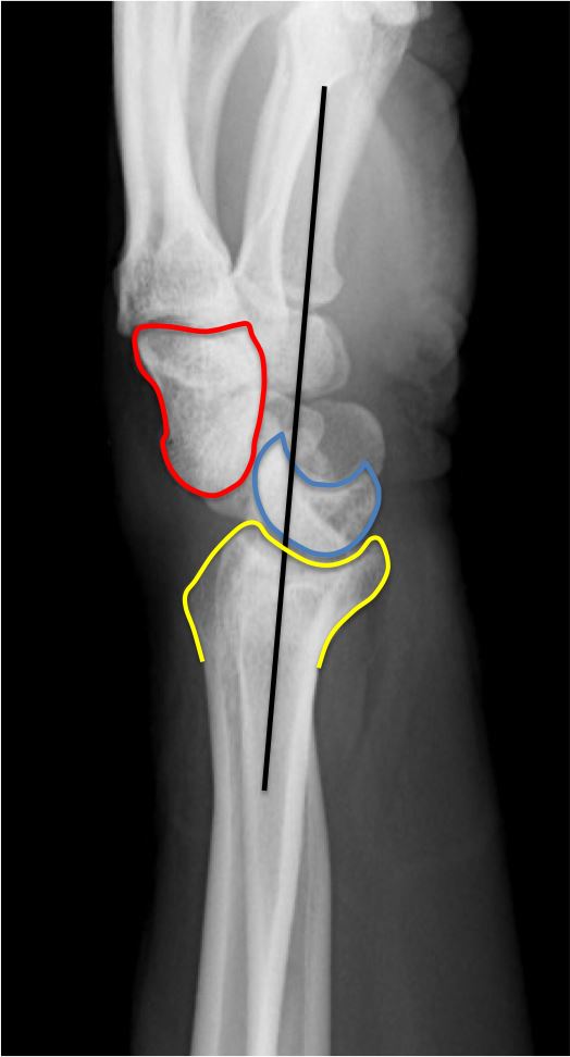
A Radiologist S Guide To Wrist Alignment
 Scaphoid View Radiograph Of The Left Wrist The Bmj
 Human Wrist Anatomy Xray View Medically Accurate 3d Illustration
 The Radiology Assistant Wrist Carpal Instability
A Radiologist S Guide To Wrist Alignment
A Radiologist S Guide To Wrist Alignment
 Wrist Spc X Ray Google Search Radiology Movie Posters

 The Radiology Assistant Wrist Carpal Instability
 The Wrist
 Radiographic Anatomy Hand Ap Radiology Medical Anatomy

 Wrist Dislocation An Overview Sciencedirect Topics
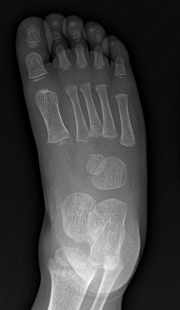
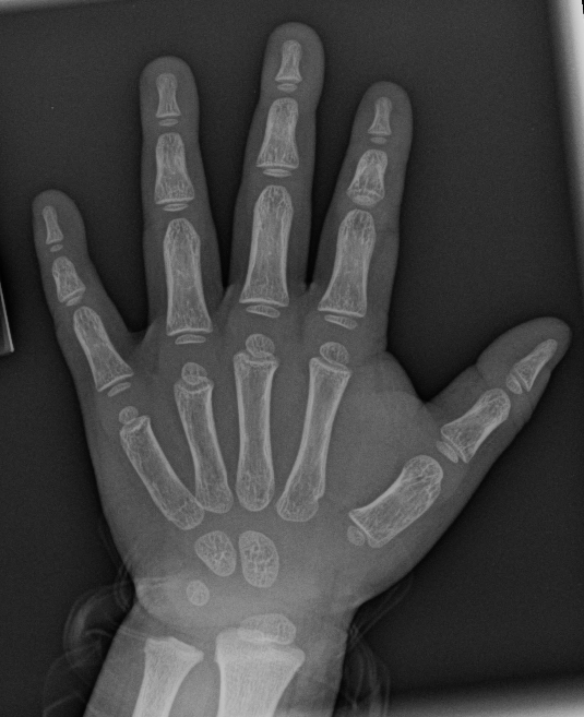
 Carpal Ossification Radiology Case Radiopaedia Org
A Radiologist S Guide To Wrist Alignment
 Pa Wrist Anatomy X Ray Diagram Quizlet
 Wrist X Ray Anatomy Radiology Radiographic Stock Photo Edit

 The Wrist
 Presentation2 Pptx Wrist Joint
 X Wrist Startradiology
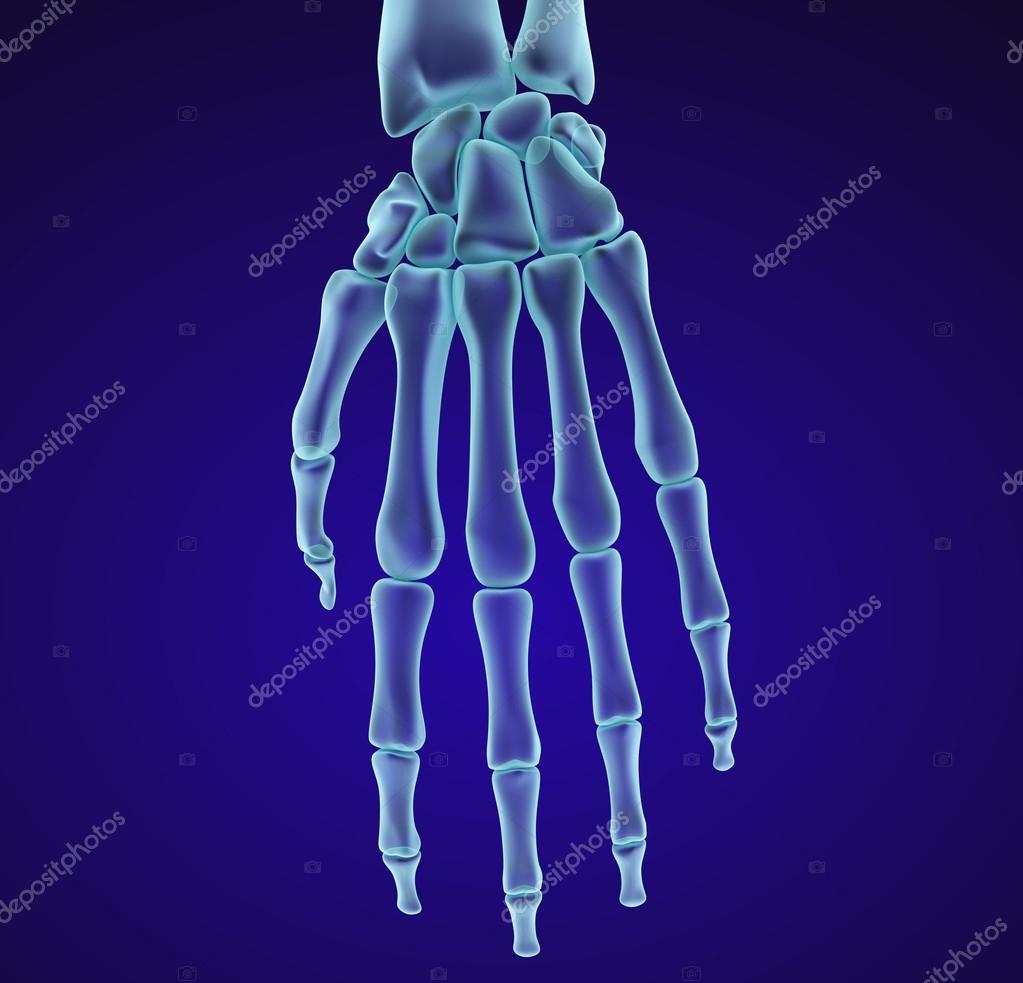 Human Wrist Anatomy Xray View Medically Accurate 3d
 The Wrist Joint Teachmeanatomy
![]() Colles Fracture Lateral A And Ap B Wrist Radiographs
Colles Fracture Lateral A And Ap B Wrist Radiographs
 Colles Fracture Physiopedia
 X Wrist Startradiology
 Wrist Wikipedia


Posting Komentar
Posting Komentar