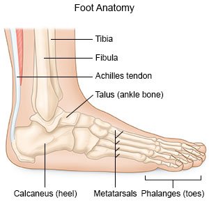
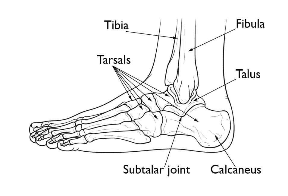
The talus bone supports the leg bones. The feet are divided into three sections.
 Anatomy Of Small Joints Of The Foot
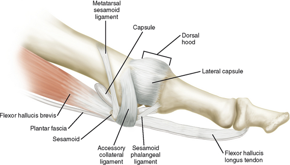
Loss or migration of the plantar fat pad from callus from diseases such as rheumatoid arthritis and diabetes or from excessive dorsiflexion ie high heeled shoes can leave the sesamoid bones relatively unprotected.

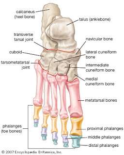
Forefoot anatomy. The calcaneus is the largest of the tarsal bones located in the heel of the foot and bears the weight of the body as the heel hits the ground. Tarsal bones gross anatomy. As in many other joints symptoms guide the ultrasound approach.
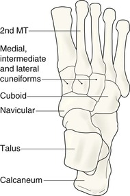
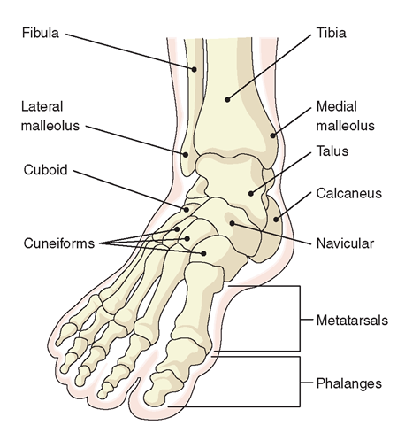
The sesamoid bones are also part of the flexor complex of the first ray. The tarsometatarsal joints tmtj joins the midfoot to the forefoot. They are named the calcaneus talus cuboid navicular and the medial middle and lateral cuneiforms.
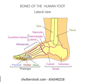
The forefoot is the portion of the foot distal to the midfoot and is composed of the metatarsals and the phalanges. The tarsal bones are 7 in number. The superficial location of most structures means that ultrasound plays an important role in the management of many patients with painful conditions of the foot.
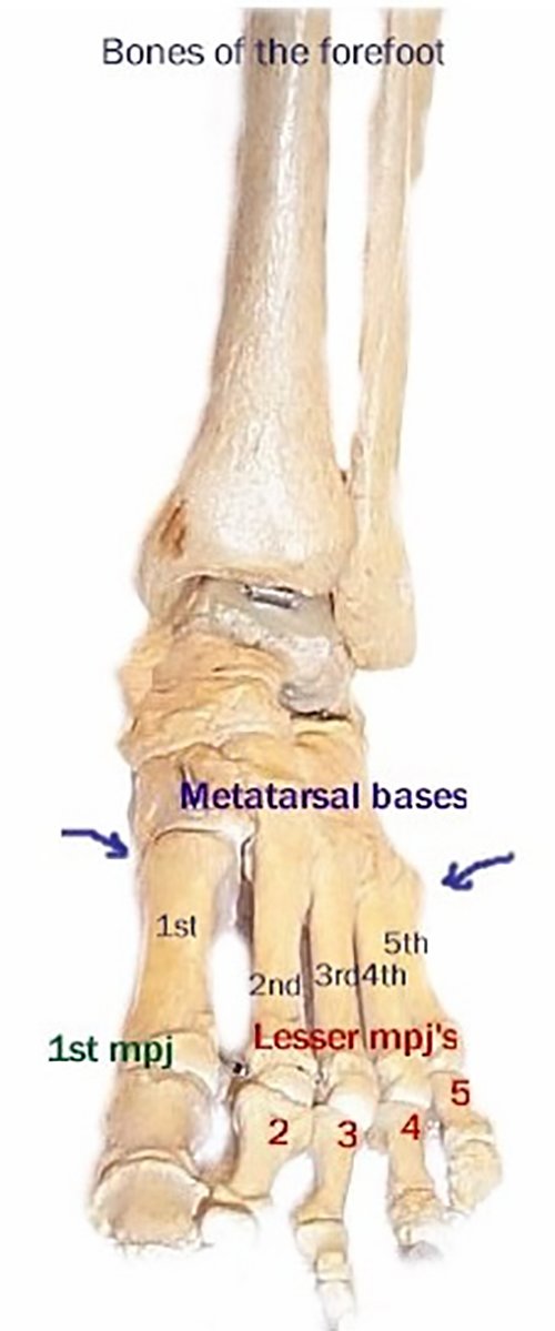
Forefoot bones anatomy 1. The hindfoot forms the heel and ankle. The forefoot contains the five toes phalanges and the five longer bones metatarsals.
Ultrasound examination of the ankle and foot is one of the most common examinations in musculoskeletal ultrasound. The midfoot is a pyramid like collection of bones that form the arches of the feet.
 Forefoot Contains Five Image Photo Free Trial Bigstock
 Longitudinal Arch Load Sharing System Of The Foot
 The Newborn Foot American Family Physician
 Ankle Foot Anatomy
 Forefoot Pain
 Schematic Diagram Of Forefoot Anatomy In The Short Axis View
 Anatomy Of The Forefoot Your Forefoot Is Made Up Of 4
 Forefoot Pain Treatment Springfield Metatarsalgia Chicopee
 Forefoot Varus Footmaxx
 Pronation Of The Foot Wikipedia
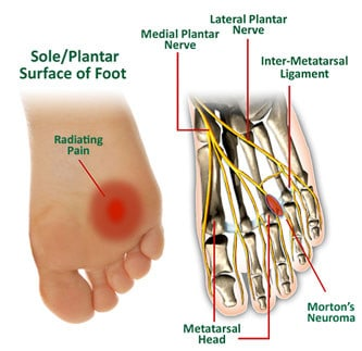 Morton S Neuroma Physiopedia
 Transmetatarsal Amputation What You Need To Know
 Foot And Ankle Patient Education
 Midfoot Forefoot Radiology Key
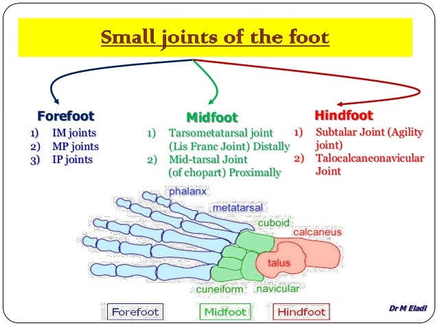 Anatomy Of Small Joints Of The Foot
 Forefoot Images Stock Photos Vectors Shutterstock
 Phalanges Approach Dorsal Approach To P1 Phalanges
 Foot Anatomy East Texas Foot Associates
 Imaging Of The Forefoot And Midfoot Radiology Key
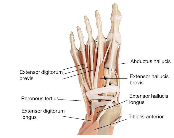
 Ankle Foot Atlas Of Anatomy
 Ankle Foot Atlas Of Anatomy
 521 Forefoot Canvas Prints And Canvas Art Barewalls
 A Review Of The Ankle And Foot Part 1 Anatomy Ptbraintrust
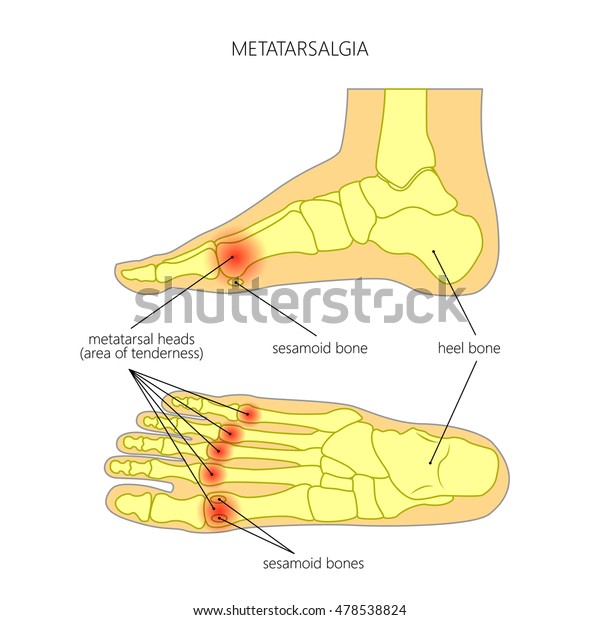 Vector Diagram Forefoot Pain Metatarsalgia Symptom Stock

 Parasagittal Section Of The Forefoot Of Horse Horses
Carpus And Forefoot Anatomy Lateral Radiograph Illustration
 Calcaneus Heel Bone Fractures Orthoinfo Aaos
 Foot Bones Foot Pain Anatomy Info


Posting Komentar
Posting Komentar