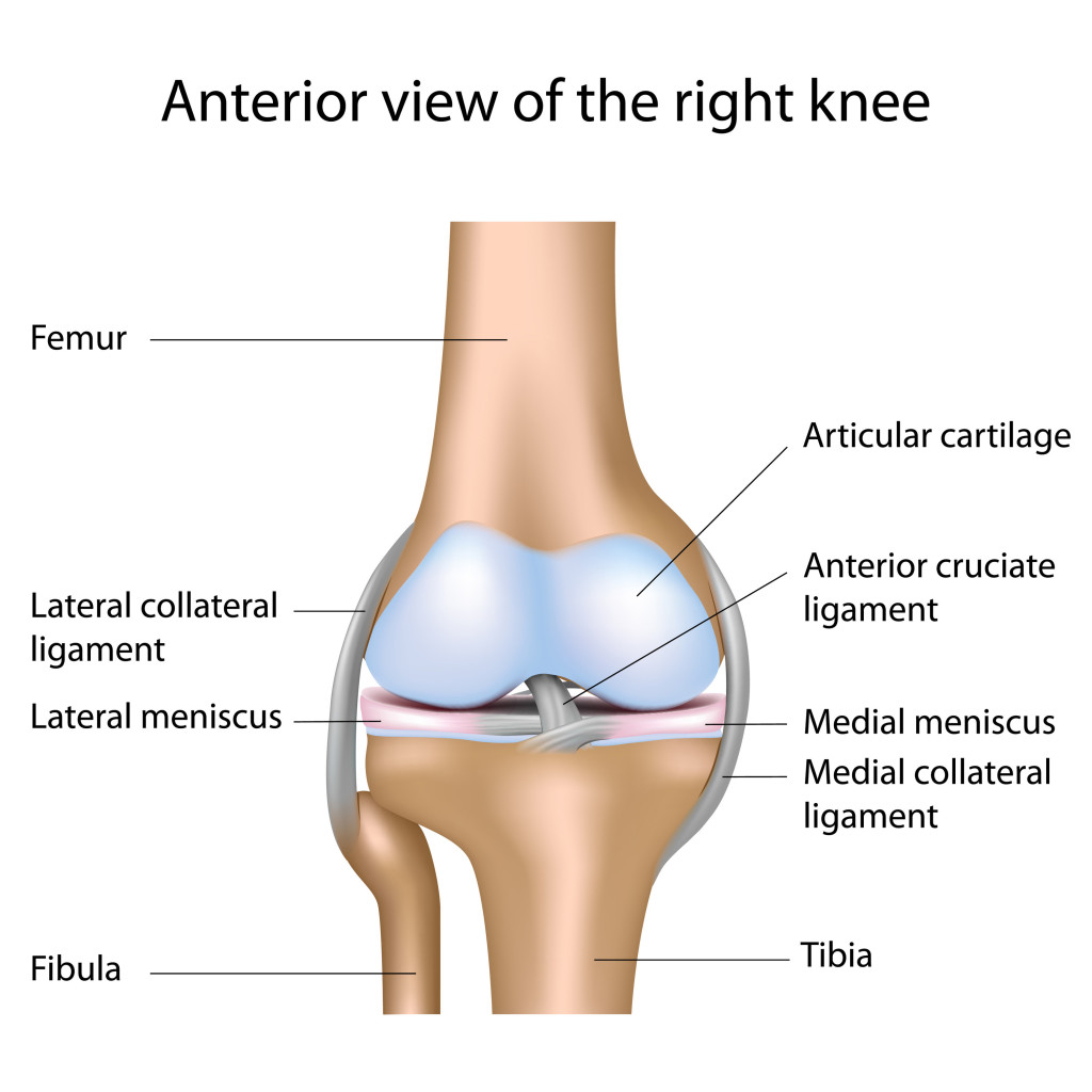
In knee joint anatomy they are the main stabilising structures of the knee acl pcl mcl and lcl preventing excessive movements and instability. An acl tear often leads to the knee giving out and may require surgical repair.
 Deep And Superficial Mcl And Acl Double Bundle Anatomy
The anterior cruciate ligament acl is a band of dense connective tissue which courses from the femur to the tibia.

Knee anatomy acl. The acl is a key structure in the knee joint as it resists anterior tibial translation and rotational loads. Anterior cruciate ligament acl is one of the two cruciate ligaments that stabilize the knee joint. Pcl posterior cruciate ligament strain or tear.
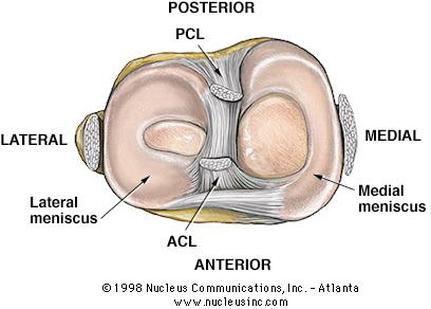
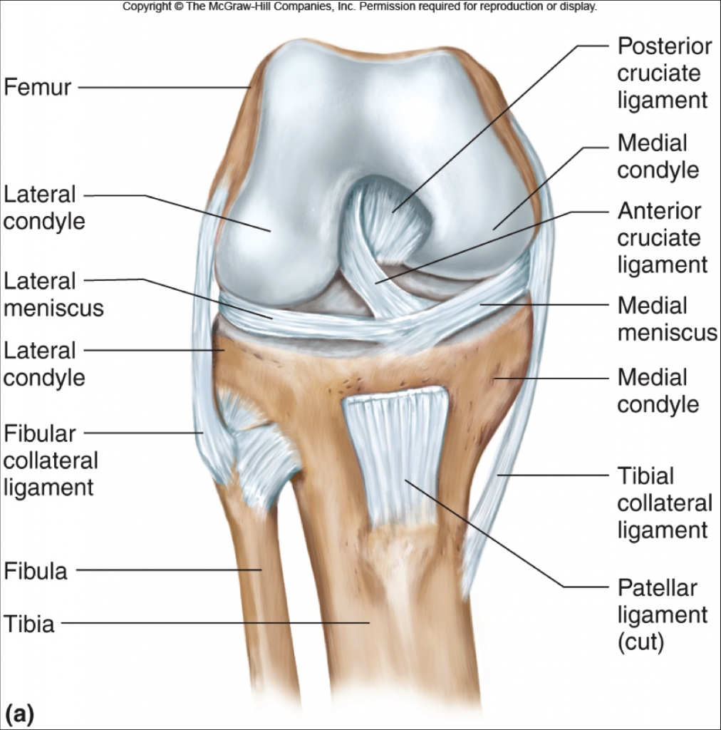
The acl and pcl are called cruciate ligaments because they form a cross in the middle of the knee joint. The acl is one of the primary ligaments providing stability to the knee and therefore is one of the most commonly injured knee ligaments. There is both an anterior cruciate ligament acl and a posterior cruciate ligament pcl.
Acl anterior cruciate ligament strain or tear. Ligaments are tough fibrous connective tissues which link bone to bone made of collagen. Gross anatomy the acl arises from the anteromedial aspect of the intercondylar area on the tibial plateau and passes upwards and backwards to.
Anterior cruciate ligament acl resists anterolateral displacement of the tibia on the femur resists varus displacement at 0 degrees of flexion posterior cruciate ligament pcl. The anterior cruciate ligament acl is one of a pair of cruciate ligaments the other being the posterior cruciate ligament in the human knee. When the knee is fully extended both cruciate ligaments are taut and the knee is locked.
During movement of the knee the anterior cruciate ligament acl prevents anterior sliding of the tibia. The acl is responsible for a large part of the knees stability. The 2 ligaments are also called cruciform ligaments as they are arranged in a crossed formation.
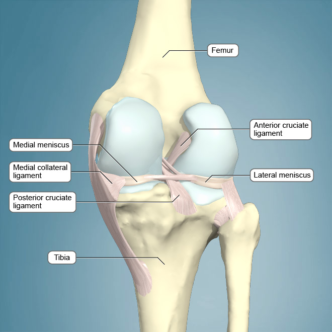
The cruciate ligament in the front of the knee is called anterior cruciate ligament or acl and the cruciate ligament in the back of the knee is called posterior cruciate ligament or pcl. The most common ligament injuries are acl tears mcl tears. Cruciate ligaments this group of ligaments present inside the knee joint control the back and forth motion of the knee.
The posterior cruciate ligament prevents posterior sliding of the tibia. The anterior cruciate ligament acl is one of a pair of ligaments in the center of the knee joint that form a cross and this is where the name cruciate comes from. Pcl tears can cause pain swelling and knee instability.

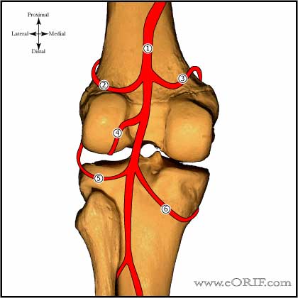 Acl Anatomy Eorif
 Is An Acl Tear Worse Than A Meniscal Tear
 Acl Solutions Acl Knee Anatomy And Diagram Images
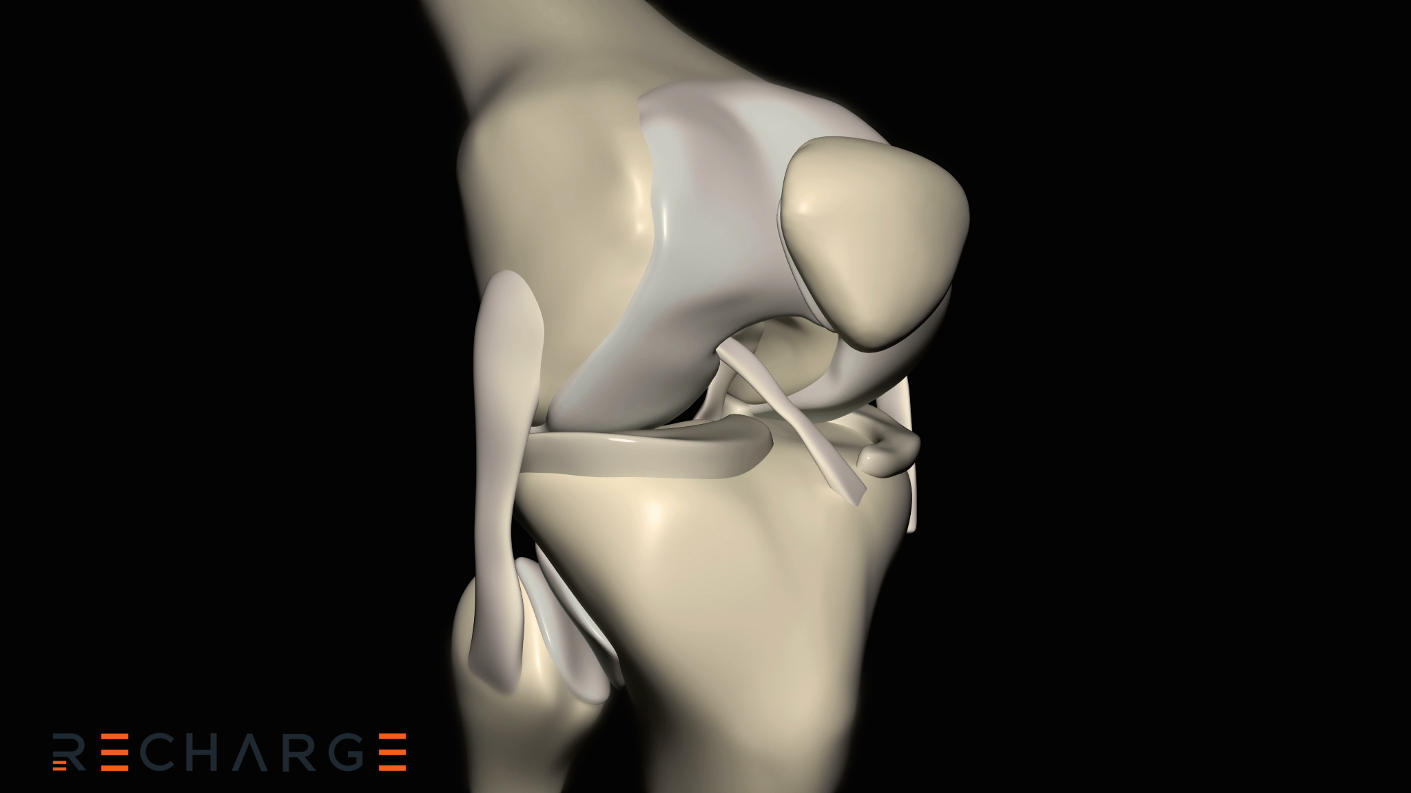 Understanding Knee Anatomy For Acl Injury Management Video
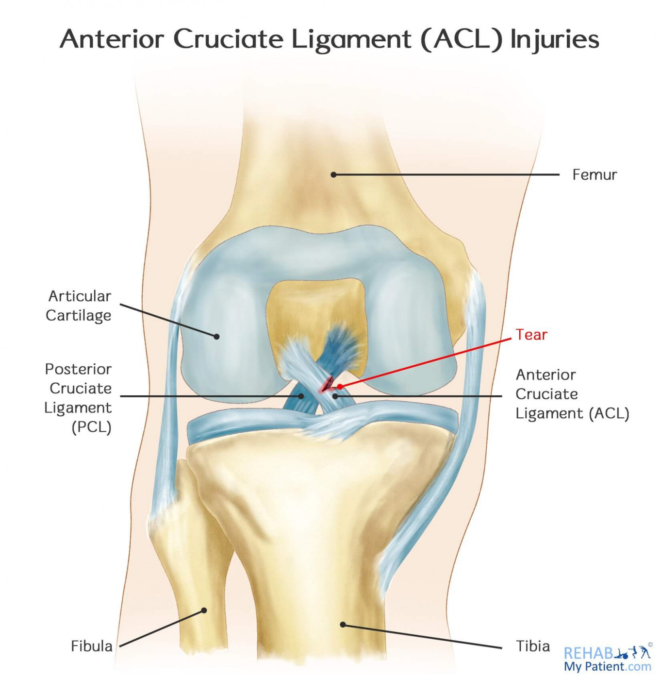 Anterior Cruciate Ligament Acl Injuries Rehab My Patient
 Acl Solutions Acl Knee Anatomy And Diagram Images
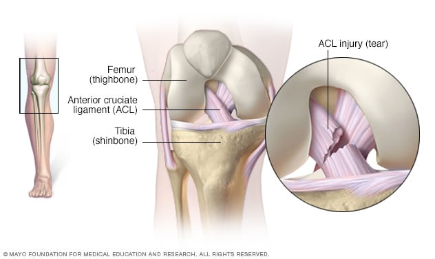 Acl Injury Symptoms And Causes Mayo Clinic
 Anterior Cruciate Ligament Acl Injury Medlineplus Medical
 Deep And Superficial Mcl And Acl Double Bundle Anatomy
Mendmeshop Knee Anatomy There Are 4 Ligaments Of The Knee
 Anatomy And Gender Disparity Of Acl Injuries Maximum

 Acl Injuries University Orthopaedic Associates
 Anterior Cruciate Ligament Acl Injuries Core Em
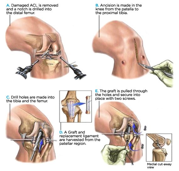
Graft Choices In Acl Surgery
 Knee Injuries Flashcards Quizlet
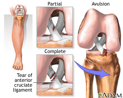
 Anatomy Of An Injury Acl Anterior Cruciate Ligament Tear
 Knee Acl Anatomy Century City Los Angeles Ca Millstein
 Anterior Cruciate Ligament Wikipedia

 Acl Solutions Acl Knee Anatomy And Diagram Images
 Acl Reconstruction The Noyes Knee Institute
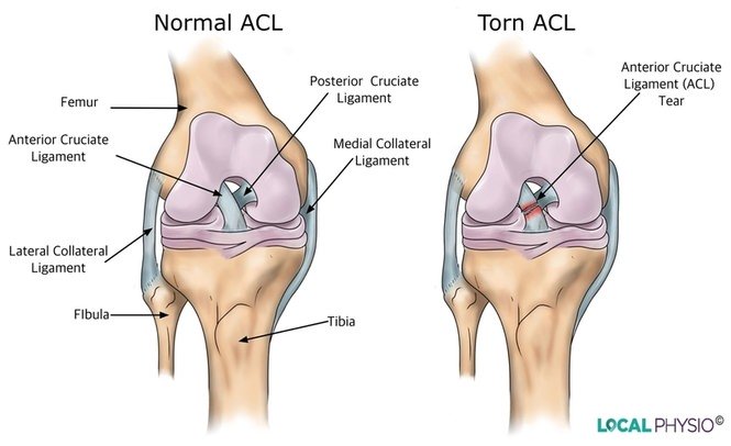 Anterior Cruciate Ligament Acl Injury Local Physio
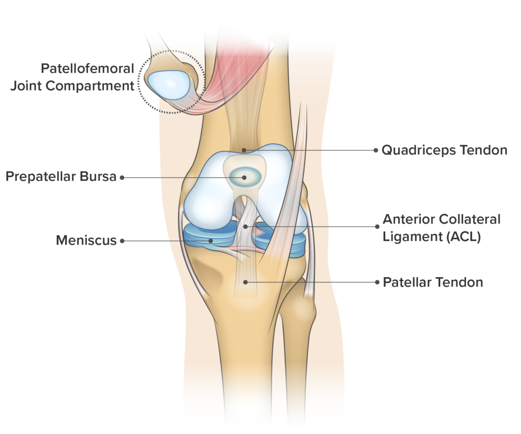 Knee Pain On The Front Of Your Joint Learn Why Spring


Posting Komentar
Posting Komentar