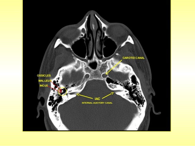
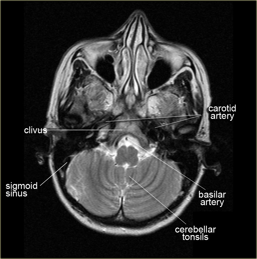
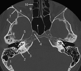
Ct demonstrates the bony anatomy best while mri has superior soft tissue resolution. Anatomy of the head on a cranial ct scan.
 Ct Scan Of Brain And Base Of Skull Stock Photo Download
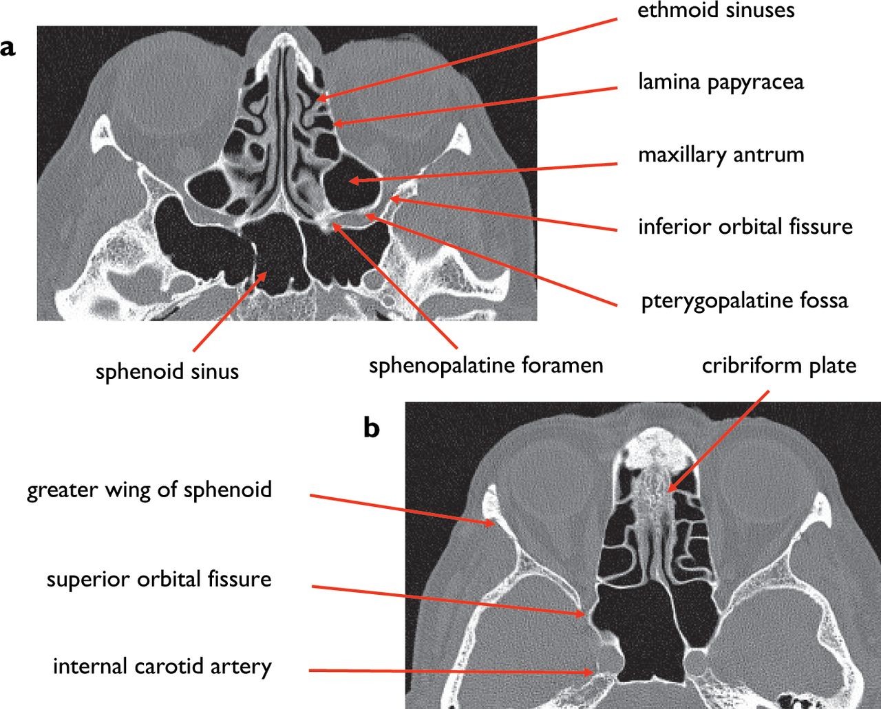
2 superior orbital fissure.

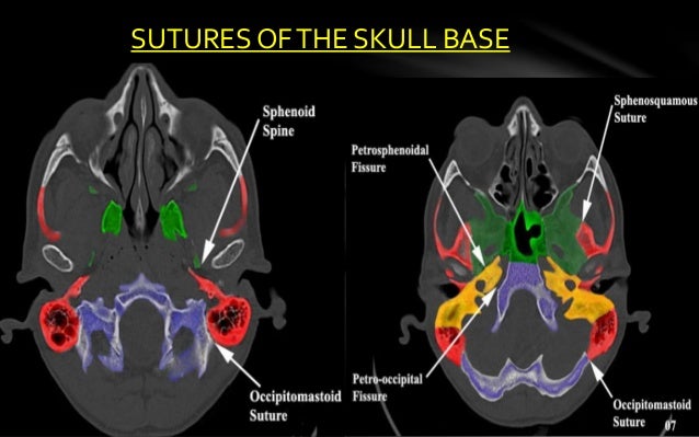
Skull base ct anatomy. The module interface is meant to mimic a radiology workstation with adjacent image scrolling via arrow keys and or mouse wheel button. Basic anatomy review the bones sutures and fissures that comprise the skull base. The skull base can be evaluated by computed tomography ct which will demonstrate the bony structures of the skull base with its foramina and fissures for vessels and cranial nerves the temporal bone and sinonasal cavities.
3 anterior clinoid process. Navigating the skull base identify the petro occipital fissure to navigate the major structures of the skull base. Ct is more sensitive in detecting fibro osseous skull base lesions calcification and sclerosis.
Detailed anatomy enter this module for a more detailed review of skull base anatomy. Brain bones of skull paranasal sinuses. Skull ct anatomy the sagittal suture is the line where the right and left parietal bone are in contact.
Basic skull base anatomy. Gross anatomy the base of the skull is a bony diaphragm composed of a number of bones from anterior. Interactive anatomy atlas.
Brain and face ct. Ct anatomy of skull base. Blue central skull base csb purple posterior skull base teal anterior skull base asb.
A axial three dimensional reconstructed ct image with color coded overlay shows the skull base sections. Ct is the modality of choice in defining the bony anatomy of the skull base and to depict the thin cortical margins of skull base neurovascular foramina. Cross sectionnal anatomy of the head on a cranial ct scan.
B axial ct image with color coded overlay shows the skull base bones. The coronal suture is the line where the parietal bone frontal bone and are in contact. The skull base is made of the paired frontal and temporal bones as well as the ethmoid and occipital bones and these bones form the floors of the anterior middle and posterior cranial fossa.
To load the skull base ct anatomy module in a new window click on its image above. The base of the skull or skull base forms the floor of the cranial cavity and separates the brain from the structures of the neck and face. Given that the file is large loading may take a few minutes.
Brain bones of cranium sinuses of the face. Head and neck ct ct.
 Skull Base Imaging
 Posterior Cranial Fossa Anatomy In Mri Ct
 The Radiology Assistant Brain Anatomy
 Foramina Of The Skull Base Ct
 The Body Online Stony Brook University Department Of Anatomy
 Normal Anatomy Of The Base Of The Skull Orbit Pituitary
 Head Ct Anatomy Radiology Key
 Self Test
Brain And Face Ct Interactive Anatomy Atlas
 Imaging Of Skull Base
 Radiologic Anatomy Of The Skull Base Radiology Key
 Imaging Of Skull Base
 North Jersey Brain Spine Center Computerized Tomography
 Brain And Face Ct Interactive Anatomy Atlas
 Imaging Of Skull Base Pictorial Essay Raut Aa Naphade Ps
 Ct Anatomy Of Skull
 Foramina Of The Skull Base Ct
 The Canine Head And Skull Ct Atlas Of Veterinary Clinical
 Brain And Face Ct Interactive Anatomy Atlas
 Skull Base Osteomyelitis Radiology Case Radiopaedia Org
 Ecr 2015 C 0264 Imaging Of The Anterior And Central
 Head Ct Anatomy Radiology Key
 Temporal Bone And Skull Base Anatomy Ct
 Ao Surgery Reference
 A 3d Stereotactic Atlas Of The Adult Human Skull Base
 Ecr 2015 C 0264 Imaging Of The Anterior And Central
 Skull Base Anatomy By Dr Aditya Tiwari
 Cranial Vault Skull Base Diagnosis Skull Base
 Skull Base Development And Anatomy
 Endoscopic Endonasal Approach For Mass Resection Of The


Posting Komentar
Posting Komentar