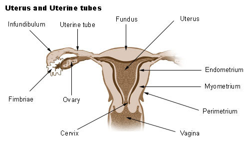
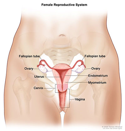
The uterus can be divided anatomically into four regions. The inner layer called the endometrium is the most active layer and responds to cyclic ovarian.
 What Are The Three Layers Of The Uterine Wall From The
Is triangular in shape in coronal section.

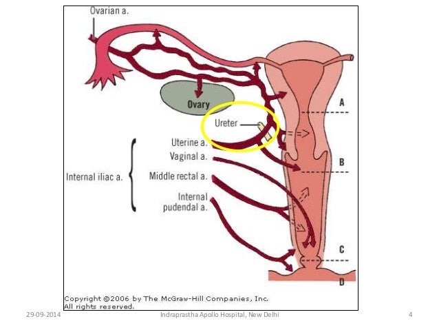
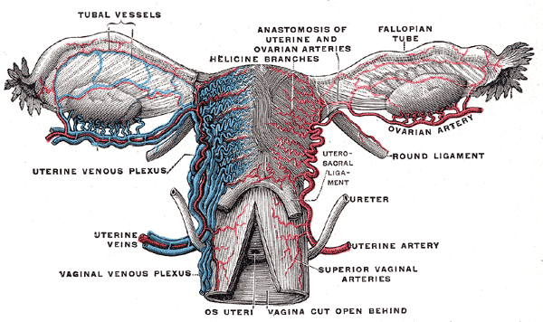
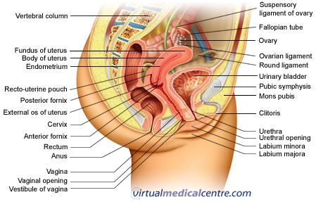
Uterine anatomy. The uterus is a secondary sex organ. Treatment depends on the cause. R elations of the body of uterus.
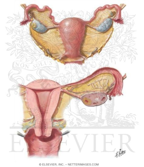
Secondary sex organs are components of the reproductive tract that mature during puberty under the influence of sex hormones produced from primary sex organs the ovaries in females and the testes in males. What are the relations of uterus. It is important to dissect the anatomy of the human uterus.
In sagittal plane it is merely a slit. The cavity communicates with the cervical canal via internal os. Uterus also called womb an inverted pear shaped muscular organ of the female reproductive system located between the bladder and rectum.
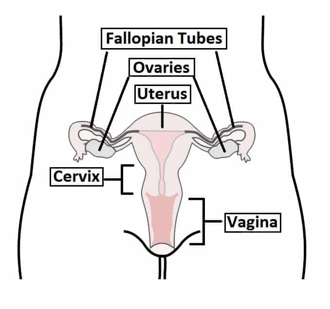
The fundus the uppermost rounded portion of the uterus the corpus body the cervix and the cervical canal. The uterus itself is a hollow organ that is shaped in the form of a pear and interestingly enough measures about that size. The uterus or womb is the place where a baby grows when a woman is pregnant.
The middle layer or myometrium makes up most of the uterine volume and is the muscular layer. It functions to nourish and house the fertilized egg until the unborn child or offspring is ready to be delivered. The cervical canal opens into vagina via external os.
Causes can include hormones thyroid problems fibroids polyps cancer infection or pregnancy. The uterus or womb is shaped like an inverted pear. The first sign of a problem with the uterus may be bleeding between periods or after sex.
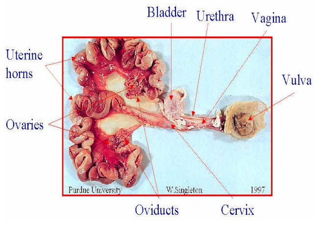
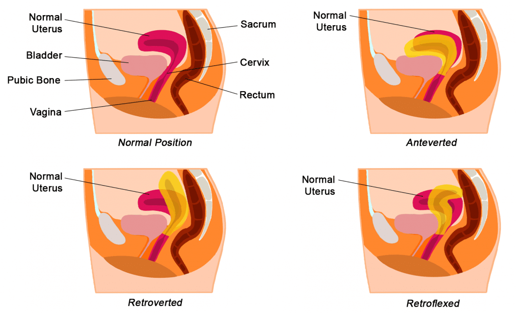
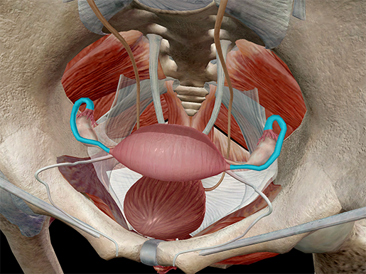
It is neatly tucked into the pelvic area of most mammals and of course in humans. The uterus is held in position within the pelvis by ligaments which are called endopelvic fascia. In the female body the upper end of the uterus called the fundus will join the fallopian tubes at either side while the lower end will open into the vagina.
The anatomy of the uterus consists of the following 3 tissue layers see the following image. The cervix protrudes into the vagina.
 Female Reproductive Anatomy Reproductive Medbullets Step 1
 Ch27 Vaginal Anatomy
 Uterus Anatomy Definition And Function Human Anatomy Kenhub
 Uterine Anatomy Stock Illustrations 815 Uterine Anatomy
 Uterine Ligaments Diagram Human Uterus Anatomy
 Applied Anatomy 2
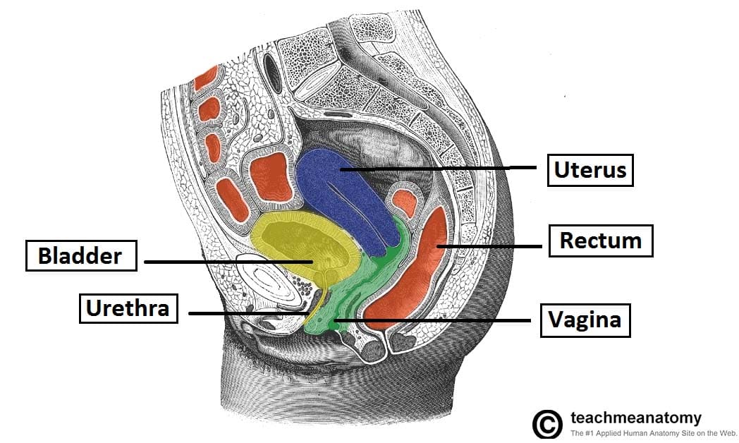 The Uterus Structure Location Vasculature Teachmeanatomy
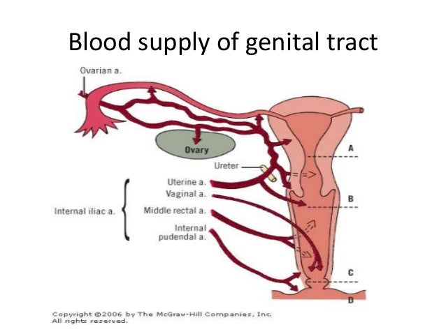
 Seer Training Genital Tract
The Female Reproductive System Boundless Anatomy And
 Ovarian Uterine Anatomy At University Of Cincinnati Studyblue
 Ovarian Uterine Anatomy At University Of Cincinnati Studyblue
 Uterine Fibroids Pain Symptoms Causes Surgery Treatment
 Cureus Comprehensive Review Of The Cardinal Ligament
 Anatomy For Anaesthesia Nerve Supply Of Uterus Cervix
 Ovarian Uterine Anatomy At University Of Cincinnati Studyblue
 Uterine Anatomy 3d Anatomy Tutorial
 Reproductive Physiology And Anatomy Of The Sow
 Uterine Anatomy Exhibits
 The Uterus Structure Location Vasculature Teachmeanatomy
 Uterus Wikipedia
 Seer Training Salpingo Ovarian Peritoneal Functional Anatomy
 Schematic Drawing Of Various Types Of Uterine Fibroids Subserosal
 Uterus Ovaries And Uterine Tubes

 The Uterus Structure Location Vasculature Teachmeanatomy
 Female Reproductive System Urogenital System Anatomy
 Foetal Fetal Development Illustrations Heart Vascular


 Anatomy And Physiology Internal Female Reproductive Anatomy


Posting Komentar
Posting Komentar