Posteromedial vein of the thigh accessory vein of the thigh drains the superficial aspect. Document the normal anatomy and any pathology found including doppler images demonstrating flow.
 Thigh Knee Thumb Human Leg Vein Others Free Png Pngfuel
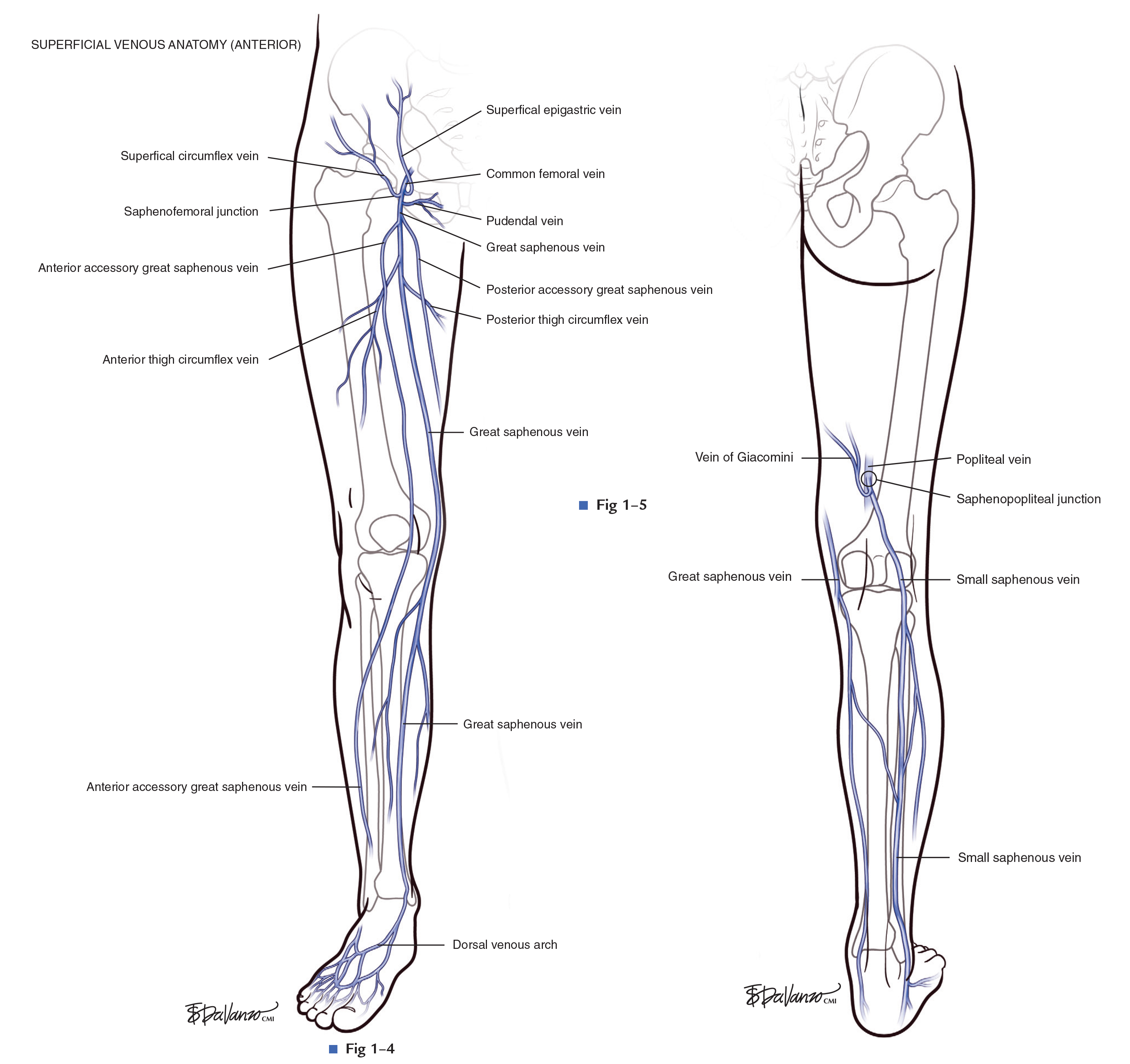
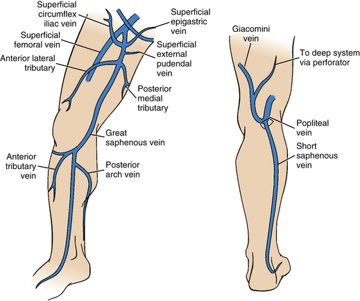
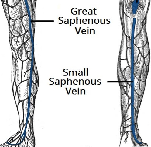
The great saphenous vein is formed by the dorsal venous arch of the foot and the dorsal vein of the great toe.

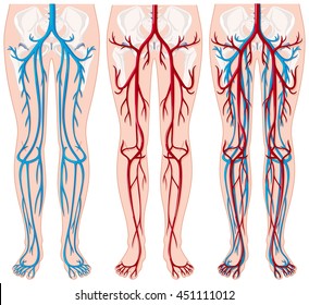
Leg vein anatomy. The deep veins of the leg accompany the arteries of the same name and are generally paired in the calf. There are three main deep veins in the lower leg. Place the probe transversely at the knee crease in the popliteal fossa.
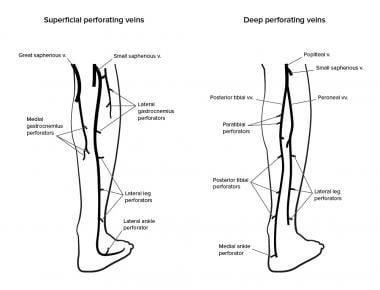
In the context of diagnosing a deep vein thrombosis the posterior tibial and peroneal veins are the most frequently affected. Anterior femoral cutaneous vein a continuation of anterior veins in the distal thigh. Superficial veins of the lateral leg and thigh form the lateral venous system.
Deep veins of the foot form two divisions. As the vein moves up the leg it receives tributaries from other small superficial veins. Seat the patient on the side of the bed to help dilate the veins for easier visualisation.
However their junctions are not paired and their locations are variable. Posterior tibial vein and fibular vein also known as the peroneal vein which form from the medial and lateral plantar veins. The lateral venous system is drained through multiple small tributaries into the gsv and ssv.
It ascends up the medial side of the leg passing anteriorly to the medial malleolus at the ankle and posteriorly to the medial condyle at the knee. There are six of such tributaries. Superficial epigastric vein drains the inferior abdominal wall and opens.
The plantar and the dorsal veins. Continue to follow the vein sequentially compressing down to the distal thigh. The sural nerve courses along the ssv in the distal calf.
Anterior tibial vein which receives blood from the dorsal venous arch.

 Detailed Anatomy Of The Venous System Of The Leg In View Of

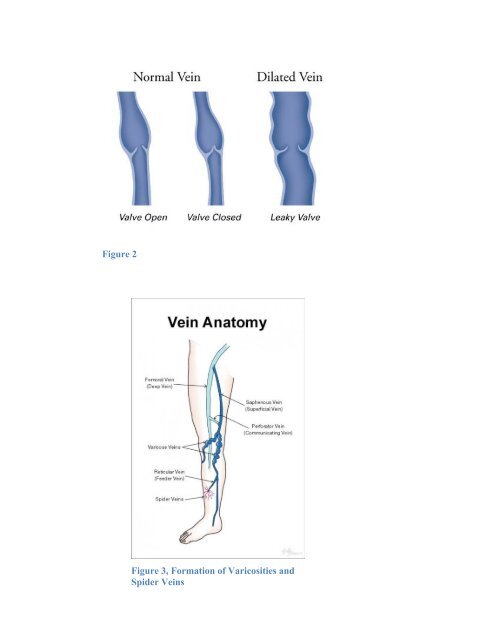
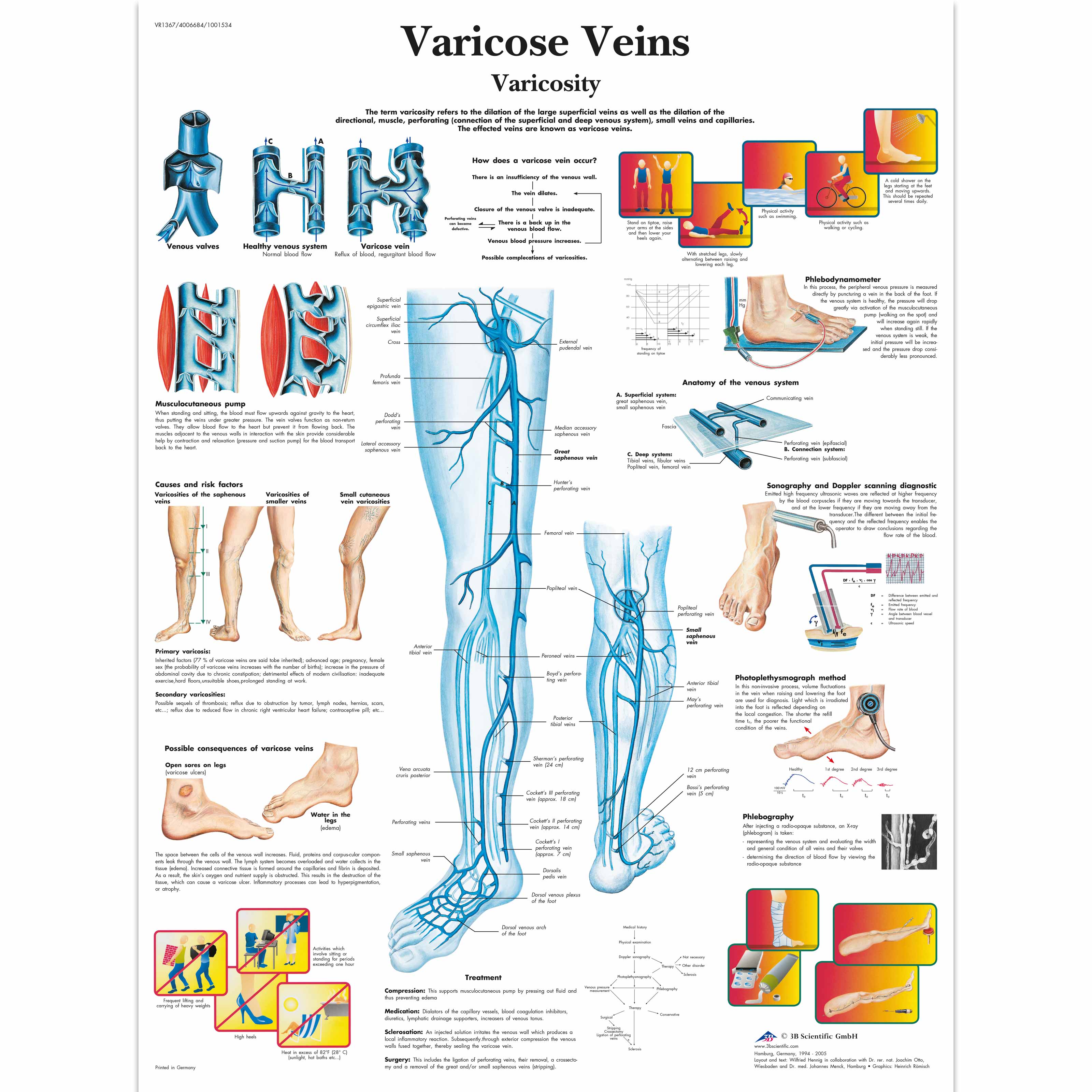 Varicose Veins Chart
 Venous Lymphatic Drainage Of Lower Limb
 Ultrasonography
 Leg Veins Stock Illustrations 316 Leg Veins Stock
 The Care Of Patients With Varicose Veins And Associated
 Location Of Venous Reflux In Primary Chronic Venous Disease

Deep And Superficial Leg Veins Tasmeemme Com
 13101 01x Arteries And Veins Anatomy Exhibits
 Lower Extremity Veins Radiology Key
 Perforator Vein An Overview Sciencedirect Topics

 Glossary Of Terms Medtronic
 Leg Vein Treatment Basics
 Varicose Vein Symptoms

 Venous Insufficiency Background Anatomy Pathophysiology
 Leg Veins Anatomy Images Stock Photos Vectors Shutterstock

 Lower Extremity Venous Anatomy Dallas Tx Venous System
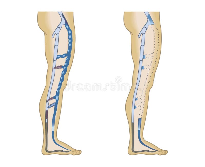
 Varithena Treats Bulging Veins That Occur In The Leg S
 Treatment Of Varicose Veins And Telangiectatic Lower
 Varicose Veins Clinical Features Management
 Deep Vein Thrombosis Harvard Health


Posting Komentar
Posting Komentar