Movements at the knee joint are essential to many everyday activities including walking running sitting and standing. It is formed by articulations between the patella femur and tibia.
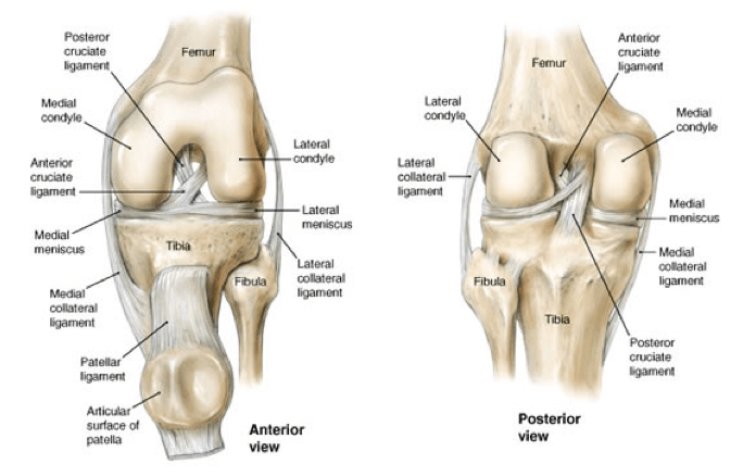 Knee Anatomy And Significance Bone And Spine
Tibia the bone at the front of the lower leg or shin bone.
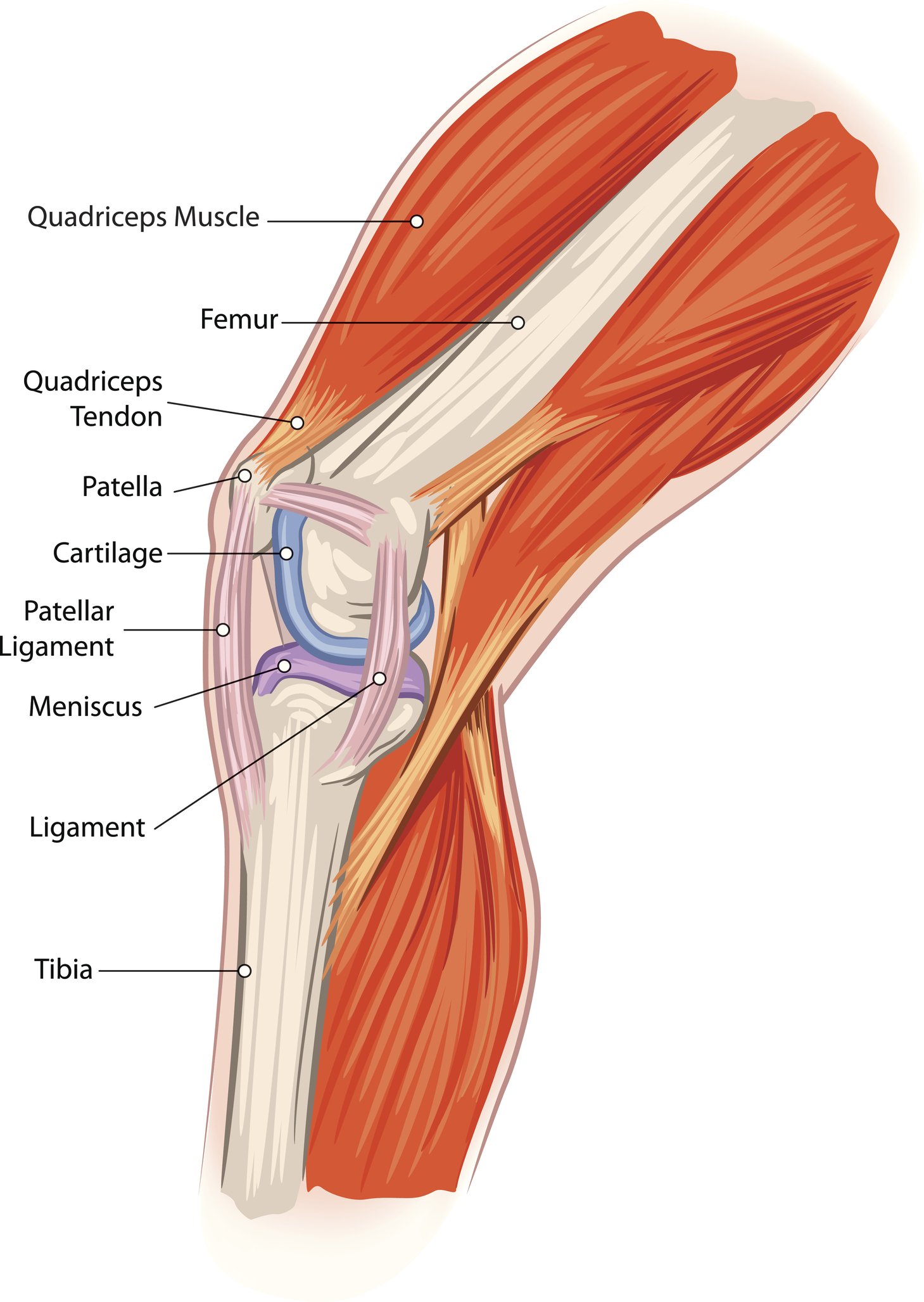
Anatomy knee joint. Tendons connect the knee bones to the leg muscles that move the knee joint. The knee joint is a synovial joint which connects the femur our thigh bone and longest bone in the body to the tibia our shinbone and second longest bone. Femur the upper leg bone or thigh bone.
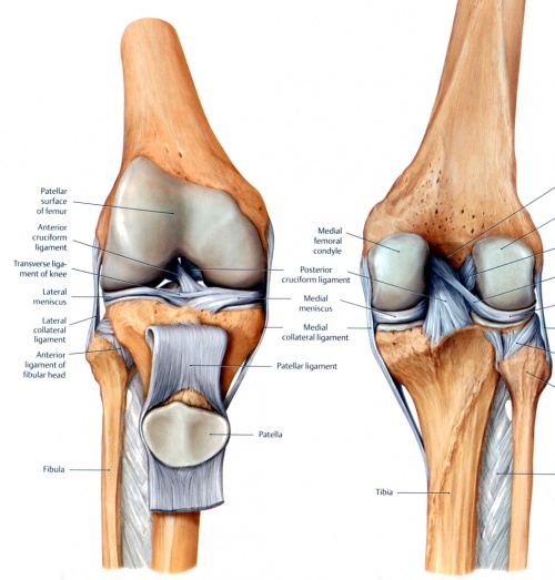
The knee joint is a complex structure that involves bones tendons ligaments muscles and other structures for normal function. The knee is one of the largest and most complex joints in the body. Damage to any structure of the knee anatomy will impact normal movement of the leg.
The knee is a complex joint that flexes extends and twists slightly from side to side. The knee is the meeting point of the femur thigh bone in the upper leg and the tibia shinbone in the lower leg. Medically reviewed by healthline medical team on april 6 2015.
The most common ligament injuries are acl tears mcl tears pcl tears and knee sprains which occur when the ligaments are overstretched. The knee joint is one of the strongest and most important joints in the human body. The hip and knee.
In knee joint anatomy they are the main stabilising structures of the knee acl pcl mcl and lcl preventing excessive movements and instability. Gracilis is the only adductor of the thigh that crosses and acts on two joints. Learn more about the anatomy of this muscle at kenhub.
The knee is also a very common area for injury. The knee is the joint where the bones of the lower and upper legs meet. It allows the lower leg to move relative to the thigh while supporting the bodys weight.
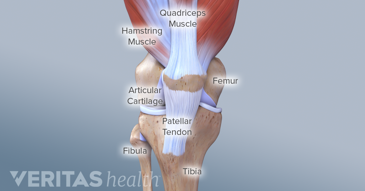
The largest joint in the body the knee moves like a hinge allowing you to sit squat walk or jump. It is designed to support the full weight of the body allowing us to stand walk run or dance with ease grace and fluidity. The smaller bone that runs alongside the tibia fibula and the kneecap patella are the other bones that make the knee joint.
There are two joints in the kneethe tibiofemoral joint which joins the tibia to the femur and the patellofemoral joint which joins the kneecap to the femur. The knee joins the thigh bone femur to the shin bone tibia. The knee joint is the largest joint in the human body.
The knee consists of three bones. The knee joint is a hinge type synovial joint which mainly allows for flexion and extension and a small degree of medial and lateral rotation. When there is damage to one of the structures that surrounds the knee joint this can lead to discomfort and disability.
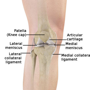 Normal Anatomy Of The Knee Joint Middletown Knee Treatment
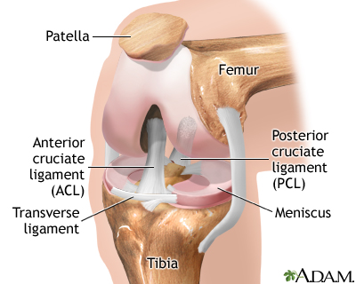 Knee Arthroscopy Series Normal Anatomy Medlineplus
 Amazon Com Emvency Mouse Pads Pain Patella Knee Joint
 The Knee Anatomy Injuries Treatment And Rehabilitation
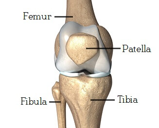 Knee Joint Anatomy Motion Knee Pain Explained
Fractures Of The Proximal Tibia Shinbone Orthoinfo Aaos
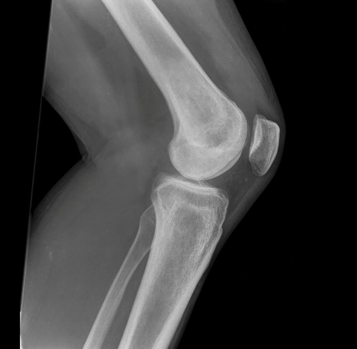 Anatomy Of The Knee Joint Owlcation
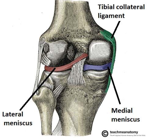 The Knee Joint Articulations Movements Injuries
Functional Anatomy Of The Knee Movement And Stability
 Anatomy Of The Knee Ct Arthrography
 Anatomy Knee Joint Cross Section Showing The Major Parts Which
 Anterior And Posterior Aspects Of The Knee Netter
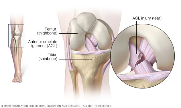 Knee Pain Symptoms And Causes Mayo Clinic
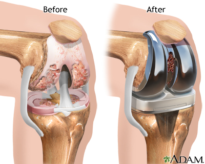 Knee Joint Replacement Medlineplus Medical Encyclopedia
 Knee Joint Part 1 3d Anatomy Tutorial
Knee Advanced Orthopedics Sports Medicine
 Knee Joint Anatomy Pictures And Information
 The Knee Joint Laminated Anatomy Chart
 Anatomy Of The Knee Central Coast Orthopedic Medical Group
 Anatomy And Function Of The Knee Skagit Northwest Orthopedics
 Amazon Com Semtomn Canvas Wall Art Print Patella Knee Joint
Collateral Ligament Injuries Orthoinfo Aaos
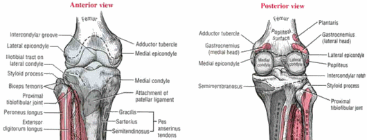 Applied Anatomy Of Knee Joint Epomedicine
Common Knee Injuries Orthoinfo Aaos



Posting Komentar
Posting Komentar