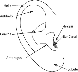
Picture of the ear. Anatomy of the auricle pinna.
The primary division of a leaf.
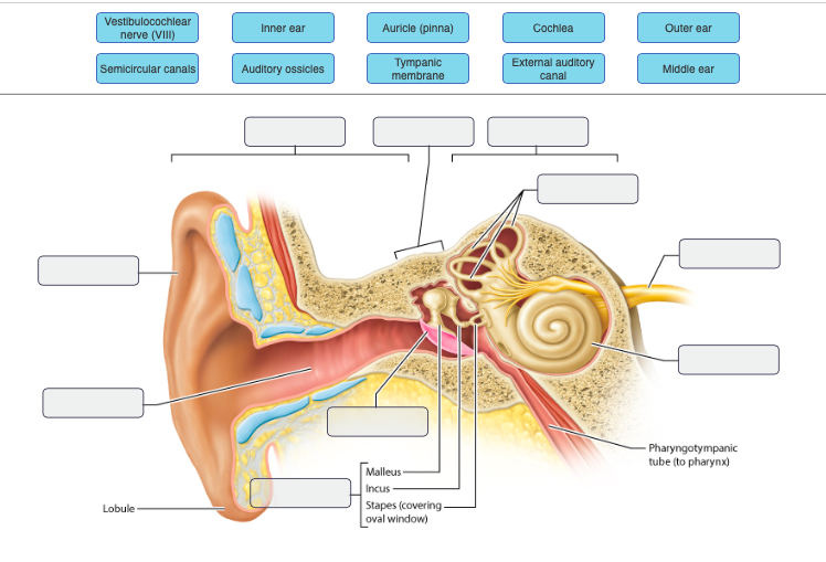
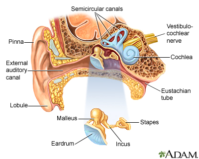
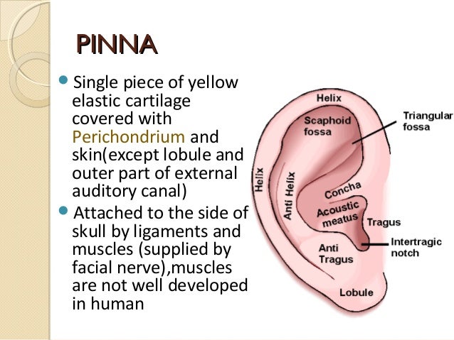
Anatomy of ear pinna. External auditory canal or tube. The external ear can be divided functionally and structurally into two parts. The auricle or pinna is a relatively large appendage on the side of the head.
Also known as the auricle. The former projects from the side of the head and serves to collect the vibrations of the air by which sound is produced. The pinna is the only visible part of the ear the auricle with its special helical shape.
The function of the pinna is to act as a kind of funnel which assists in directing the sound further into the ear. The external ear consists of the expanded portion named the auricula or pinna and the external acoustic meatus. Auricle anatomy the auricle or auricula is the visible part of the ear that resides outside the head.
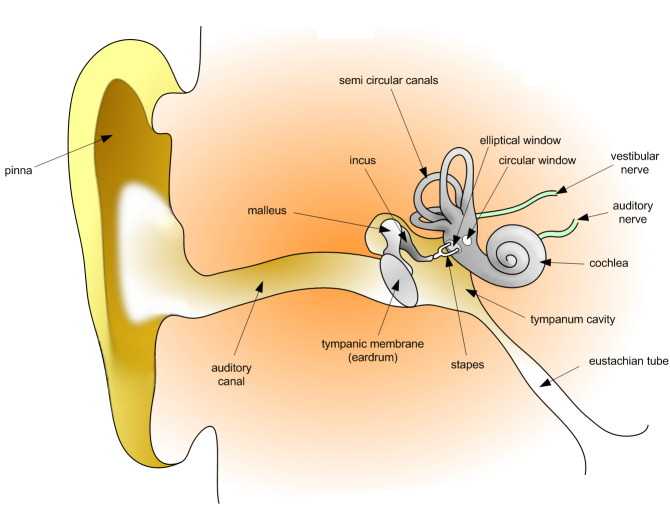
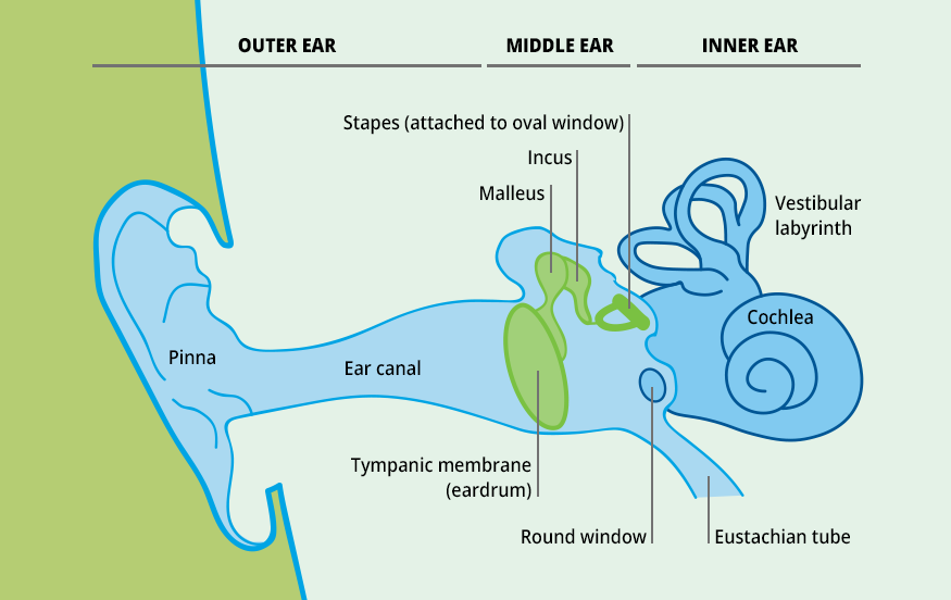
Sound causes the eardrum and its tiny attached bones in the middle portion of the ear to vibrate. This article will focus on the anatomy of the external ear its structure neurovasculature and its clinical correlations. A wing or fin.
It is the first part of the ear that reacts with sound. It has comparatively little effect on the acuity of hearing. It is prominent in most people and plays a very important role in hearing it directs sound waves into the ear canal.
The projecting part of the external ear of mammals. Anatomy of the auricle. Pinna the visible external ear.
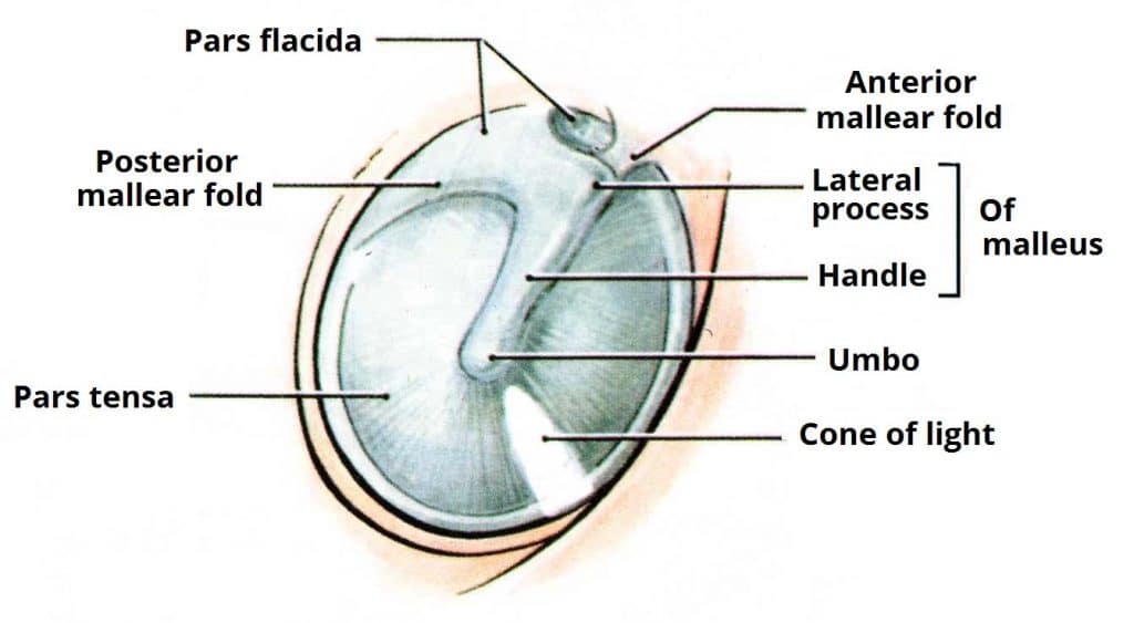
The latter leads inward from the bottom of the auricula and conducts the vibrations to the tympanic cavity. This is the outside part of the ear. Without this funnel the sound waves would take a more direct route into.
It is also called the pinna latin for wing fin plural pinnae a term that is used more in zoology. Sound funnels through the pinna into the external auditory canal a short tube that ends at the eardrum tympanic membrane. For more anatomical pictures of the auricle external auditory canal and tympanic membrane click here.
The auricle is composed of cartilage which is covered by skin. This is the tube that connects the outer ear to the inside or middle ear. The auricle or pinna and the external acoustic meatus which ends at the tympanic membrane.
The pinna consists of a skin flap on a skeleton of cartilage. The outer ear is called the pinna and is made of ridged cartilage covered by skin. External or outer ear consisting of.
 Picture Of The Ear Ear Conditions And Treatments
 15 10 Hearing Medicine Libretexts
 External Ear An Overview Sciencedirect Topics
 How We Hear Introduction To Psychology
 Ear Pinna Images Stock Photos Vectors Shutterstock
 Ear Anatomy Outer Ear Mcgovern Medical School
 The External Ear Structure Function Innervation
 Biology For Kids Hearing And The Ear
 Auricle Anatomy Google 검색 External Ear Anatomy Ear
 The Human Ear The Auricle Part 2 Wayne Staab Wayne S World
What Is Otoplasty Procedure Surgery And Recovery Sutured
Ear Anatomy Causes Of Hearing Loss Hearing Aids Audiology
 Anatomy Of The Ear Inner Ear Middle Ear Outer Ear
Ear Anatomy Causes Of Hearing Loss Hearing Aids Audiology
Anatomy Of The Ear Diagnosis 101
 Solved Vestibulocochlear Nerve Vlll Outer Ear Inner Ear
 Ear Anatomy Medlineplus Medical Encyclopedia Image
 Artificial Pinna For Ear Simulation Tme Systems
 Audition And Somatosensation Anatomy And Physiology I
 2 Anatomy Of The Pinna Of External Ear And Iannarelli S
 Anatomy And Landmarks Of The Auricle Download Scientific


Posting Komentar
Posting Komentar