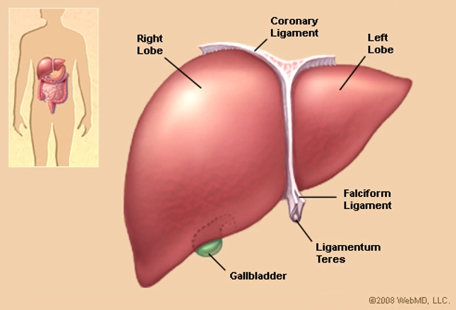
Each lobe is separated into many tiny hepatic lobules the livers functional units figure 3. The falciform ligament runs inferiorly from the diaphragm across the anterior edge of.
 Liver Anatomy Picture Function Conditions Tests
Each lobule is made up of millions of hepatic cells that are the basic metabolic cells of the liver.
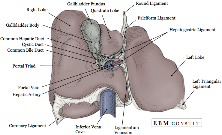
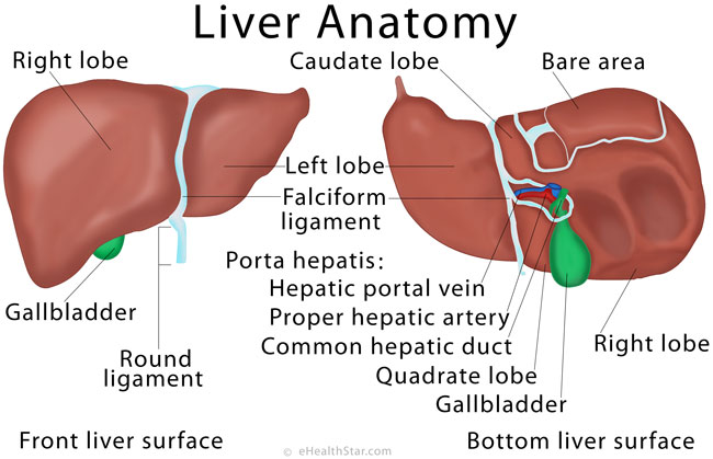
Lobes of liver anatomy. The liver has deep interlobular fissures and a large amount of interlobular connective tissue. It is mostly on the right of the. Right hepatic vein divides the right lobe into anterior and posterior segments.
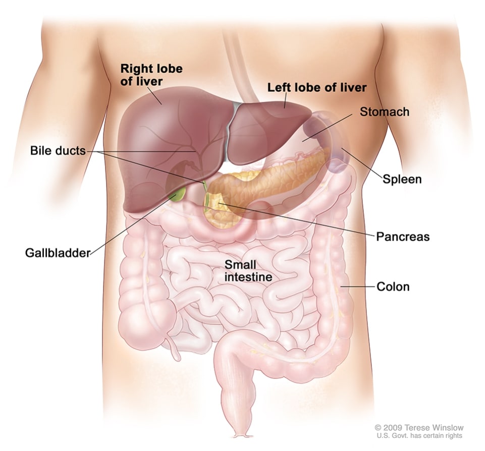
These lobules are connected to small ducts tubes that connect with larger ducts to form the common hepatic duct. The liver has two large sections called the right and the left lobes. Traditionally the liver is divided into four lobes.
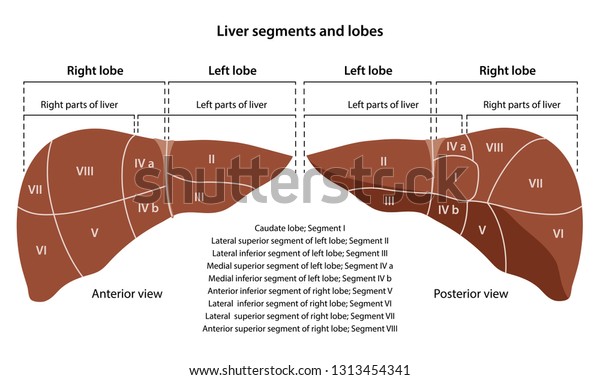
Left right caudate and quadrate. A fibrous capsule encloses the liver and ligaments divide the organ into a large right lobe and a smaller left lobe figure 2. Both are made up of 8 segments that consist of 1000 lobules small lobes.
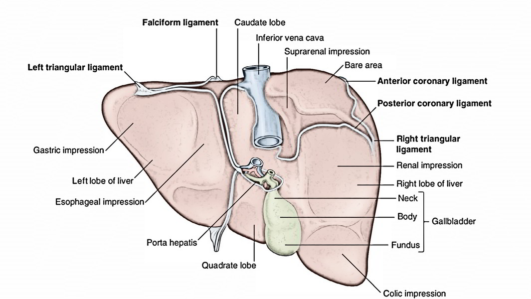
It is divided into a right lobe and left lobe by the attachment of the falciform ligament. Located on the lateral borders of the left and right lobes respectively the left and right triangular ligaments. There is a small caudate lobe which does not contact the kidney so no renal impression.
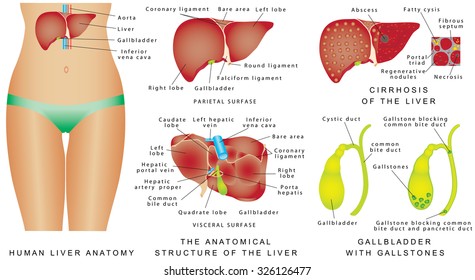
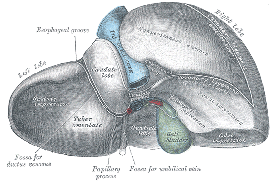
The liver consists of 2 main lobes. The liver is grossly divided into two parts when viewed from above a right and a left lobe and four parts when viewed from below left right caudate and quadrate lobes. The liver and these organs work.
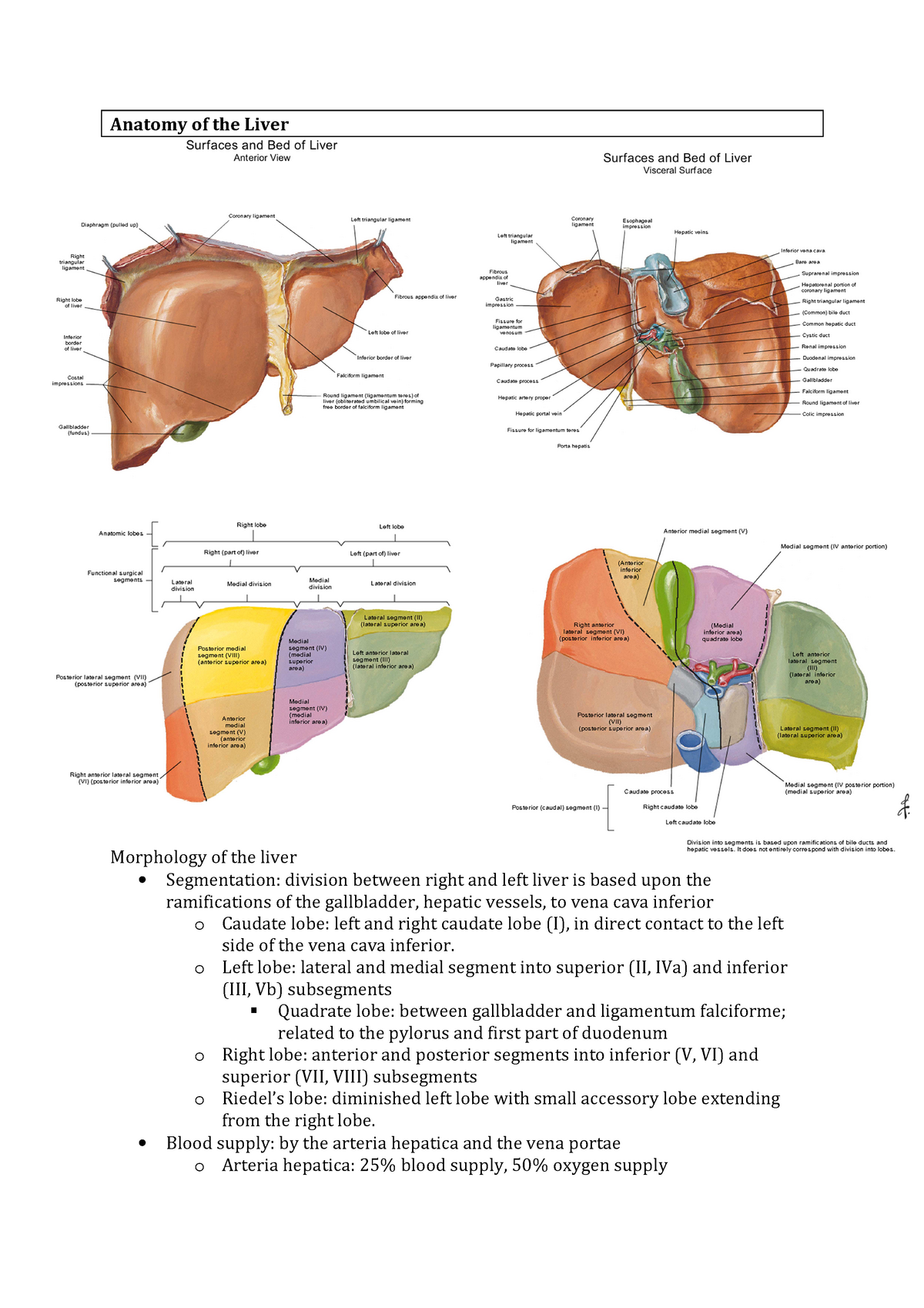
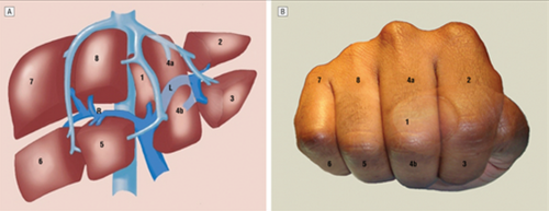
The falciform ligament divides the left lobe into a medial segment iv and a lateral part segment ii and iii. From below the two additional lobes are located between the right and left lobes one in front of the other. Middle hepatic vein divides the liver into right and left lobes or right and left hemiliver.
The liver also has two minor lobes the quadrate lobe and the caudate lobe. This plane runs from the inferior vena cava to the gallbladder fossa. It lies between the inferior vena cava and a fossa produced by the ligamentum venosum a remnant of the fetal ductus venosus.
There are two further accessory lobes that arise from the right lobe and are located on the visceral surface of liver. The falciform ligament divides the liver into a left and right lobe. Caudate lobe located on the upper aspect of the visceral surface.
The wide coronary ligament connects the central superior portion of the liver to the diaphragm. It has a mottled appearance. A deep interlobular fissure divides the liver into 4 lobes the left right medial and lateral.
The gallbladder sits under the liver along with parts of the pancreas and intestines. The lobes are further divided into lobules the functional units of the liver. Lobes of the liver.
:background_color(FFFFFF):format(jpeg)/images/library/11954/anterior-view-of-liver_english.jpg) Liver And Gallbladder Anatomy Location And Functions Kenhub
Liver And Gallbladder Anatomy Location And Functions Kenhub
 Liver Normal Anatomy And Examination Techniques Radiology Key
 Liver Lobe Images Stock Photos Vectors Shutterstock
 Notes Learning Stage Surgery Liver Anatomy Lecture 1 Vu
 Liver Anatomy Histology And Its Functions In Detail
 Easy Notes On Liver Learn In Just 4 Minutes Earth S Lab
Gastroenterology Education And Cpd For Trainees And
 Human Liver Lobes Anatomy Liver Lobes Medical Science Vector
 4 Lobes Of Liver Png Cliparts For Free Download Uihere
 Human Liver Anatomy Front Back And Two Lobes Location Of The
![]() Human Liver Infographic Poster With Chart Diagram And Icon
Human Liver Infographic Poster With Chart Diagram And Icon
 Liver Lobe Images Stock Photos Vectors Shutterstock
 Trauma Residents How To Remember Liver Anatomy The Trauma Pro
 Anatomy Human Liver Description Segments Lobes Stock Image
 Figure Anatomy Of The Liver The Pdq Cancer




Posting Komentar
Posting Komentar