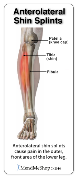
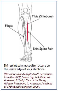
The muscles of the calf also work subtly to stabilize the ankle joint and foot and to maintain the bodys balance. Shin splint pain most often occurs on the inside edge of your tibia shinbone.
 Shin Splints Cramps Thrombosis Oh My
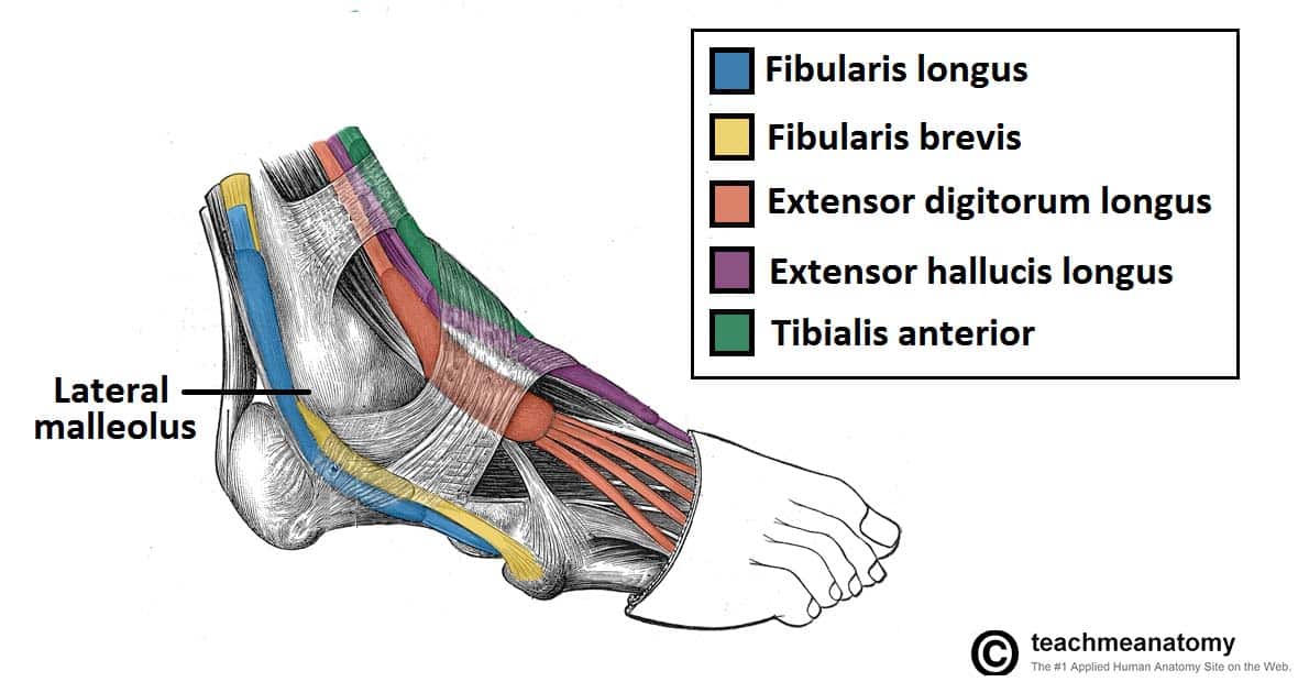
Shin muscles such as the tibialis anterior and extensor digitorum longus dorsiflex the foot and extend the toes.
Shin muscles anatomy. Muscles of the foot. Pain typically occurs along the inner border of the tibia where muscles attach to the bone. The hamstring muscles also known as the rear thighs make up the backside of the upper leg anatomy.
Biceps femoris long head. Like the quadriceps the hamstring muscle group also contains four separate muscles. Muscles of the gluteal region.
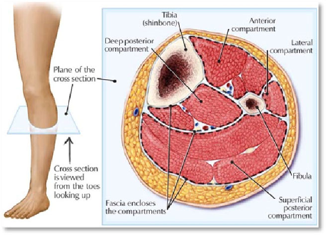
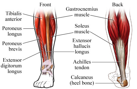
The deep posterior compartment. The medical information on this site is provided as an information resource only and is not to be used or relied. The gastrocnemius and soleus muscles taper and merge at the base of the calf muscle.
During walking running or jumping the calf muscle pulls the heel up to allow forward movement. The posterior compartment holds the large muscles that we know as the calf muscles. Some nerves of the sacral plexus innervate this area namely the superficial fibular nerve the deep fibular nerve and the tibial nerve.
The achilles tendon inserts into the heel bone calcaneus. It mainly serves as an attachment point for the muscles of the lower leg. Part of the teachme series.
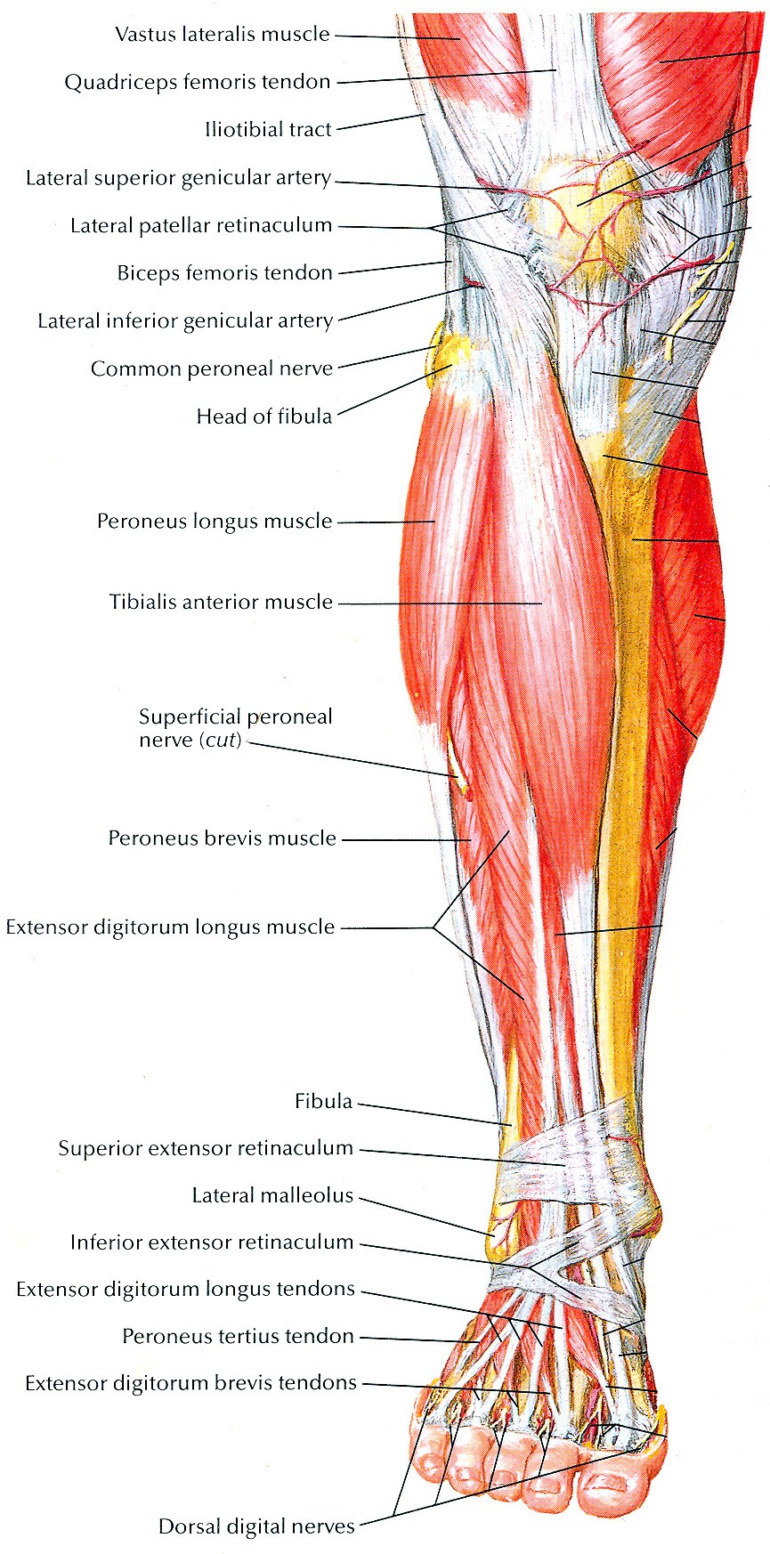
It acts as the main weight bearing bone of the leg. The lateral compartment is along the outside of the lower leg. The anterior tibial posterior tibial and the fibular arteries supply blood to the lower leg.
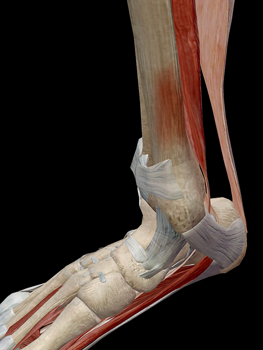
The fibula is located next to the tibia. This muscle is mostly located near the shin. The muscles of the lower leg the anterior compartment in the front of the shin holds the tibialis anterior.
Start now for free. Muscles of the thigh. Tough connective tissue at the bottom of the calf muscle merges with the achilles tendon.
The tibialis anterior is a muscle in humans that originates in the upper two thirds of the lateral outside surface of the tibia and inserts into the medial cuneiform and first metatarsal bones of the foot. It acts to dorsiflex and invert the foot. The main muscle in this area of the leg is the gastrocnemius which gives the calf a bulging muscular appearance.
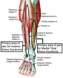
Muscles of the leg. Also called the shin bone the tibia is the longer of the two bones in the lower leg. Shin splints medial tibial stress syndrome is an inflammation of the muscles tendons and bone tissue around your tibia.
![]() Tibia Anatomy And Clinical Notes Kenhub
Tibia Anatomy And Clinical Notes Kenhub
 Muscular Function And Anatomy Of The Upper Leg

 Muscles Of The Anterior Leg Attachments Actions
:max_bytes(150000):strip_icc()/StandingAnteriorTibialisShinStretch_annotated-9e92ebc36e0441ebaf326dbe74a126e5.jpg) Stretch Your Anterior Tibialis To Prevent Shin Pain
Stretch Your Anterior Tibialis To Prevent Shin Pain
 Shin Pain Physio Podiatry Massage
 Iliopsoas Wikipedia
 Hold On To Your Tibias The Anatomy And Causes Of Shin Splints
 Leg Knee Anatomy
 The Simple Bump That Could Turn Serious High Country
 Muscles That Lift The Arches Of The Feet
 Hold On To Your Tibias The Anatomy And Causes Of Shin Splints

 Shin Splints Thermoskin Supports And Braces For Injury
 What Is Compartment Syndrome Surgery Symptoms Treatment
 Anatomy Of The Knee Howstuffworks
 Human Anatomy Leg Muscles Human Anatomy Leg Muscles Diagram
 Shin Splints And How To Treat Them Ice Rest Stretch
 Shin Splints Medial Tibial Stress Syndrome Zion Physical
Shin Splints Orthoinfo Aaos
 Anatomy Of Shin Splints Treatment And Prevention
 Tibial Stress Syndrome Shin Splints Knee Sports
 Symptom Of The Month March


Posting Komentar
Posting Komentar