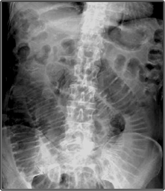
Lung bases on abdominal x ray. This involves assessment of the bowel gas pattern soft tissue structures and bones.
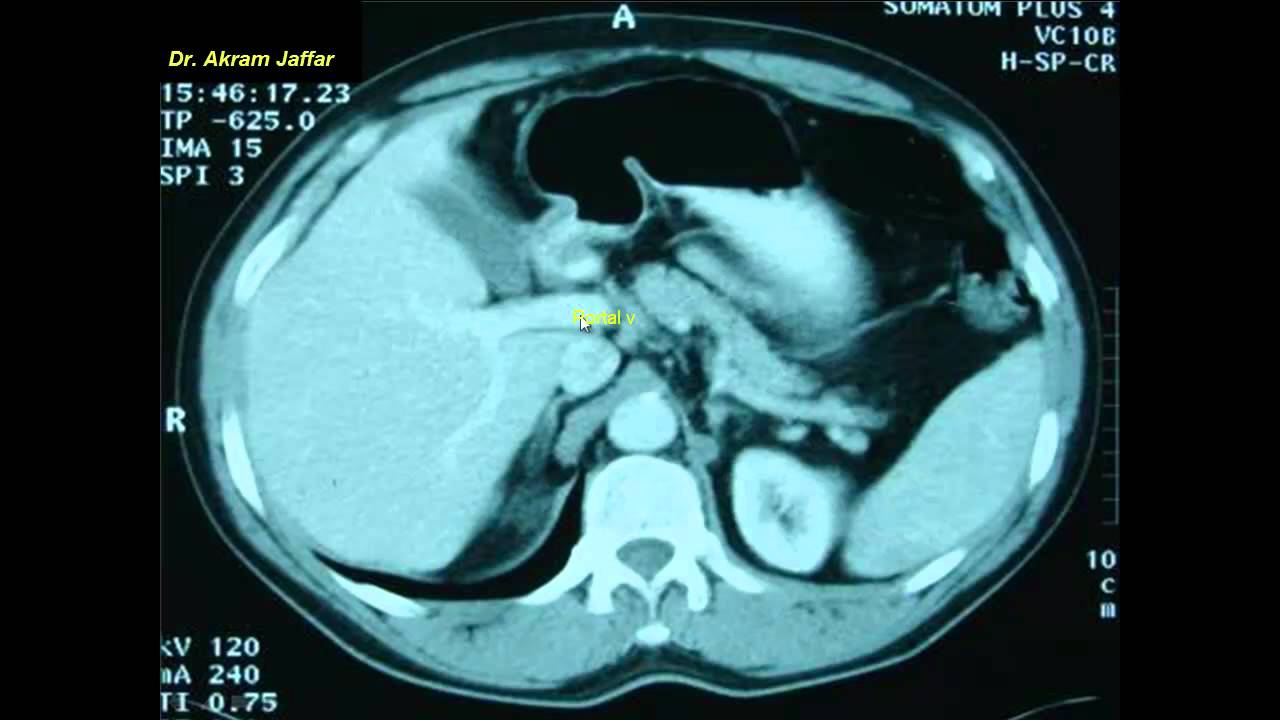 Cross Sectional And Imaging Anatomy Of The Abdomen
Other games by same author.
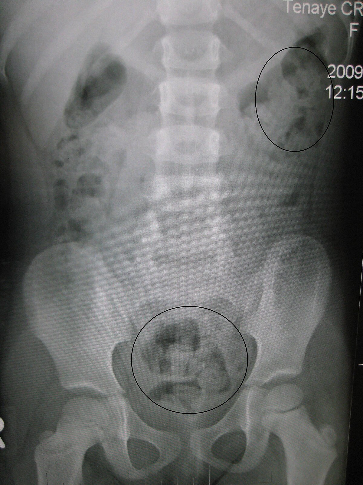
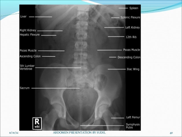
X ray abdomen anatomy. When an abdominal x ray is performed to provide pictures of the kidneys ureters and bladder its called a kub x ray. Tap onoff image to showhide findings. Click image to align with top of page.
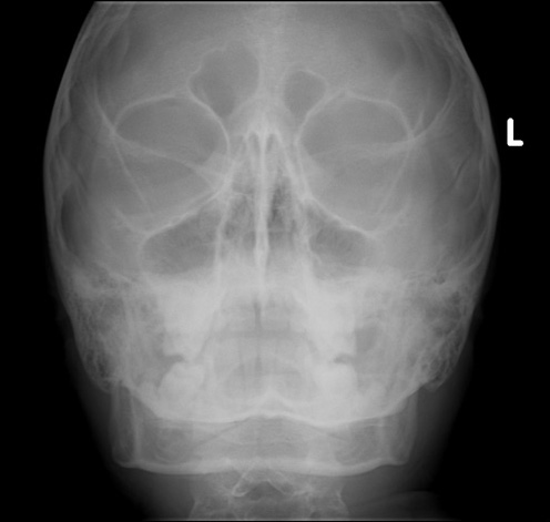
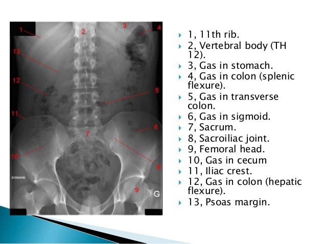
The teeth is the most important accessory organs of the digestive systems in radiology because it use to masticate the food. A plain x ray of the abdomen can help see the organs and conditions in the belly including intestinal obstruction or perforation. Abdominal x ray is a commonly performed diagnostic x ray examination that produces images of the organs in the abdominal cavity including the stomach liver intestines and spleen.
Ap knee x ray anatomy. Play this quiz called abdomen x ray anatomy and show off your skills. The parenchymal organs within the abdomen absorb x rays as they pass through the patient and therefore alter the appearance of the radiograph.
Abdominal radiography abdomen basic anatomy the digestive system consist of the alimentarty tract and certain organs that contribute to the digestive process. Anatomy abdomen xray abdomen xray anatomy supine abdomen xray supine abdomen xray supine xray kub kub anatomy. The lung bases which pass behind the liver and diaphragm in the posterior sulcus of the thorax may be visible on some abdominal x rays.
A systematic approach to abdominal x ray interpretation is therefore relatively straightforward. Login register free. Exposure will need to be adjusted according to the imaging system cr or dr and patient size.
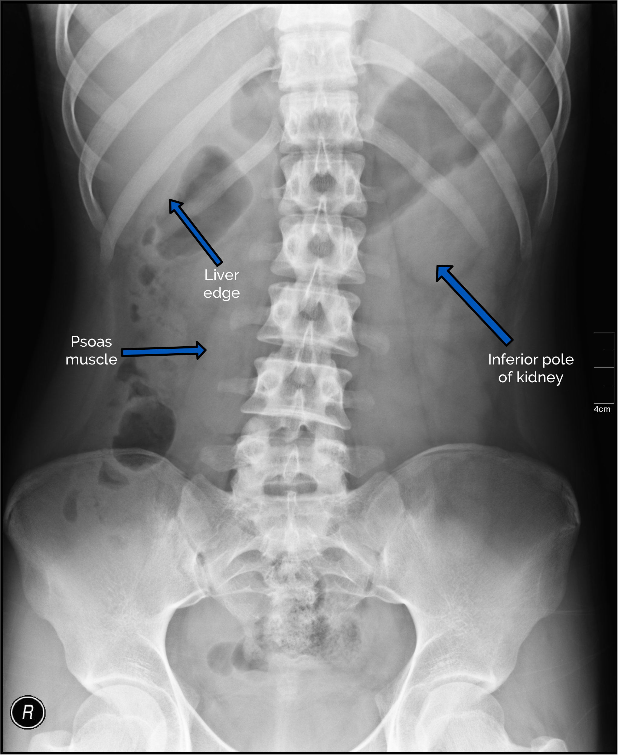
This is a quiz called abdomen x ray anatomy and was created by member desimichelle. These changes are subtle but with practice you should be able to make out several organs and muscles. Practical points for larger patients it may be necessary to perform two x rays using a landscape orientation of the detector to include the entire abdomen.
Full assessment includes a check of patient data image quality and checking for artifact and abnormal calcification. Lung bases on abdominal x ray. Hover onoff image to showhide findings.
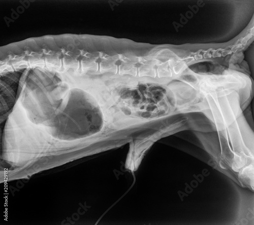
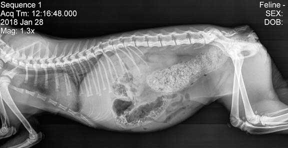
 Radiographs Of The Cat
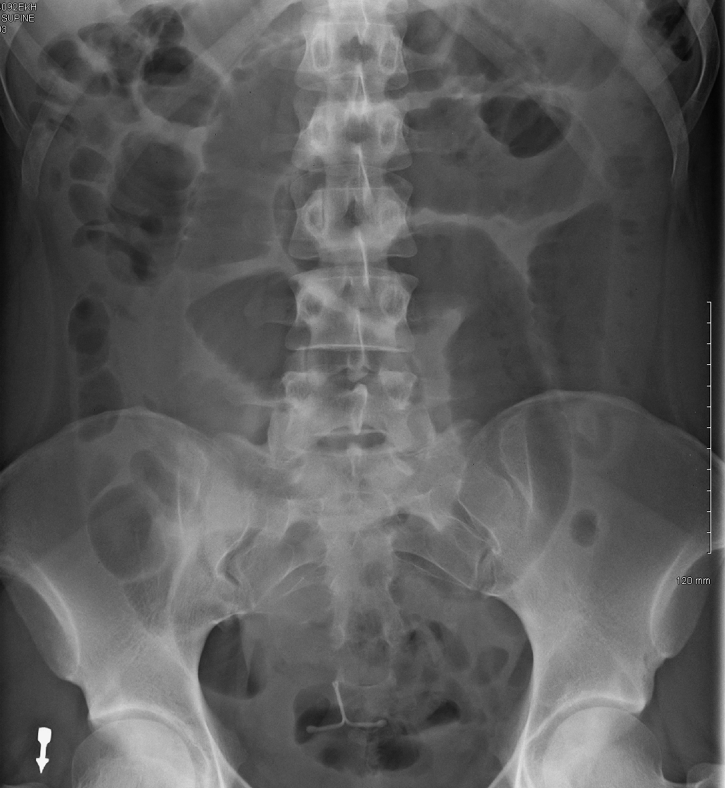
 Film Xray Abdomen Supine Show Small Stock Photo Edit Now
 Film Critique Part 2 Abdomen
 Abdominal X Ray Interpretation Axr Geeky Medics
 X Ray Film Of Dog Anterior View Closed Up In Thorax Standard
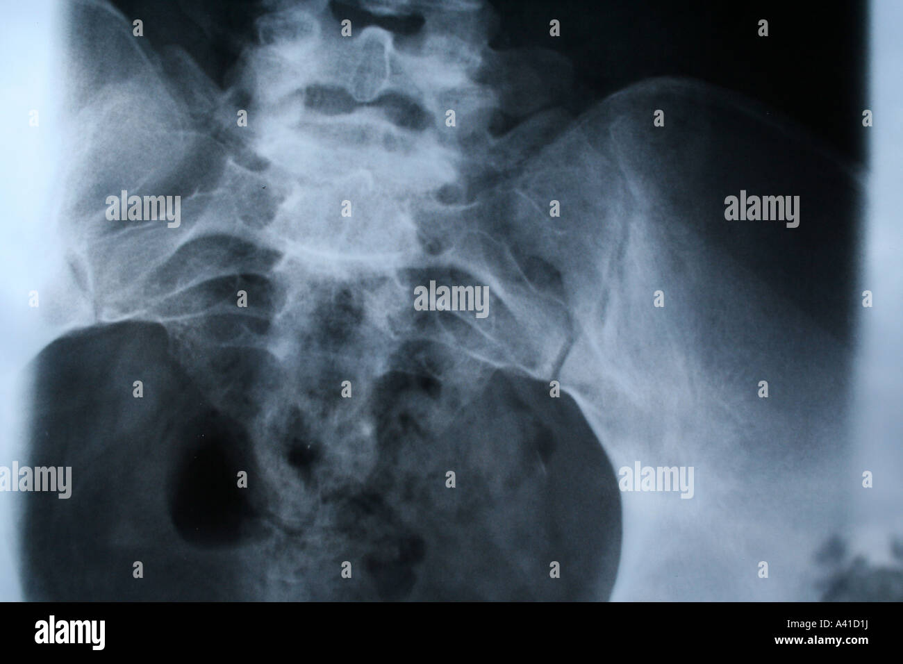 Grain Grainy Health Healthcare Medical Medicine Abdomen
 Abdominal X Ray Startradiology
X Ray Film Of Dog Lateral View Closed Up In Thorax Standard
 Untitled Document

 Learn To Read An X Ray Long Beach Animal Hospital
 Constipation Wikipedia
 The Radiology Assistant Chest X Ray Basic Interpretation
 Radiographic Anatomy Abdomen Ap Supine Medical

 Abdominal X Ray Interpretation Axr Geeky Medics
 Abdomen Radiography
 Abdominal X Rays Normal Anatomy On Axr
 Abdominal X Ray Startradiology
 Normal Abdominal Radiograph Annotated X Ray Radiology
 Radiographic Anatomy Of Gastrointestinal Tract


Posting Komentar
Posting Komentar