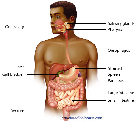
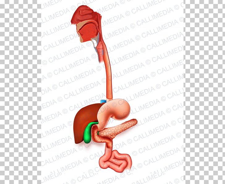
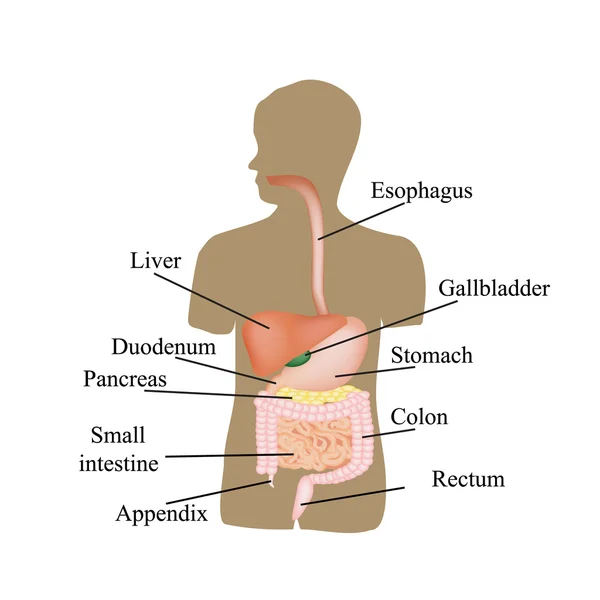
The diaphragm forms the upper surface of the abdomen. The alimentary canal is made up of the oral cavity pharynx esophagus stomach.
 Understanding Ulcers Chart 20x26 Anatomy Gastroenterology
In human anatomy the intestine bowel or gut.
Gastro anatomy. Gastrointestinal system anatomy introduction to the gastrointestinal system. This layer is responsible for creating the motion. The endoscope allows examination of the esophagus stomach and duodenum the first part of the small intestine.
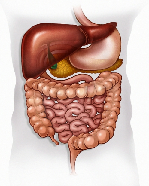
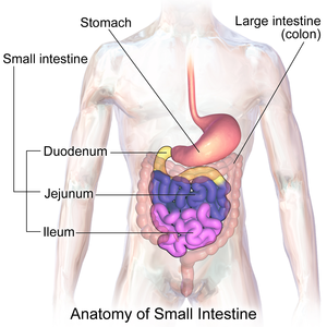
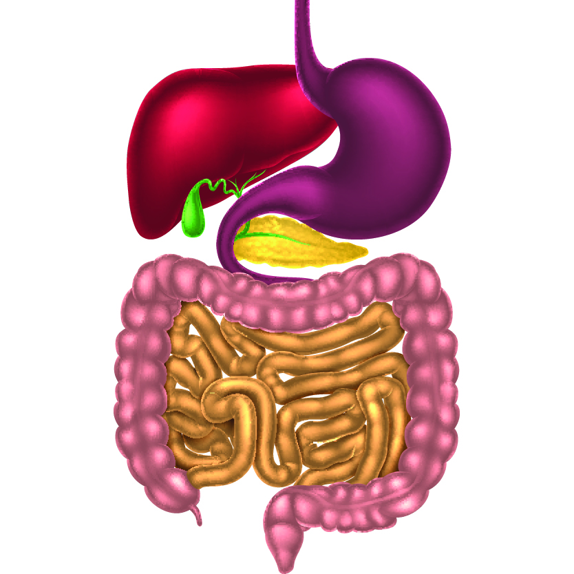
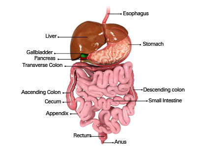
The abdomen contains all the digestive organs including the stomach small and large intestines pancreas liver and gallbladder. The anatomical structures of the gastrointestinal system work together to achieve three major goals. The digestive system is a group of organs working together to convert food into energy and basic nutrients to feed the entire body.
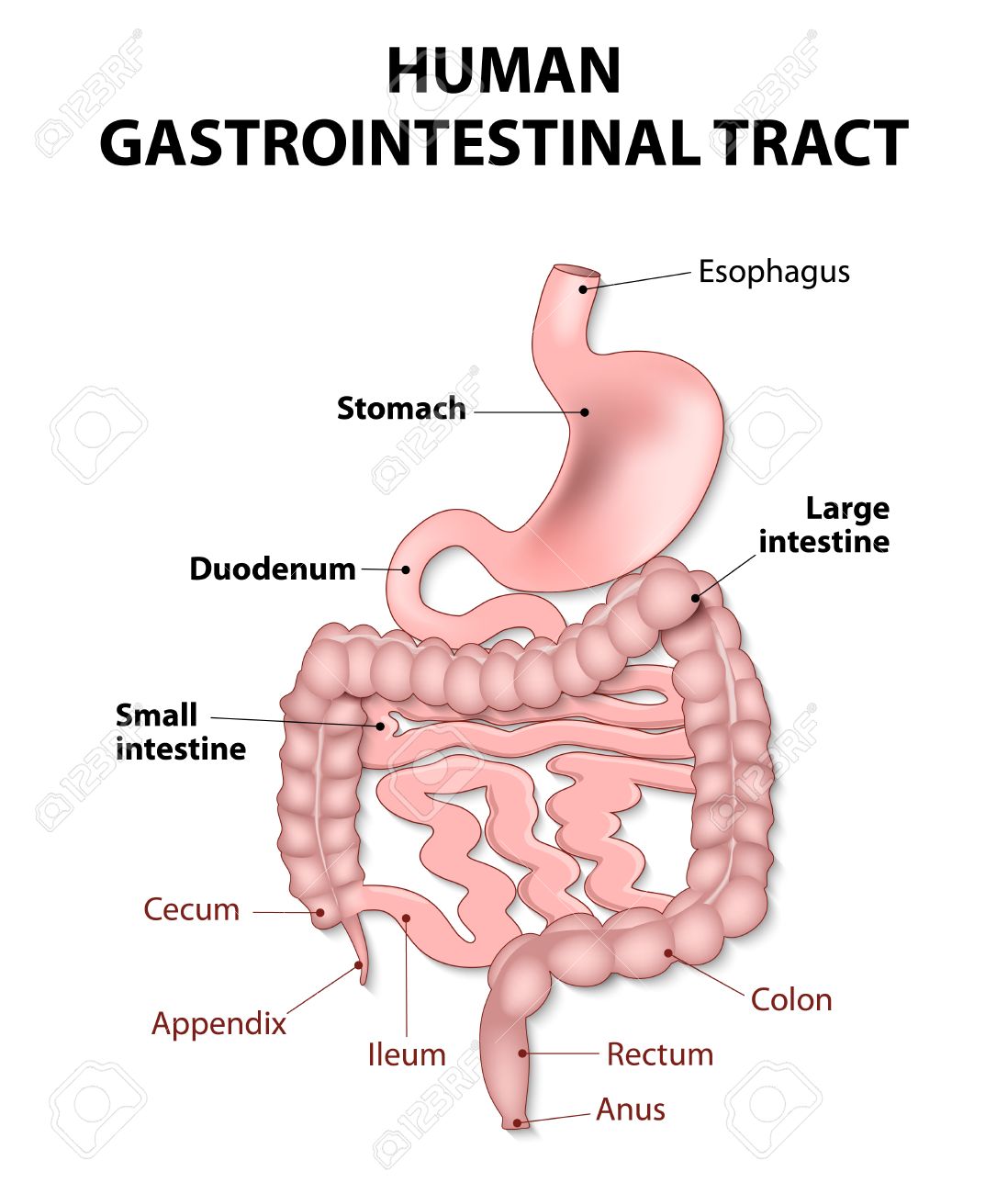
Gastrointestinal anatomy and physiology the major function of the gastrointestinal gi tract is digestion. The focus of this gastrointestinal anatomy and physiology course is to teach you about the structures and functions of the gastrointestinal system and its accessory organs. The anal canal is the final segment of the gastrointestinal tract and is involved in defecation and maintaining faecal continence.
The gastrointestinal tract is a muscular tube lined by a special layer of cells. At this layer the pylorus is surrounded by a thick. At the level of the pelvic bones the abdomen ends and the pelvis begins.
The muscularis externa lies beneath the submucosa and is unique from other organs of the gastrointestinal tract consisting of three layers. Its main function is to store and break down the foods and liquids that we consume before those. Computed tomography ct scan.
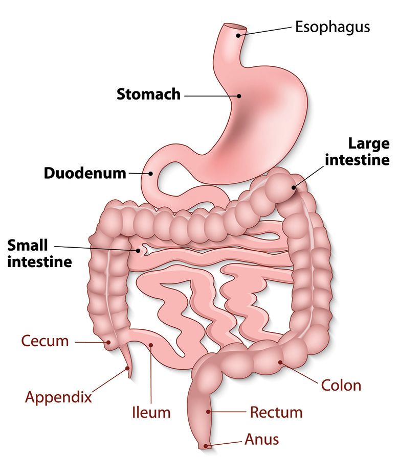
Individual components of the gastrointestinal system. Food passes through a long tube inside the body known as the alimentary canal or the gastrointestinal tract gi tract. The middle circular layer.
The inner oblique layer. éntera is the segment of the gastrointestinal tract extending from the pyloric sphincter of the stomach to the anus and as in other mammals consists of two segments the small intestine and the large intestine. A ct scanner uses x rays and a computer to create images of the stomach and abdomen.
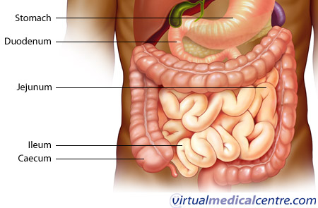
In this section learn more about the anatomy of the gastrointestinal tract the oesophagus stomach small intestine appendix cecum colon rectum and anal canal. It converts ingested nutrients into simpler forms that can be transported from the tracts lumen to the portal circulation and then used in metabolic processes. The stomach is located in the upper left area of the abdomen below the liver and next to the spleen.
 How Your Gastrointestinal Tract Works Mu Health Care
 Vessel Anatomy Abdominopelvic Organs And Their Arteries
 Gastrointestinal Stock Vector Illustration Of Medical
 Gastro Intestinal Tract
 Gastrointestinal System Anatomy Healthengine Blog
 Human Digestive System Digestion Gastrointestinal Tract
 Anatomy And Physiology Of Gastrointestinal System Tutorial
 Gastroenterology And Digestive Health
 The Gastrointestinal Tract Complete Anatomy
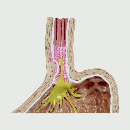
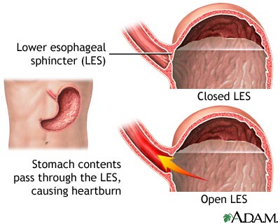 Gastroesophageal Reflux Disease Wake Gastroenterology
 Book An Appointment Today Gastro Specialist Dr Jimmy
![]() Lined Organs By S Leonov
Lined Organs By S Leonov
 Gastro Intestial Tract Alimentary Canal
 Gastro Stock Vectors Royalty Free Gastro Illustrations

 Gastro Intestinal Tract
![]() Ligament Of Treitz Suspensory Ligament Of Duodenum Kenhub
Ligament Of Treitz Suspensory Ligament Of Duodenum Kenhub
 Gastrointestinal Tract Includes All Structures Between The Esophagus
 Blind Loop Syndrome Wikipedia
Gastrointestinal System Anatomy Healthengine Blog
 4 Pc Gerd Gastro Esophageal Reflux Disease
 Gastroenterology Budge Clinic

 Pacific Gastroenterology Center For Digestive Health
 Anatomy Of Pelvic Floor Notes S2 S4 Represent Sacral Nerve


Posting Komentar
Posting Komentar