Where are the paranasal sinuses situated. The maxillary sinus the largest of the sinuses.
 Ct Anatomy Of Para Nasal Sinuses
Your cheekbones hold your maxillary sinuses the.

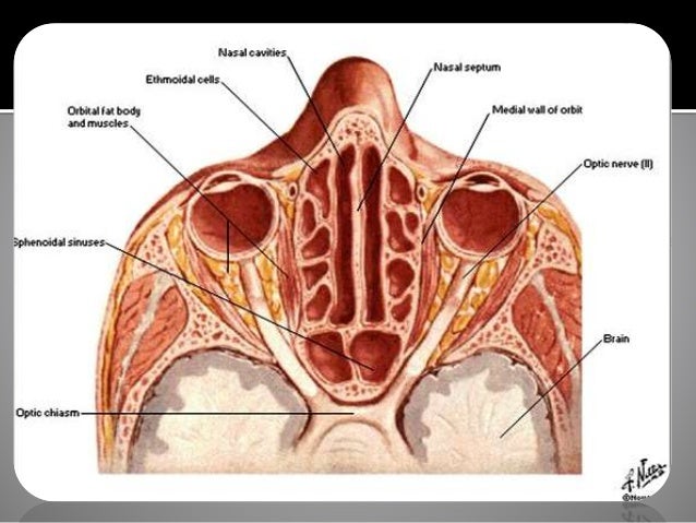
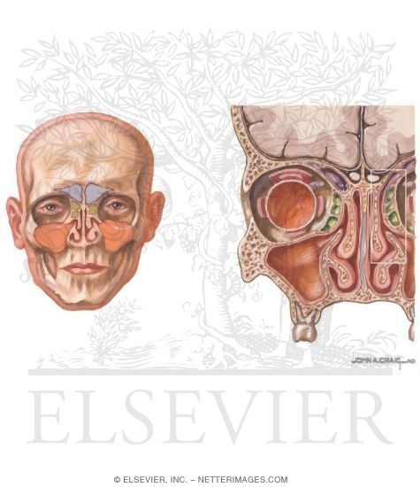
Paranasal sinuses anatomy. My interpretation of the anatomy and literature leads me to recognise seven paired paranasal sinuses. The superior border of this sinus is the bony orbit the inferior is the maxillary alveolar bone and corresponding tooth roots. They have thin walls which are often penetrated by the long roots of the posterior maxillary teeth.
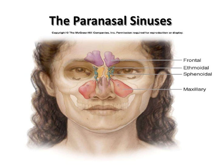
In the frontal ethmoid and sphenoid of the cranium and the maxillary bones of the face. The maxillary and ethmoid sinuses are present at birth starting to form around the 3rd or 4th month of gestational development 10. They further develop over the first few years of life 11.
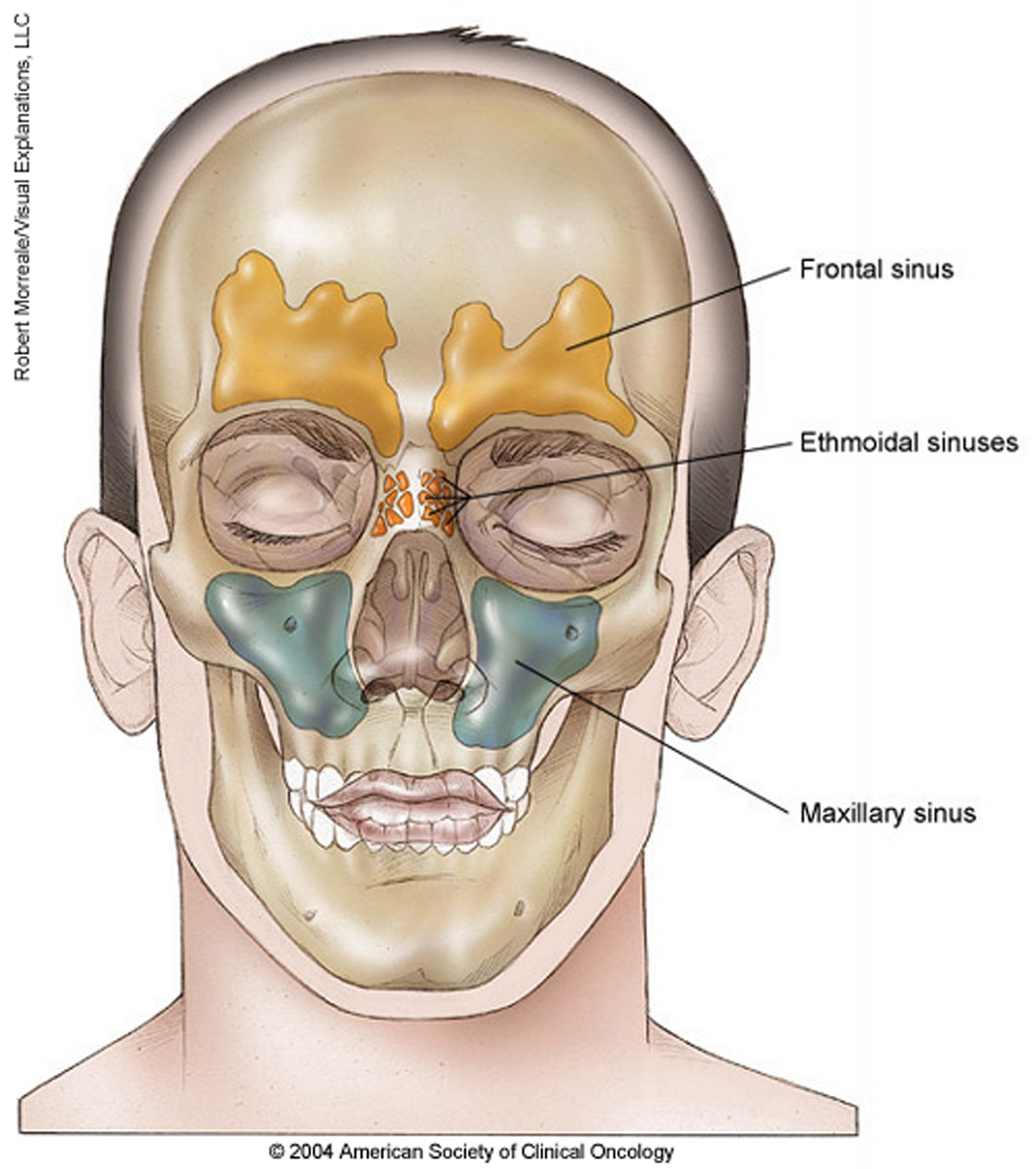
Humans possess four paired paranasal sinuses divided into subgroups that are named according to the bones within which the sinuses lie. Paranasal sinuses are air filled cavities in the frontal maxilae ethmoid and sphenoid bones. The largest sinus cavities are about an inch across.
Sphenoidal sinus see figs. The maxillary sinuses are the largest of the all the paranasal sinuses. The maxillary sinuses the largest of the paranasal sinuses are under the eyes.
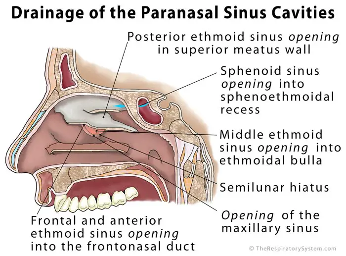
Anatomy of the paranasal sinuses development. 52 6 maxillary sinus see fig. As the paranasal sinuses are continuous with the nasal cavity an upper respiratory tract infection can spread to the sinuses.
From the nasal mucosa and their continued communication with the nasal fossae. Paranasal sinuses frontal sinuses. Ethmoid air cells sinuses.
Infection of the sinuses causes inflammation particularly pain and swelling of the mucosa and is known as sinusitis. These sinuses which have the same names as the bones in which they are located surround the nasal cavity and open into it. Rudimentary sphenoid sinuses are there at birth forming pneumatizing completely by the age of 5 years 6.
Others are much smaller. If more than one sinus is affected it is called pansinusitis. The frontal sinuses superior to the eyes in the frontal bone.
42 4 42 5 and 53 4. The frontal sinus the rostral maxillary sinus the caudal maxillary sinus the sphenopalatine sinus the dorsal conchal sinus the ventral conchal sinus and the ethmoidal sinus also known as the middle conchal sinus. Nose hairs at the entrance to the nose trap large inhaled particles.
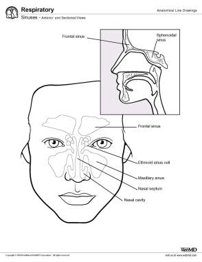
What are the paranasal sinuses a formation from. Frontal sinus see figs. The sinuses are a connected system of hollow cavities in the skull.
42 18 52 2 and 53 4.
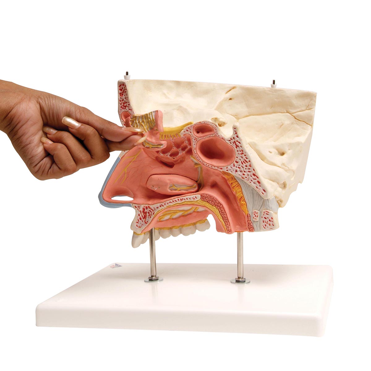 Human Nose Model With Paranasal Sinuses 5 Part 3b Smart
 Paranasal Sinus An Overview Sciencedirect Topics
 Paranasal Sinuses 14
 Anatomy Physiology Of Nose Nasal And Paranasal Sinus
 Overview And Topographic Anatomy Of The Paranasal Sinuses
 Nasal Cavity And Paranasal Sinus Cancer Cancer Net
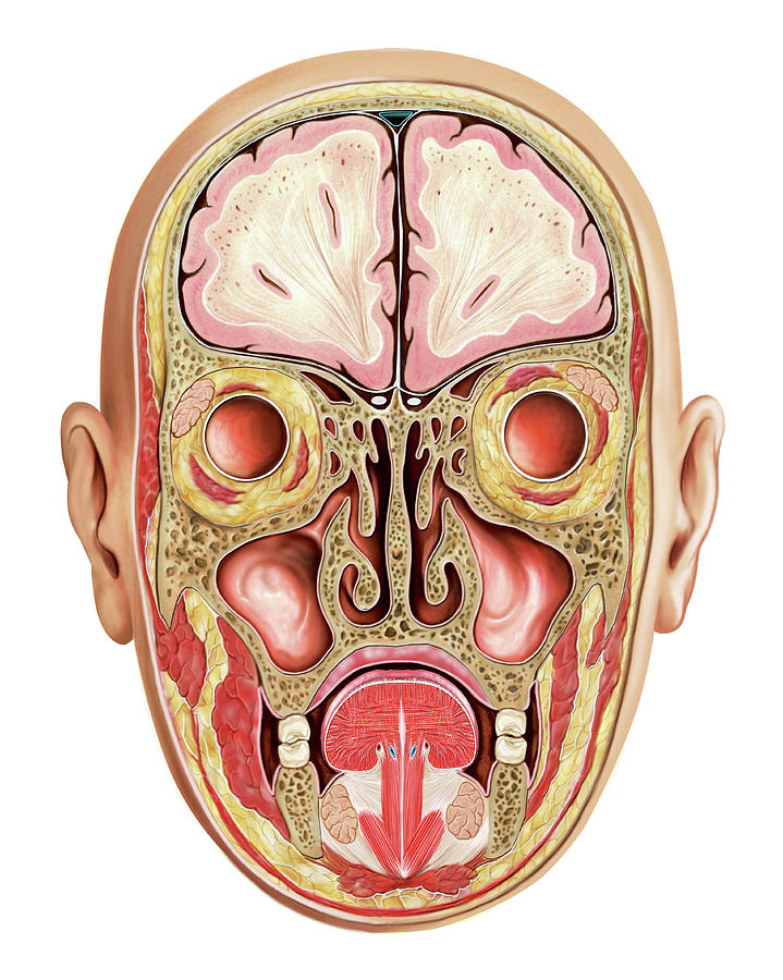
 Paranasal Sinuses Male Face Stock Illustration Download
Ga08 Nasal Cavity Paranasal Sinuses Pterygopalatine New
 Amazon Com Emvency Wall Tapestry Sinuses Of Nose Human
 Paranasal Sinus Definition Location Anatomy Function
 Anatomy Of The Sinuses Otolaryngology Houston
 Sinuses Of Nose
 Nose Useful Notes On Human Nose And Para Nasal Sinuses
 Paranasal Sinus Anatomy Overview Gross Anatomy
![]() Paranasal Sinuses Anatomy And Clinical Aspects Kenhub
Paranasal Sinuses Anatomy And Clinical Aspects Kenhub
 Ethmoid Sinus Ethmoid Bulla Paranasal Sinuses Anatomy Png
 Computed Tomography Anatomy Of The Paranasal Sinuses And
 Ear Paranasal Sinuses Pituitary Gland Bone Anatomy Studying
Alison Burke Anatomy Of Paranasal Sinuses And Nasal Passages
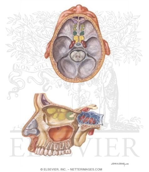
 Anatomy Of The Paranasal Sinuses
 Nose And Sinus Anatomy Thomas S Higgins Md Msph
 Ecr 2017 C 2117 Ct Anatomy Of Paranasal Sinuses Epos
 Overview And Topographic Anatomy Of The Paranasal Sinuses
 Anatomy Of Nose And Paranasal Sinus
 Paranasal Sinuses Wikipedia
 Anatomy Nasal Cavity Paranasal Sinuses Nasopharynx
 Anatomical Representation Of Paranasal Sinuses And Their Names


Posting Komentar
Posting Komentar