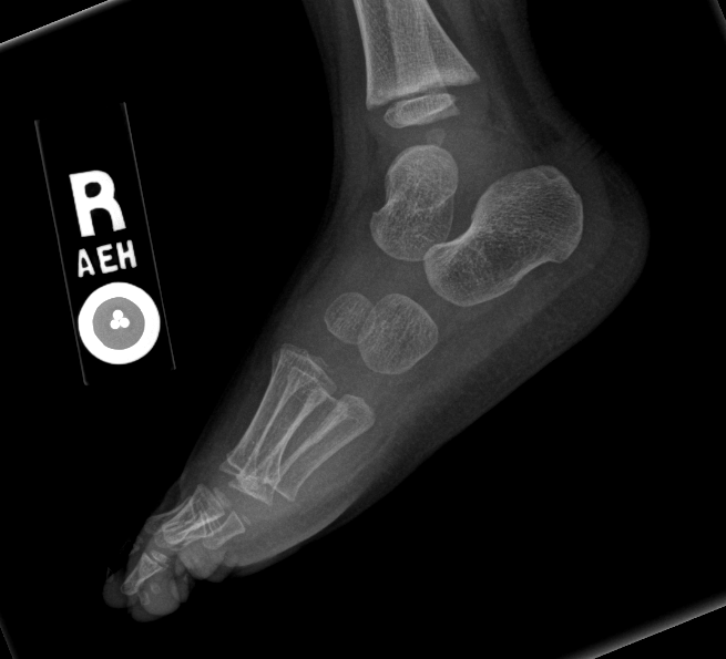
In a stress x ray the physician will apply pressure on the affected ankle while an x ray is being taken. The foot is a very stable composition of bones supported by strong ligaments.

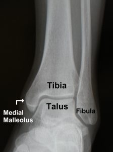
The ankle joint is comprised of the tibia fibula and talus as well as the supporting ligaments muscles and neurovascular bundles.
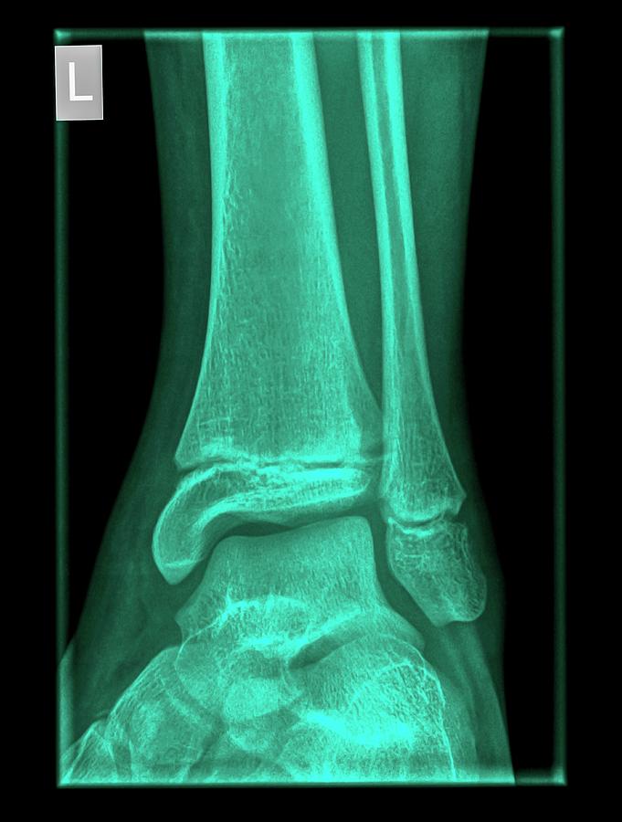
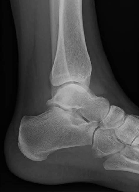
Ankle x ray anatomy. Normal foot and ankle x rays the human foot has 26 bones and 33 joints. X rays directly visualise bone injury but understanding of the anatomical position of ligaments is required to appreciate the presence of ligament injuries which are not directly visualised. The best sleeping position for back pain neck pain and sciatica tips from a physical therapist duration.
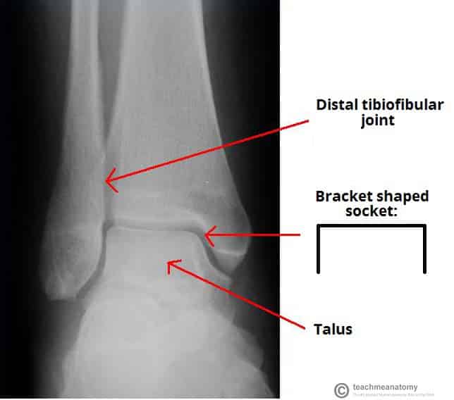
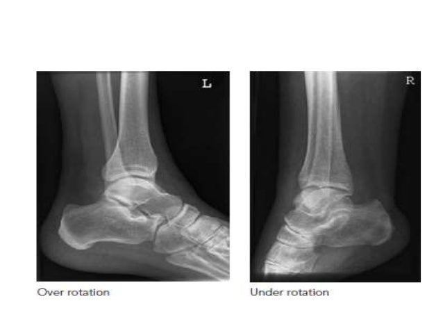
The ankle also consist of two joints the ankle joint where the tibia fibula and talus meet and the syndesmosis joint the joint between the tibia and fibula which is help together by ligaments. A doctor puts pressure on an injured ankle and takes an x ray film. Tone and tighten recommended for you.
For more anatomy content please follow us and visit our website. When calcaneal pathology is suspected an additional image can be made in axial direction. There are more than a hundred muscles tendons and ligaments.
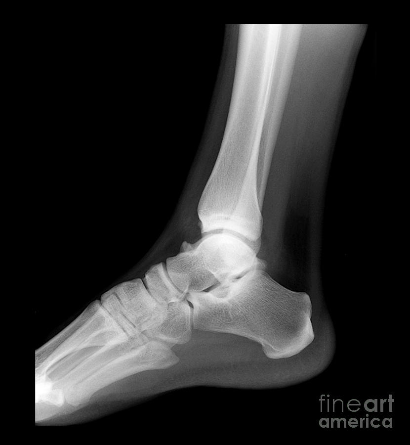
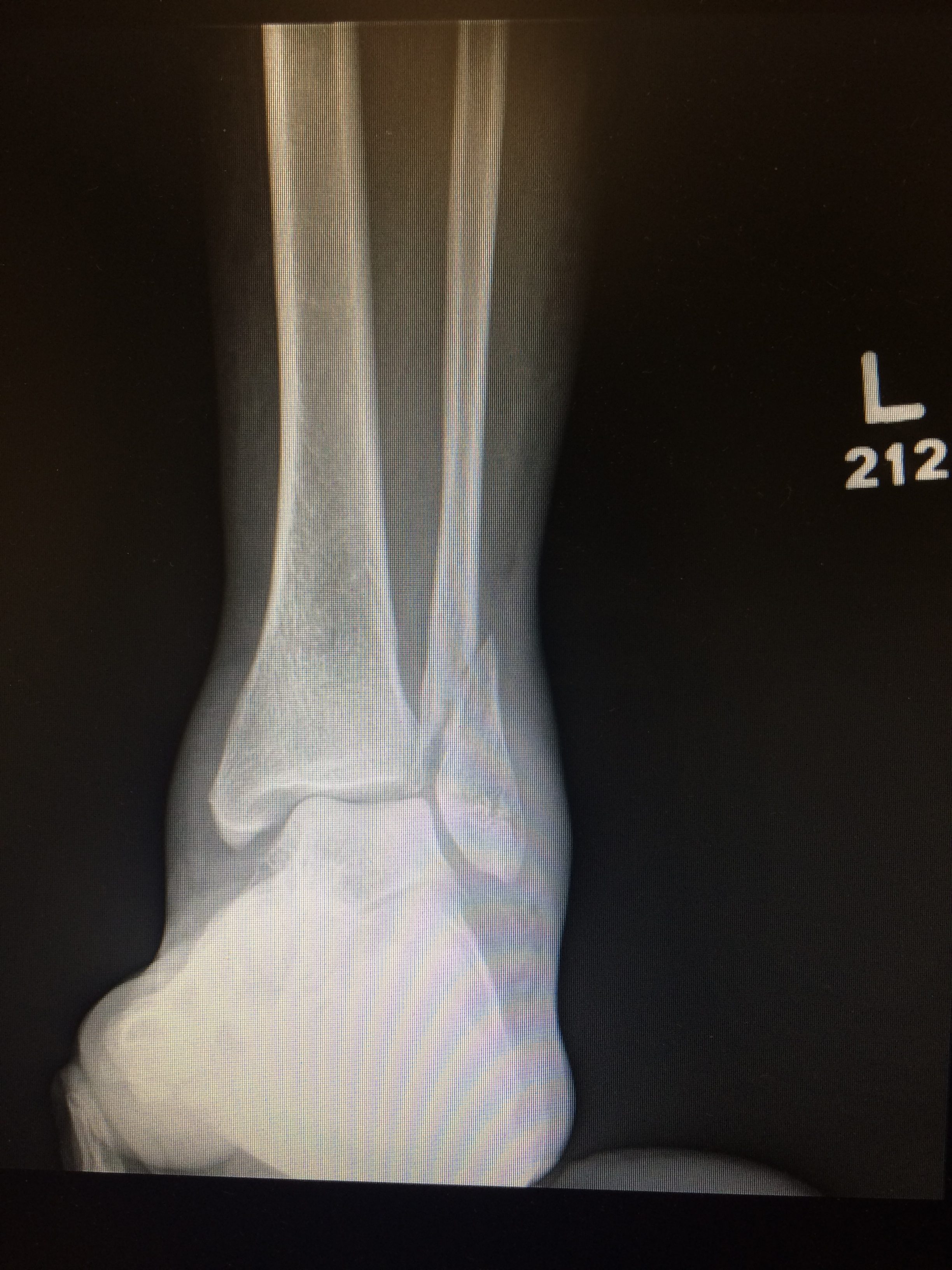
We hope this picture foot ankle x ray lateral view can help you study and research. We think this is the most useful anatomy picture that you need. Also called a stress film or a stress test this may uncover ankle problems unseen on regular x rays.
This test will be able to show any crack or break in the bones in the ankle 4 9. There are three main sets of ligaments. The x ray is the most commonly requested radiographic examination because of its availability.
Anatomy the ankle is a synovial joint composed of the distal tibia and fibula as they articulate with the talus. A standard series includes an anteroposterior ap image a mortise image and a lateral image. Ankle ligament anatomy ankle injuries may involve bones or ligaments in isolation or a combination of bones and ligaments.
This is a very complex structure due to the need to support your entire body weight. Depending on the request various images can be made. Ankle x rays the ankle consists of three bones the tibia the fibula and the talus.
The ankle x ray is used primarily to demonstrateexclude a fracture. The distal tibia and fibula articulate with each other at the distal tibiofibular joint which is more commonly referred to as the tibiofibular syndesmosis or simply the syndesmosis. It carries the weight of the body and can undergo a myriad of pathology most commonly traumatic injuries of the medial and lateral malleoli.
 The Ankle Joint Articulations Movements Teachmeanatomy
![]() Normal Chest X Ray Anatomy Tutorial Kenhub
Normal Chest X Ray Anatomy Tutorial Kenhub
 Royalty Free Foot Xray Stock Images Photos Vectors
 Radiological Anatomy Calcaneus Ankle X Ray Youtube
 Ankle X Ray Normal
 Broken Ankle Types Of Fractures Diagnosis Treatments
 Basketball Player With Left Foot Pain
 Ankle Fractures Pediatric Pediatrics Orthobullets
 Ankle X Ray By Photostock Israel
 Ankle Stress Views Why When What Core Em
 Ankle X Rays
 X Ray Of Foot And Ankle
 X Enkel Startradiology
 X Enkel Startradiology
 Skeletal Trauma
 X Ray Of Ankle Joint Anteroposterior View Showing Fracture
 Radiograph X Ray Of The Ankle Anatomy On An Anterior
 Ankle Fracture Footeducation
 The Ankle
 X Ray Anatomy Lateral Ankle Diagram Quizlet
![]() Student Corner Ottawa Ankle Rules Em Rems
Student Corner Ottawa Ankle Rules Em Rems
 The Radiology Assistant Ankle Fracture Mechanism And
5 Kinds Of Medial Malleolus Ankle Fractures
 Game Statistics Ankle X Ray Anatomy Purposegames
 Radiographic Positioning Of The Heel And Ankle Radiology
 Film Critique Of The Lower Extremity Part 3
 Imaging In Ankle Fractures Overview Radiography Computed


Posting Komentar
Posting Komentar