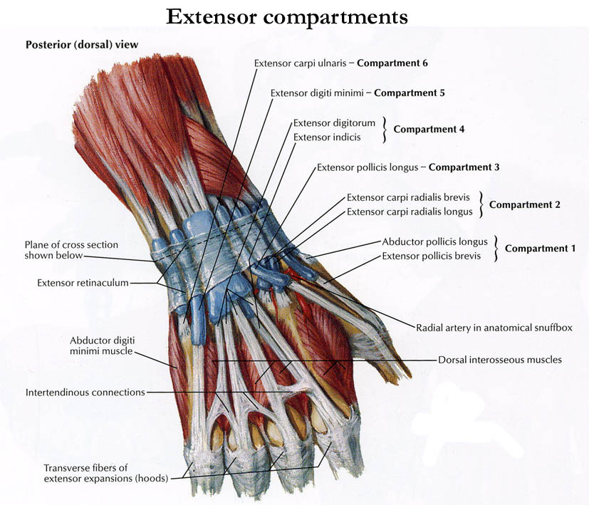
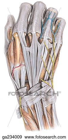
Now we can actually clearly see the two tendons that make up the lateral border of the anatomical snuff box both extensor pollicis brevis here and abductor pollicis longus. E anatomy image gallery anatomical parts download e anatomy.
 Figure 5 From The Sensory Distribution In The Dorsum Of The
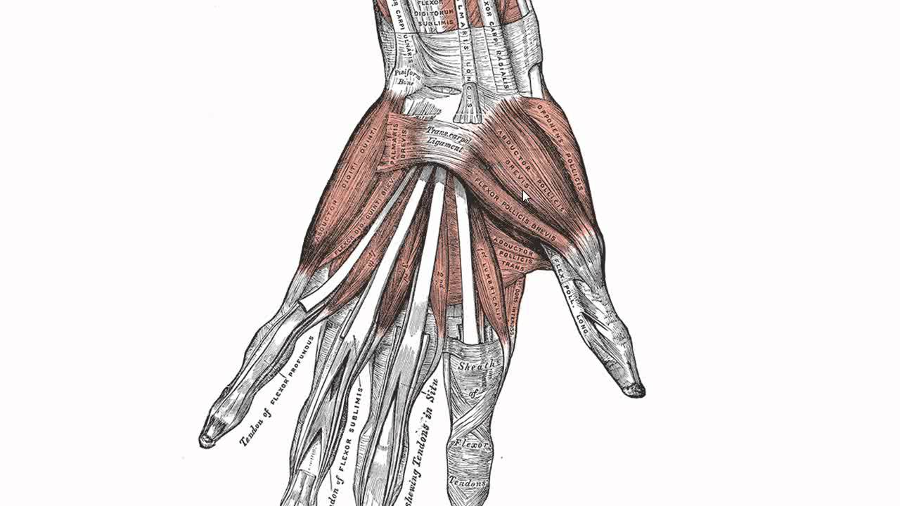
The hypothenar muscles produce the hypothenar eminence a muscular protrusion.
Dorsum of hand anatomy. Muscles of the hand thenar muscles. Anatomy of back of fore arm and dorsum of hand 1. These two terms used in anatomy and embryology refer to back dorsal and front or belly ventral of an organism.
The dorsal from latin dorsum meaning back surface of an organism refers to the back or upper side of an organism. The heel of the hand is the area anteriorly to the bases of the metacarpal bones. You can click the image to magnify if you cannot see clearly.
This image added by admin. So now we can see those two tendons. Mobile and tablet users you can download e anatomy on appstore or googleplay.
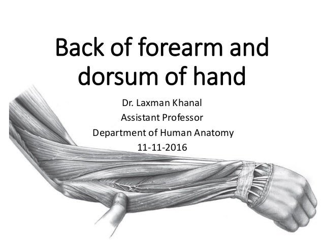
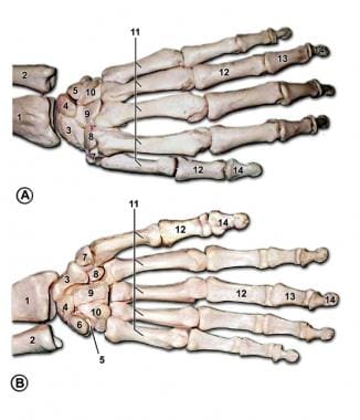
Back of forearm and dorsum of hand dr. These are four lumbricals in the hand each associated with a finger. Objectives learn the skeletal components of posterior forearm and dorsum of hand.
The thenar muscles are three short muscles located at the base of the thumb. The opisthenar area dorsal is the corresponding area on the posterior part of the hand. The dorsum of hand opisthenar area dorsal area is the corresponding area on the posterior part of the hand.
Most of what we can feel on the dorsal side of the hand is bone and most of what we can see are tendons along with a few superficial veins like the dorsal foot the dorsal hand has seven visible tendons although we dont always see them all at the same time and only one clearly visible muscle. Identify the seven superficial and five deep muscles of posterior forearm. And we can see that here if we look at a section through the wrist.
If talking about the skull the dorsal side is the top. Discover our subscription plans. Laxman khanal assistant professor department of human anatomy 11 11 2016 2.
Dorsum anatomy of the hand. The palm volar which is the central region of the anterior part of the hand. Areas of the human hand include.
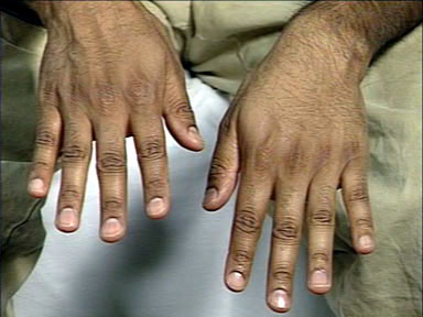
Though pellagra can present on any site the dorsum of the hands feet face neck and arms are the usual sites of involvement7 the most common site is the dorsum of hands with the lesions extending up the arm in the form of gauntlet. We think this is the most useful anatomy picture that you need.
 Anatomy Of Back Of Fore Arm And Dorsum Of Hand
 Handcare Org Anatomy Muscles
 Example Of Hand Stimuli These Are Male Left Hands For The
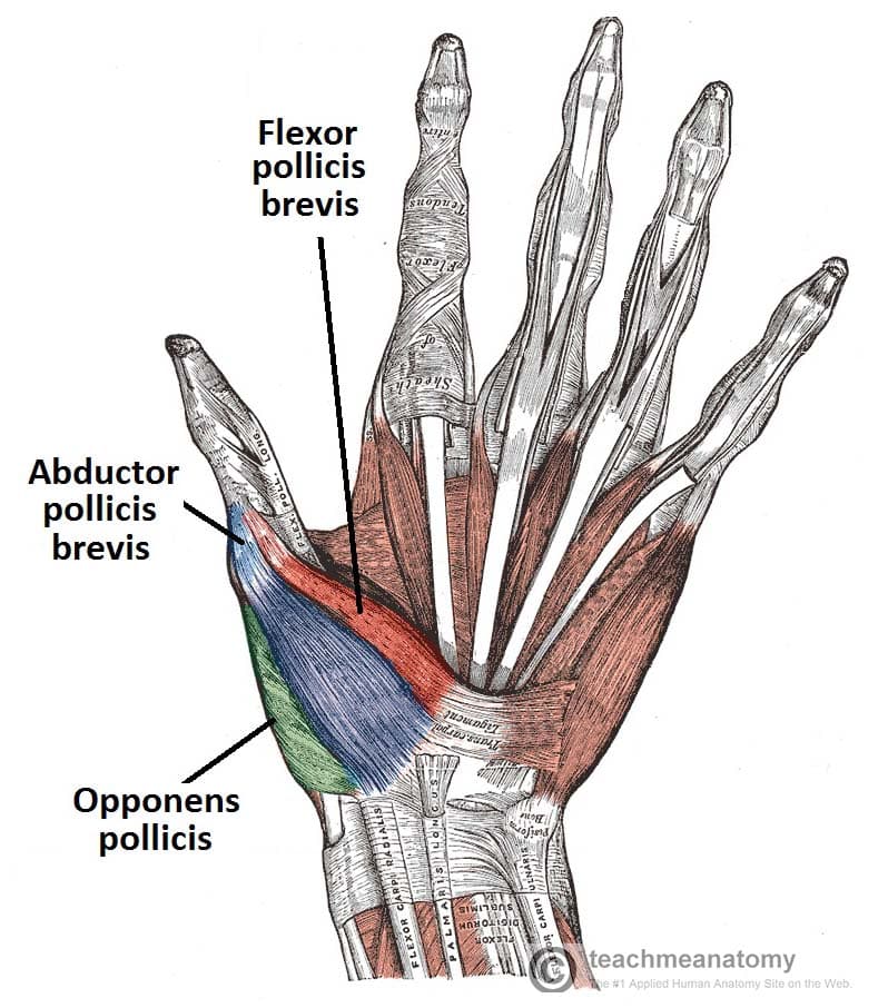 The Muscles Of The Hand Thenar Hypothenar Teachmeanatomy
 Wrist Hand Anatomy
Dorsum Of The Hand Stock Photos Dorsum Of The Hand Stock
 A Arterial Anatomy Of The Palm B Dorsum Of The Hand 1
 Hand Anatomy Overview Bones Blood Supply Muscles Geeky
 Anatomy Of Back Of Fore Arm And Dorsum Of Hand
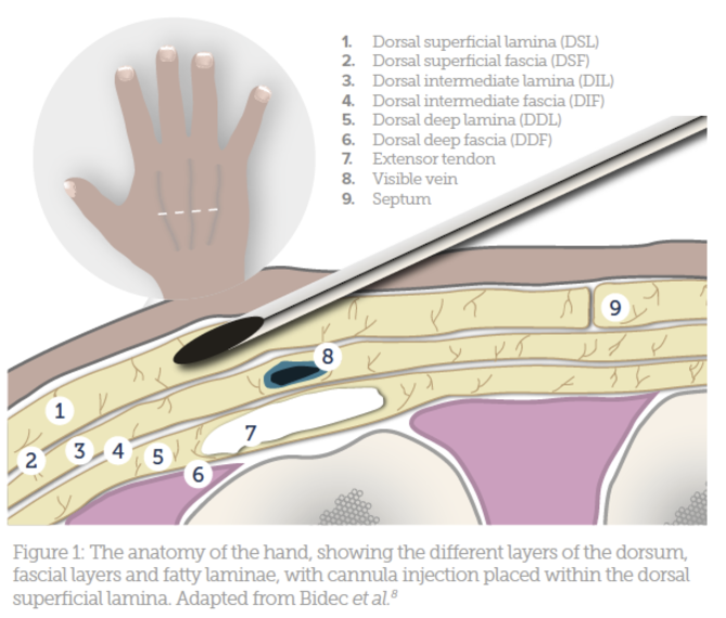 Hand Rejuvenation Using Fillers Aesthetics
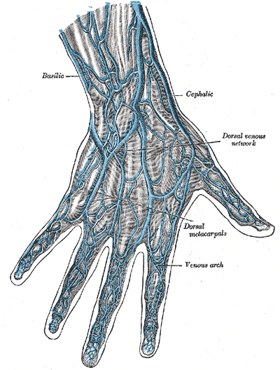 Dorsal Venous Network Of Hand Wikipedia
 Injuries To The Hand And Digits Tintinalli S Emergency
A Palm Of Left Hand B Dorsum Of Left Hand
 Hand And Wrist Anatomy Baxter Regional Medical Center
 Demystifying The Hand Exam Nuem Blog
 Palm And Dorsum Of The Hand Flashcards Quizlet
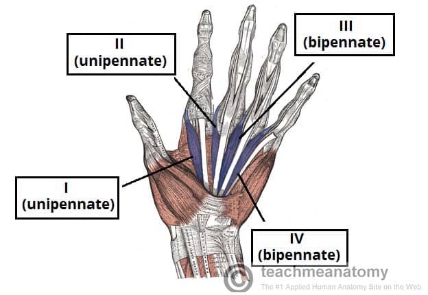 The Muscles Of The Hand Thenar Hypothenar Teachmeanatomy
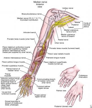 Nerve Compression Syndromes Of The Hand Overview Anatomy
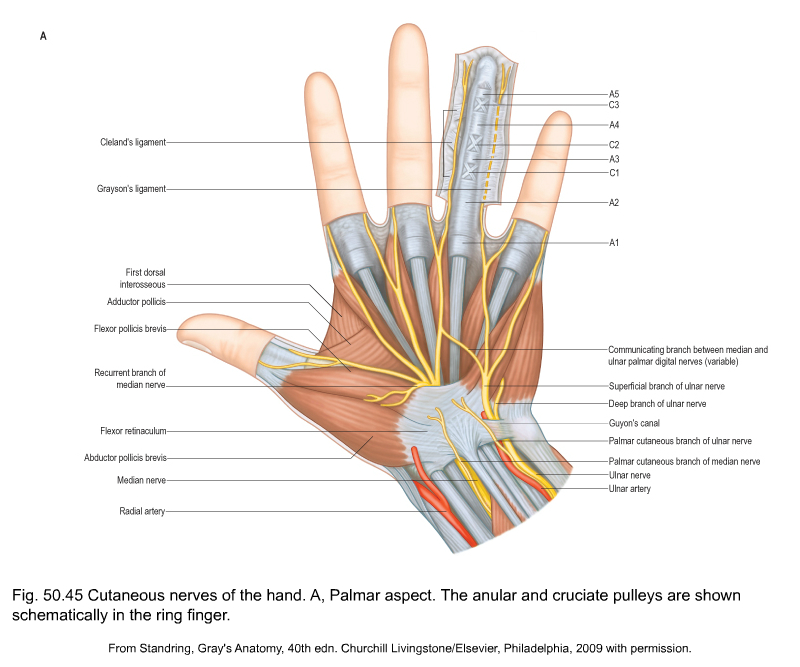
 Cutaneous Innervation Of Hand Anatomy Qa
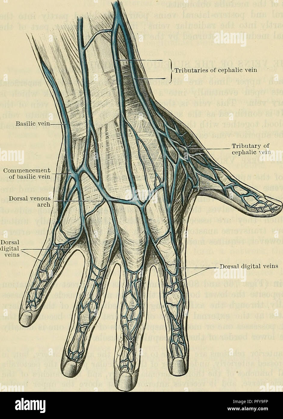
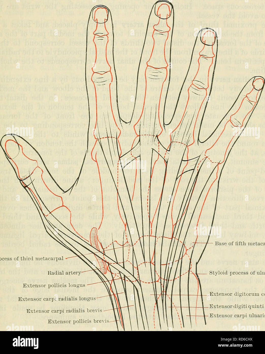 Cunningham S Text Book Of Anatomy Anatomy The Fokeakm And
 Dorsum Of Hand Anatomy Dorsum Lateral Hand Hand Anatomy
 Muscles Of The Hand Anatomy Tutorial
 Anatomy Of The Hand Team Bone
 Hand Anatomy Overview Bones Skin
 Dorsal View Of Tendons On The Dorsum Of Hand And Extensor


Posting Komentar
Posting Komentar