The hip joint can be imaged under various angles. A video tutorial in interpreting radiographs of the pelvis hip joint and femur.
 The Radiology Assistant Hip Pathology In Children
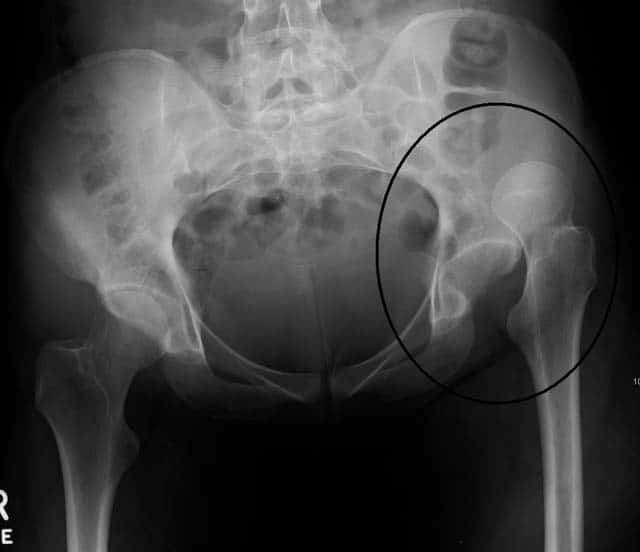
The hip x ray is used primarily to demonstrateexclude a fracture.

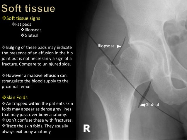
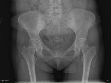
Hip anatomy xray. The second xray is of the pelvis in a 53 year old female with osteopenia. She is post menopausal and has a borderline osteoporosis of the hips. Pelvic and hip x rays are most frequently obtained when there is concern for fracture joint dislocation and effusion and several pediatric pathologies involving the pelvic girdle which are outlined below.
Shentons line is formed by the medial edge of the femoral neck and the inferior edge of the superior pubic ramus. A standard hip x ray examination generally includes an anteroposterior pa image and a lateral image. This image added by admin.
This is the fourth video in a series of five by teachmeanatomy have a loo. Notice that the bone in the area of the calcar is much thinner and the cortex of the femoral shaft is much thinner as well. The first xray is of a 35 year old male with no arthritis of the hip.
Hip x rays are also frequently opted for as initial test in chronic hip symptoms eg. Loss of contour of shentons line is a sign of a fractured neck of femur. You can click the image to magnify if you cannot see clearly.
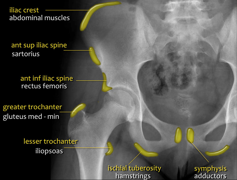
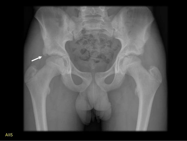
Before delving into the radiographic approach to pelvic and hip x rays let us first review some anatomy. Fractures of the femoral neck do not always cause loss of shentons line. Hip x ray anatomy normal ap.
We think this is the most useful anatomy picture that you need.
 Film Critique Of The Lower Extremity Part 1
 Trauma Image Interpretation Of The Pelvis And Hip
 Ao Surgery Reference
 Hip X Ray Anatomy Normal Ap Shenton S Line Is Formed By The
 International Hip Dysplasia Institute
 Radiography Of The Hip Joint Anatomy Of The Whole Human

Interpreting X Rays Of The Pelvis Hip Joint And Femur
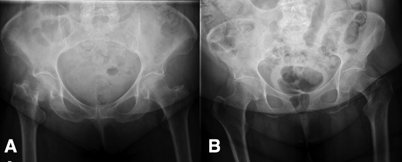
 A And 4 B Standing And Sitting Lateral Spine Pelvis Hip
 Startradiology
 The Hip Joint Articulations Movements Teachmeanatomy

 Ilium Bone Hip Bone Image Photo Free Trial Bigstock

Hip Dislocation Orthoinfo Aaos
 Trauma Image Interpretation Of The Pelvis And Hip
 Radiographic Anatomy Pelvis Ap Female My Next Practical I
 Untitled Document
 Startradiology
 Neck Of Femur Fracture Subcapital Intertrochanteric
 Separate Human Bones Of Hip And Lower Limb Healthcare X Ray
 Film Critique Of The Lower Extremity Part 1

 Hip Dislocation Background Epidemiology Functional Anatomy


Posting Komentar
Posting Komentar