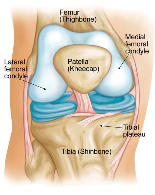
The knee is the largest and most complex joint in the body holding together the thigh bone shin bone fibula on the outer side of the shin and kneecap. Muscles tendons and ligaments connect the knee bones.
 Knee Joint Anatomy Bones Ligaments Muscles Tendons Function
Pain and swelling are the most common signs of knee injury.
Knee anatomy diagram. The knee is the meeting point of the femur thigh bone in the upper leg and the tibia shinbone in the. The knee cap actually sits inside the patellar tendon. In many cases injuries involve more than one structure in the knee.
Tendons are often overlooked as part of knee joint anatomy. An mri of the knee of a healthy subject was performed in the 3 planes of space coronal axial sagittal commonly used in osteoarticular imagery with two weightings most commonly used to explore the musculo skeletal pathology of the knee. The kneecap glides in a groove in the thighbone and adds leverage to the thigh muscles which are used to extend the leg.
Tendons at the knee. The range of motion of the knee is limited by the anatomy of the bones and ligaments but allows around 120 degrees of flexion. The femur thigh bone tibia shin bone patella knee cap fibula smaller bone next to shin bone.
The largest joint in the body the knee moves like a hinge allowing you to sit squat walk or jump. The knee is a complex joint that flexes extends and twists slightly from side to side. The knee is the joint where the bones of the lower and upper legs meet.
The main tendon found at the knee is the patellar tendon which links the quads muscles to the shin bone. The ends of the bones are covered with a layer of cartilage a slick elastic material that absorbs shock and allows the bones. The most common knee injuries include fractures around the knee dislocation and sprains and tears of soft tissues like ligaments.
Fast facts on knee anatomy. Anatomy function knee anatomy function and common problems. Knee anatomy function and common problems.
Your knee is made up of many important structures any of which can be injured. A special characteristic of the knee that differentiates it from other hinge joints is that it allows a small degree of medial and lateral rotation when it is moderately flexed. See the pictures and anatomy description of knee joint bones cartilage ligaments muscle and tendons with resources for knee problems injuries.
Webmds knee anatomy page provides a detailed image and definition of the knee and its parts including ligaments bones and muscles. They are they soft tissues found at the end of muscles which link the muscle to bone. Mri of the knee.
Spin echo t1 and proton density with fat saturation sequences. Bones of the knee.
 Knee Joint Part 2 3d Anatomy Tutorial
 Side View Of Knee Human Anatomy Medical Images For Power Point
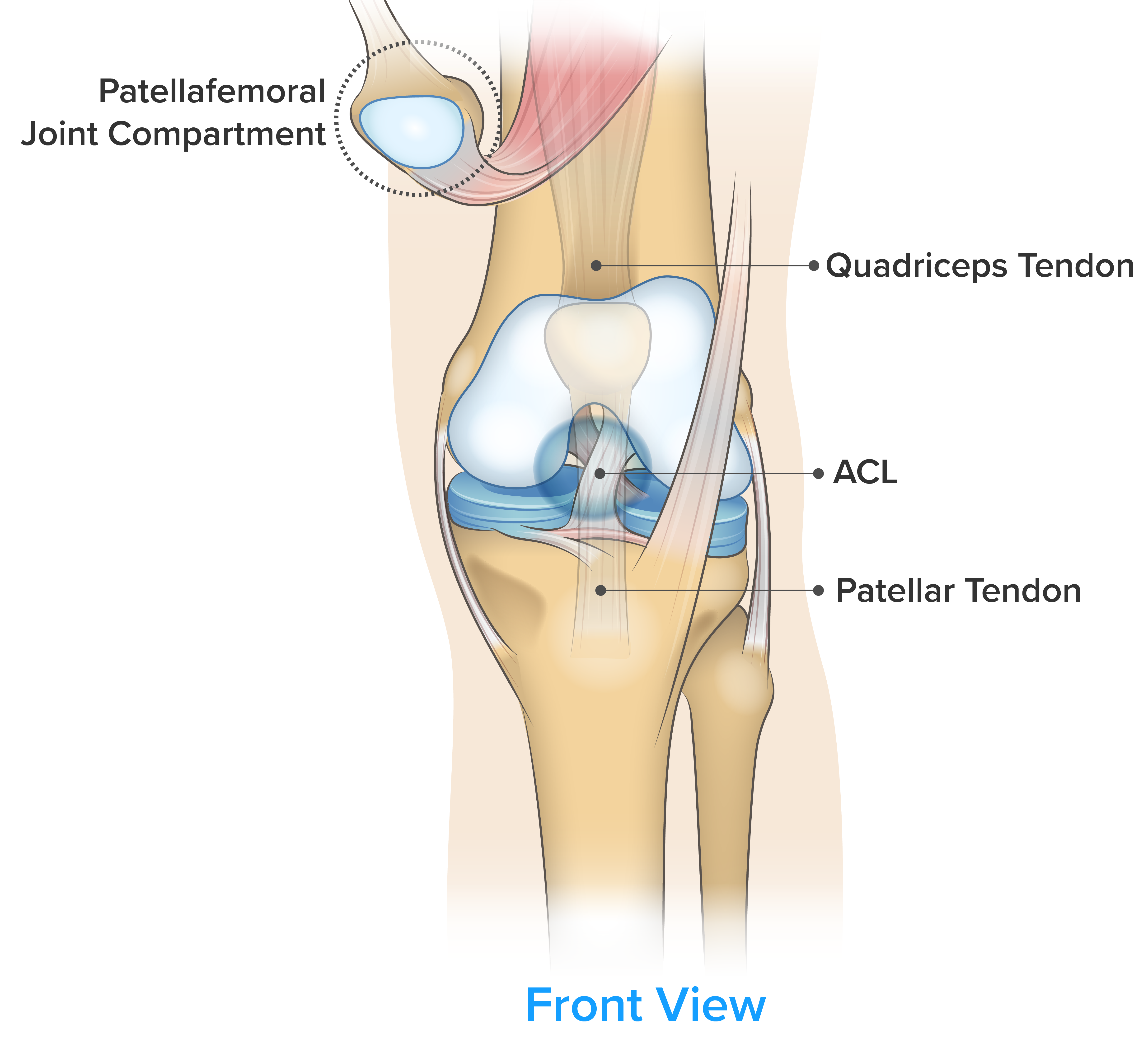
 What Are The Parts Of The Knee Joint Systems4knees
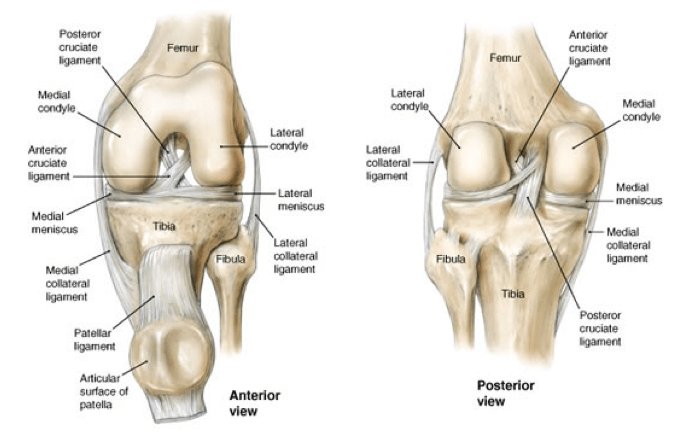
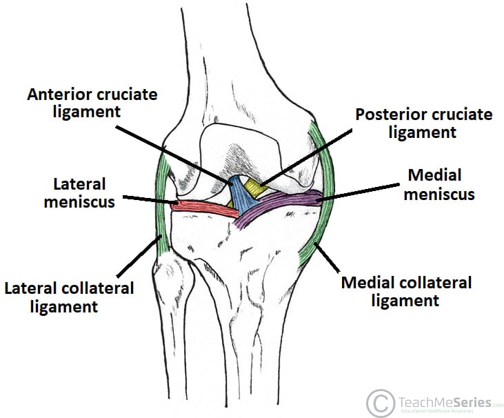
 The Knee Joint Articulations Movements Injuries
 Pcl Injury Knee Sports Orthobullets
 Total Knee Anthroplasty

 Knee Physiopedia
 Pain Behind Knee Why It Hurts In Back Of Or Under Your Kneecap
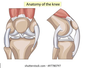
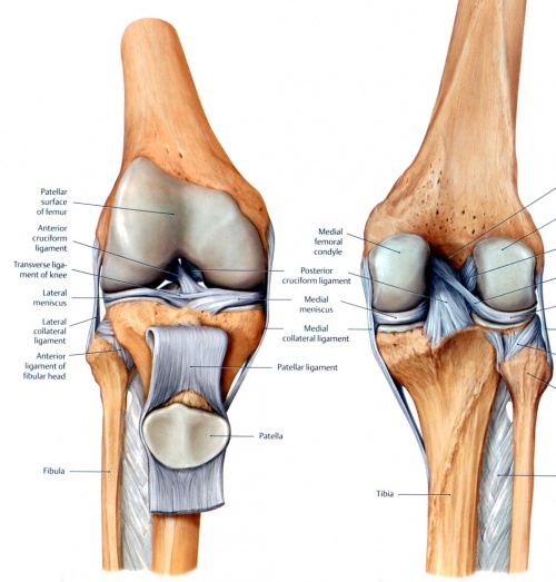
 The Human Knee Joint S Anatomy With Visible Cruciate
 Pain Behind Knee Why It Hurts In Back Of Or Under Your Kneecap
 Anatomy Knee Joint Cross Section Showing Stock Illustration
 The Knee Anatomy Injuries Treatment And Rehabilitation
![]() Leg And Knee Anatomy Bones Muscles Soft Tissues Kenhub
Leg And Knee Anatomy Bones Muscles Soft Tissues Kenhub
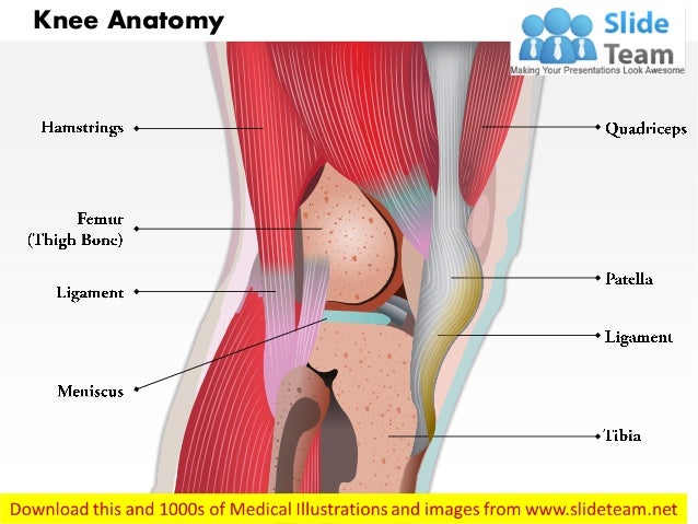
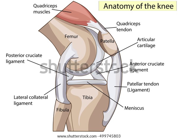
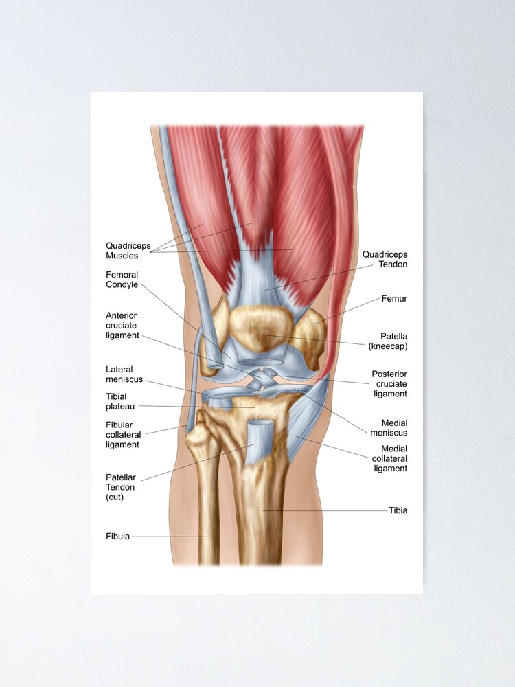
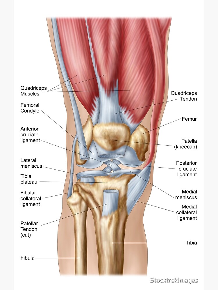
 Anatomy Of The Knee Joint
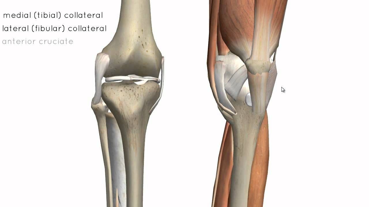
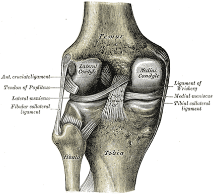
 Clinical Anatomy Knee
 Complete Guide To Knee Pain Spring Loaded Technology
 Amazon Com Semtomn Canvas Wall Art Print Patella Knee Joint
 Knee Anatomy 4 Diagram Quizlet
 2 Anatomy Of Knee Joint Adapted From 34 Download
 The Knee Joint Human Anatomy
![]() Leg And Knee Anatomy Bones Muscles Soft Tissues Kenhub
Leg And Knee Anatomy Bones Muscles Soft Tissues Kenhub
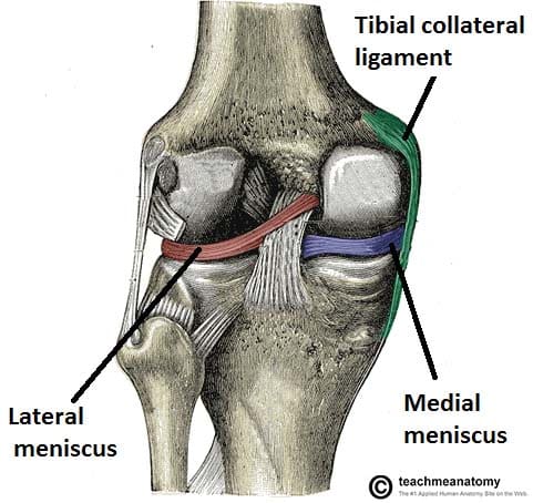
 Knee Anatomy And Significance Bone And Spine
 Anatomy Of Human Knee Joint Poster
 Anatomy Of Human Knee Joint Greeting Card
 Total Knee Replacement Omni
Common Knee Injuries Orthoinfo Aaos
 A Guide To Your Knees Well Guides The New York Times
Knee Anatomy Images Stock Photos Vectors Shutterstock
 The Knee Joint Articulations Movements Injuries
 Knee Wikipedia
 Osteonecrosis Of The Knee Orthoinfo Aaos


Posting Komentar
Posting Komentar