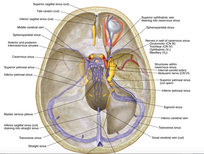
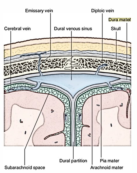
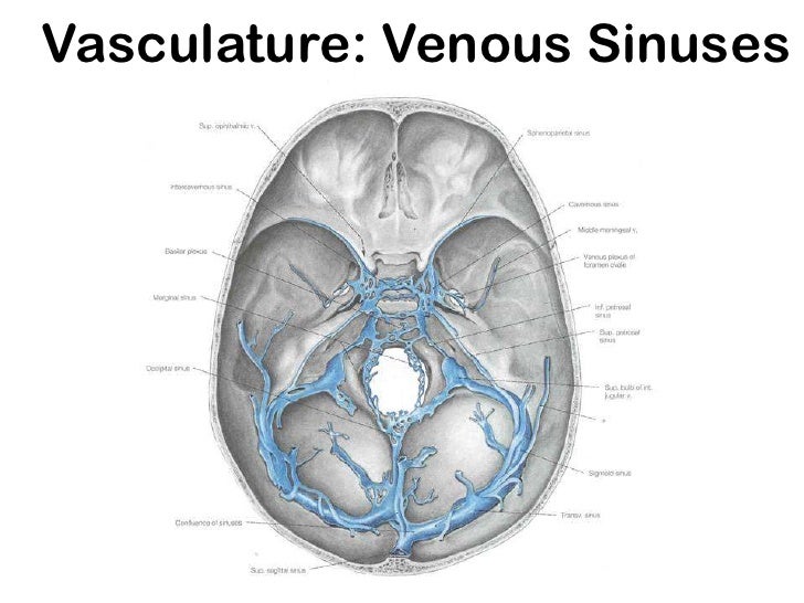
Dural venous sinuses are venous channels located intracranially between the two layers of dura mater endosteal layer and meningeal layer. The sphenoparietal sinus courses along the free border.
They do not have muscle in their walls.

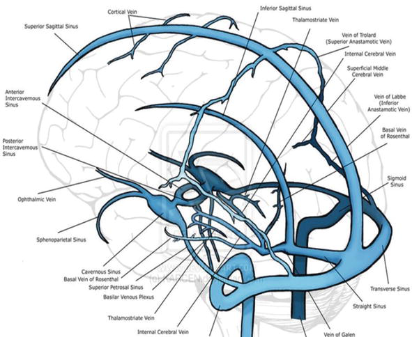
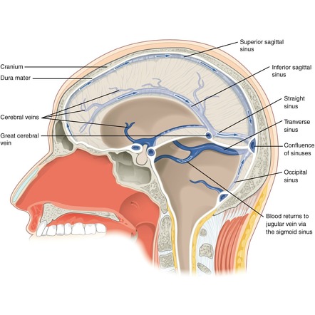
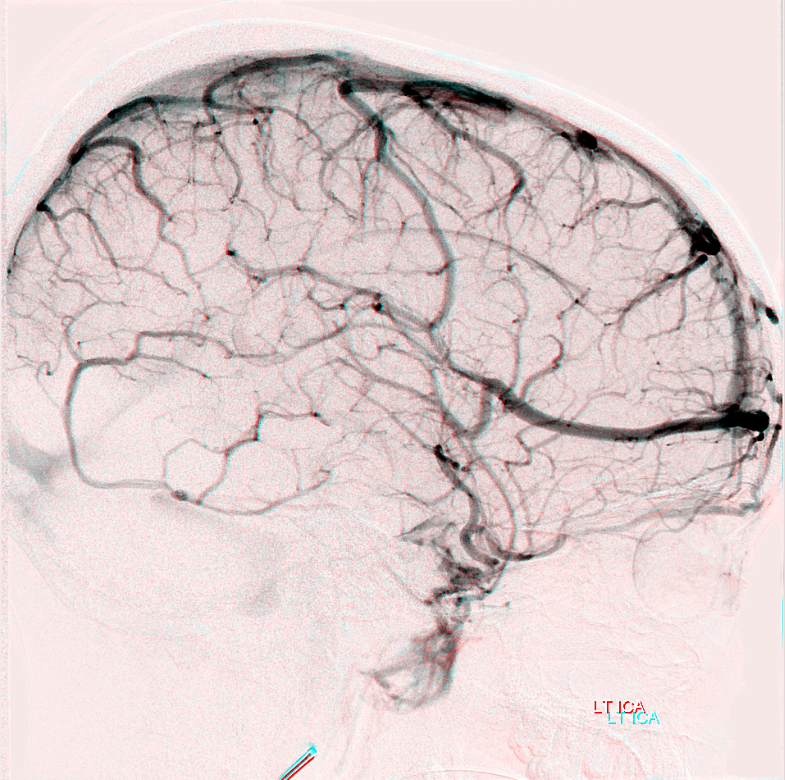
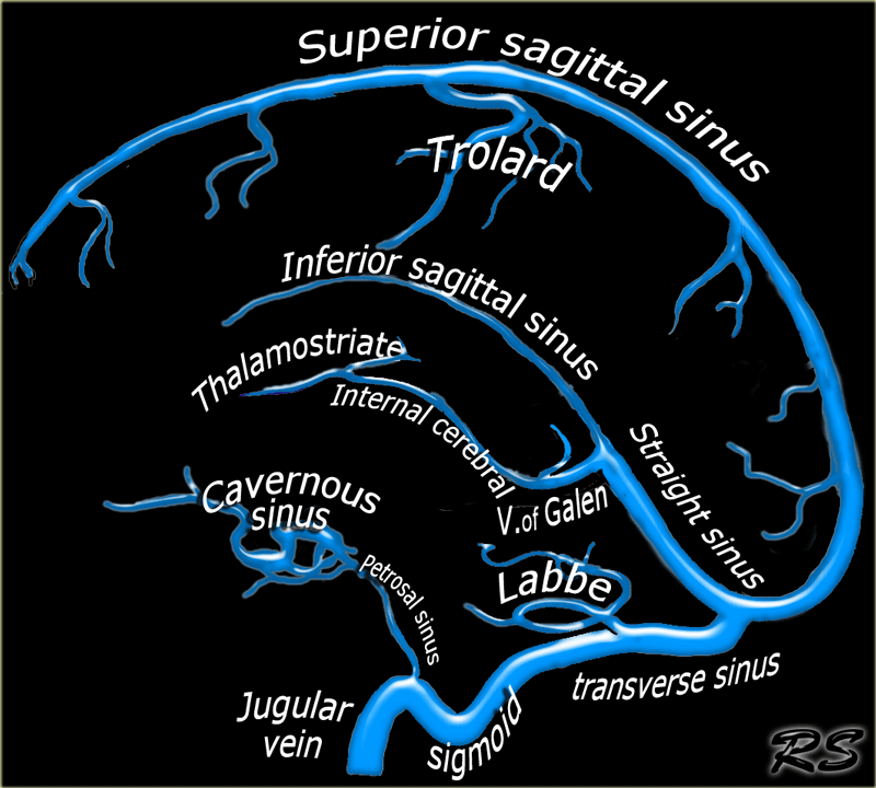
Dural venous sinuses anatomy. All the dural venous sinuses ultimately drain into the internal jugular vein. What are the characteristic features of dural venous sinuses. They receive blood from the cerebral veins receive cerebrospinal fluid csf from the subarachnoid space via arachnoid granulations and mainly empty into the internal jugular vein.
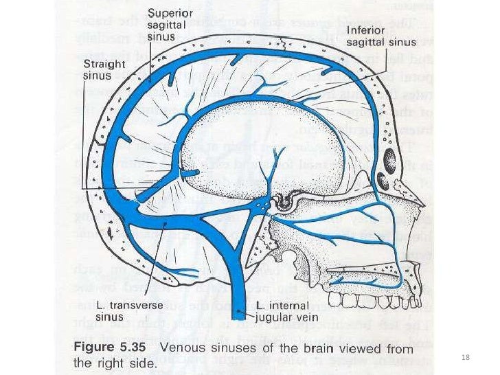
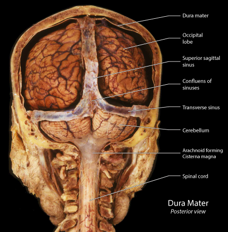
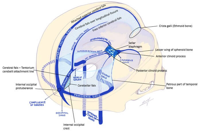
Superior vena cava and the azygos system clinical anatomy svc obstruction oncology emergency duration. It communicates with the straight sinus superior sagittal sinus and the occipital sinus at a point called the confluence of sinuses. The left and right transverse sinuses travel in the base of the tentorium cerebelli along the occipital bone.
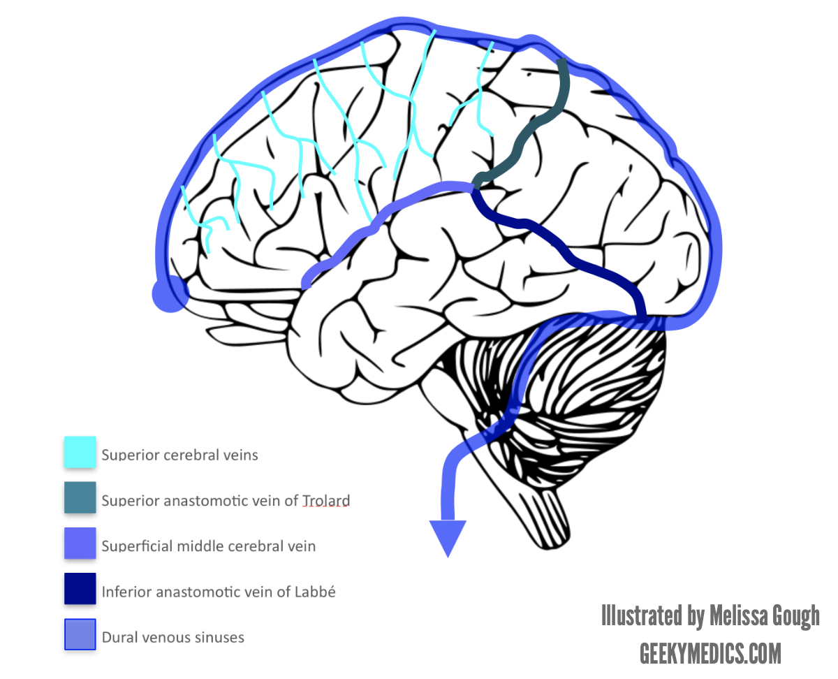
Aprof frank gaillard et al. They are lined by endothelium. Each anterior cerebral vein leaves the longitudinal cerebral fissure inferiorly.
It collectively returns deoxygenated blood from the head to the heart to maintain systemic circulation. Unlike other veins in the body they run alone not parallel to arteries. They also drain csf.
Dural venous sinuses sagittal sinuses. Dural venous sinuses are a group of sinuses or blood channels which drains venous blood circulating from the cranial cavity. They are best thought of as collecting pools of blood which drain the central nervous system the face and the scalp.
Dural venous sinuses are venous channels that are present usually the two layers of dura mater. They have no valves. They can be conceptualised as trapped epidural veins.
Unlike most veins of the body the dural venous sinuses do not have valves. Also known as the longitudinal inferior sinus. The dural venous sinuses lie between the periosteal and meningeal layers of the dura mater.
They drain blood from. Armando hasudungan 34176 views. Dural venous sinuses dvs superior sagittal sinus sss the sss is situated along the superior border of falx cerebri.
A dural venous sinus thrombosis of the transverse sinus. The dural venous sinuses also called dural sinuses cerebral sinuses or cranial sinuses are venous channels found between the endosteal and meningeal layers of dura mater in the brain. There are two sagittal sinuses that occupy the longitudinal cerebral fissure.
At the level of the internal occipital protuberance.
 Superior Sagittal Sinus An Overview Sciencedirect Topics
Anatomy 7 Cn Dural Venous Sinuses Ventricles Medicine
 Anatomy Imaging And Surgery Of The Intracranial Dural
 Venous Sinuses
 Peritorcular Meningioma Resection Ami 2018 Meeting
 Dural Venous Sinuses Cranial Nerves Brain Cranial Nerve 1
 Annals Of B Pod Dural Venous Sinus Thrombosis Taming The Sru
 Cerebral Vein And Dural Sinus Thrombosis Intechopen
 A Review Of Extraaxial Developmental Venous Anomalies Of The
 Dural Venous Sinuses Radiology Reference Article
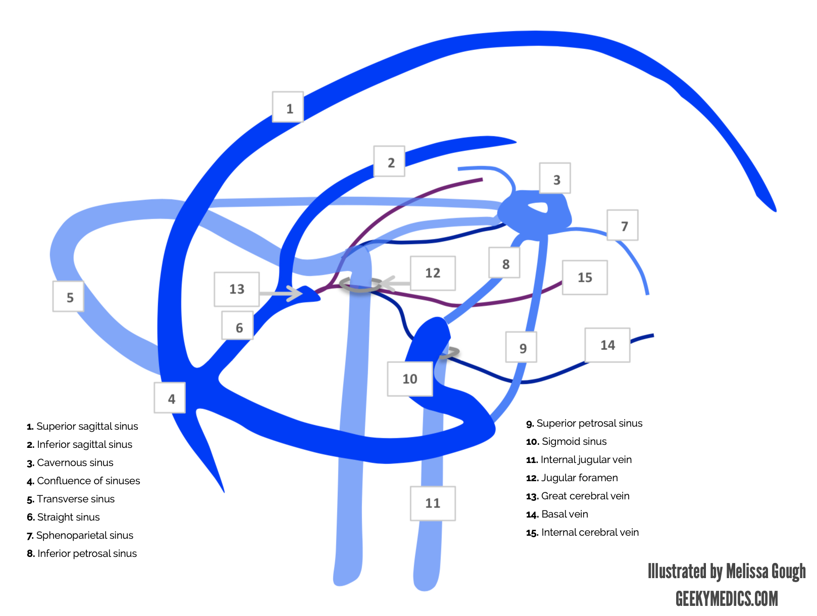 Venous Drainage Of The Brain Anatomy Geeky Medics
 Easy Notes On Dura Mater Learn In Just 4 Minutes
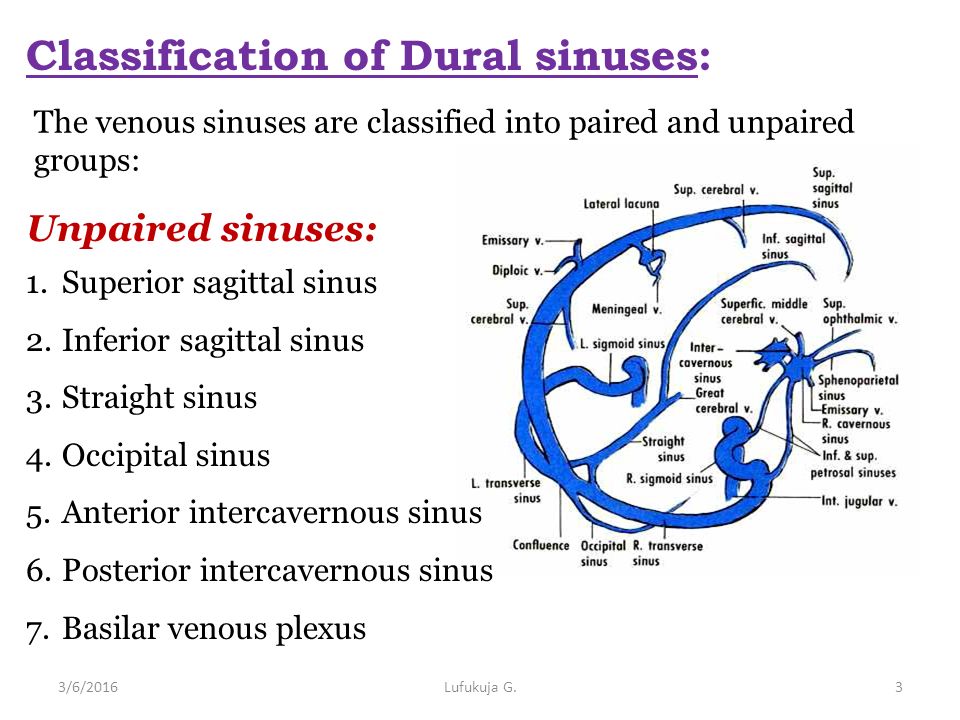 Dural Venous Sinuses 4 27 2017 Lufukuja G Ppt Video
 Venous Sinuses
 Venous Drainage Of The Brain Anatomy Geeky Medics
 Dural Reflections And Venous Sinuses Epomedicine
Plos One Mida A Multimodal Imaging Based Detailed
 Dural Venous Sinuses Anatomy Qa
 Dural Venous Sinus Connections To The Vertebral Venous
Anatomy Of The Dural Venous Sinuses The Bmj
 The Radiology Assistant Cerebral Venous Thrombosis
 Venous Drainage Of The Head And Neck Dural Sinuses
 Anatomy Imaging And Surgery Of The Intracranial Dural
![]() Dural Venous Sinuses Anatomy Kenhub
Dural Venous Sinuses Anatomy Kenhub


Posting Komentar
Posting Komentar