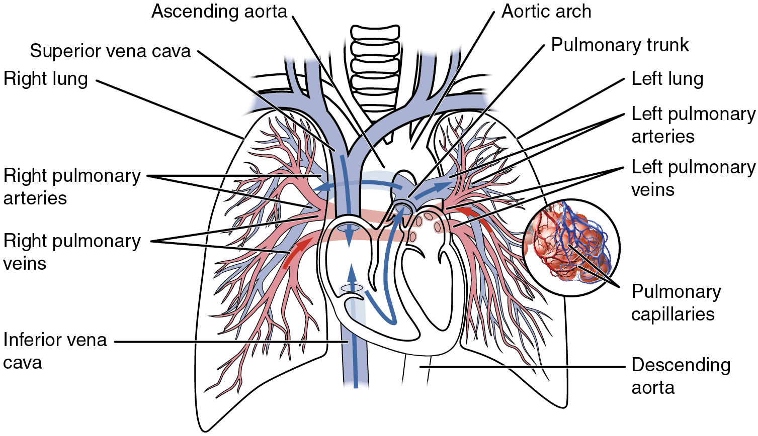
Embryologically the pulmonary arteries originate from the truncus arteriosus. The pulmonary veins along with the pulmonary arteries make up the pulmonary circulation.
Right Top Pulmonary Vein Draining The Posterior Segment Of
Histology of the arteries and veins.

Pulmonary veins anatomy. Drains the right lower lobe. Drains the left upper lobe. This differentiates the pulmonary veins from other veins in the body which are used to carry deoxygenated blood from the rest of the body back to the heart.
The largest pulmonary veins are the four main pulmonary veins two from each lung that drain into the left atrium of the heart. Typically there are four pulmonary veins with superior and inferior pulmonary veins on either side draining into the left atrium. Just like the other veins in your body your pulmonary veins arise from a network of capillaries.
Drains the left lower lobe. Drains the right upper and middle lobes. Pulmonary veins are responsible for carrying oxygenated blood from the lungs back to the left atrium of the heart.
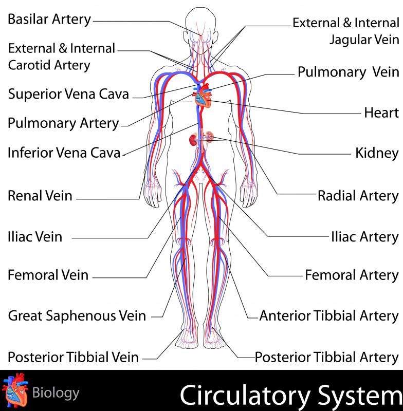
Veins are the blood vessels that carry blood to the heart. Large veins have diameters greater. The pulmonary veins are part of the pulmonary circulation.
The pulmonary veins can be affected by. The pulmonary veins serve a very important purpose of delivering freshly oxygenated blood. There are typically four pulmonary veins two draining each lung.
Pulmonary arteries and veins anatomy. The pulmonary veins are the veins that transfer oxygenated blood from the lungs to the heart. The pulmonary capillaries surround and embrace millions of tiny air sacs called alveoli in your lungs.
However the capillaries that give rise to the pulmonary veins differ from capillaries elsewhere in your body. The distal segments of the pulmonary veins are intrapericardial. The anatomy of the pulmonary vein anatomy.
Most individuals have four pulmonary veins two on the left and two on the right the inferior and the superior one but there are also different anatomical variations. The portion of myocardium extending onto the pulmonary veins is a frequent source of atrial fibrillation and the left superior pulmonary vein which has the longest segment of myocardium is the focal cause of atrial fibrillation in as many as half of the cases 12. Anatomy of the pulmonary veins.
The pulmonary veins pvs are large blood vessels that carry oxygenated blood from the lungs and drain into the left atrium la of the heart. While veins usually carry deoxygenated blood from tissues back to the heart in this case.
 Obstructed Infradiaphragmatic Total Anomalous Pulmonary
 This Figure Includes Six Ct Or Mr Images Of The Left Atrium
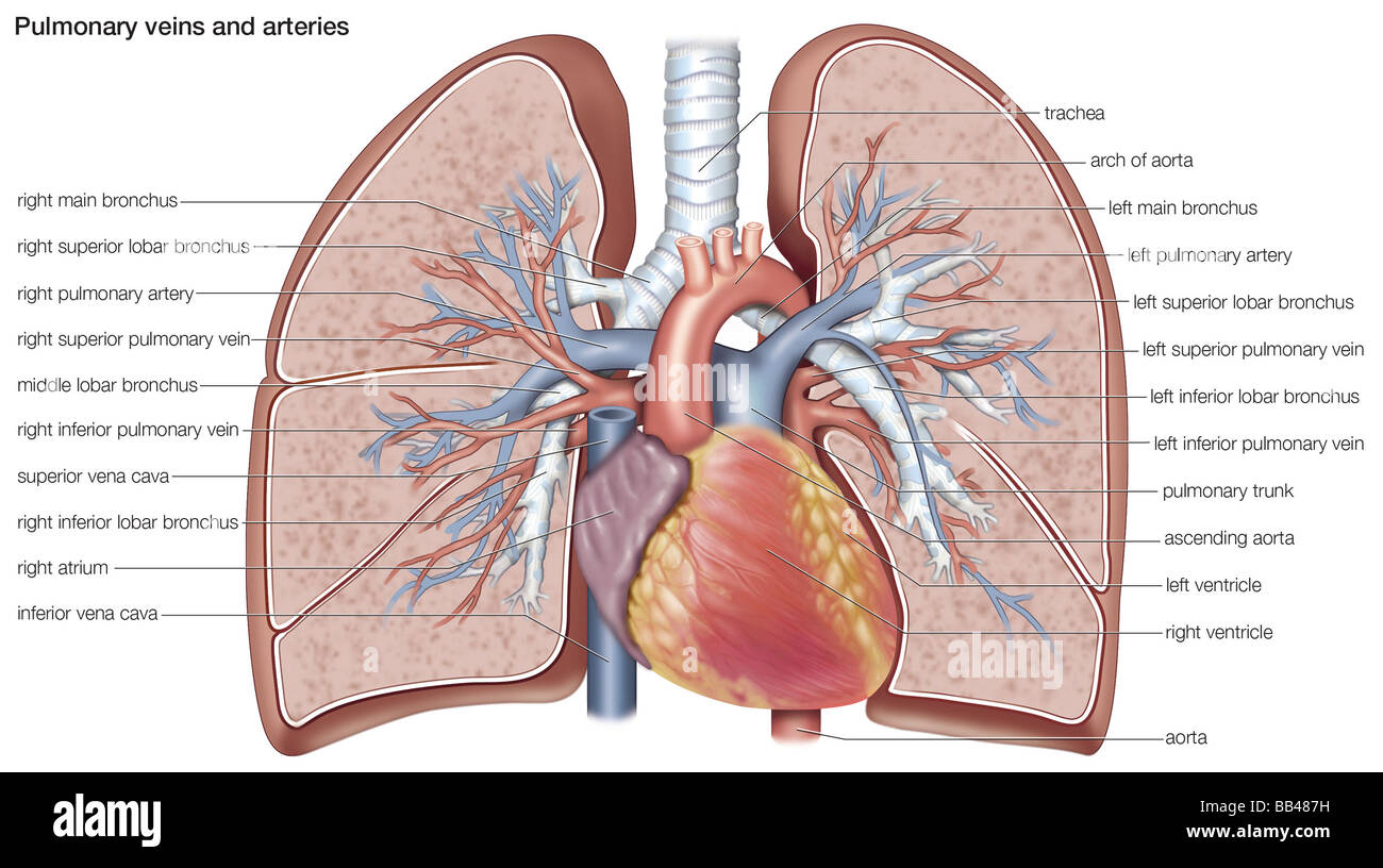 Pulmonary Veins And Arteries Stock Photo 24065877 Alamy
 Anatomical Regions Of The Left Atrium La And Pulmonary
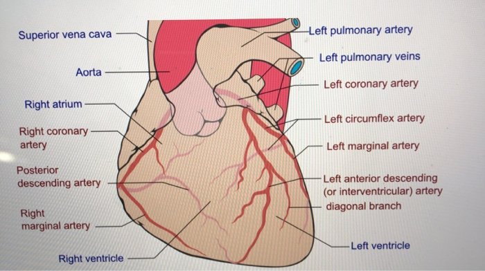 Solved Superior Vena Cava Left Pulmonary Artery Left Pu
Anatomy Of Pulmonary Veins By Real Time 3d Tee Jacc
 Figure 2 From Comprehensive Surgical Approach To Treat
 Anatomical Characteristics Of The Junction Of Left Atrium
Figure 6 Anatomical And Functional Evaluation Of Pulmonary
 Pulmonary Vein Anatomy Britannica
Paroxysmal Atrial Fibrillation Catheter Ablation Outcome
 Pulmonary Veins Right Superior Pulmonary Vein Right
Anatomy Of Pulmonary Veins By Real Time 3d Tee Jacc
 Three Dimensional Reconstructions Of The Left Atrium And
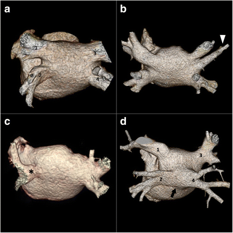 Anomalous Pulmonary Venous Drainage A Pictorial Essay With
 Anatomy Of The Respiratory System 6
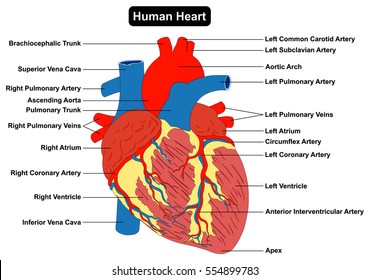 Pulmonary Veins Images Stock Photos Vectors Shutterstock
 Percutaneous Treatment Of Pulmonary Vein Stenosis Thoracic Key
 Pulmonary Veins Function And Definition Beltina Org
 How To Identify Pulmonary Veins In Transthoracic
 File Left And Right Pulmonary Veins Anatomy Function
Figure 3 Anatomy Of Pulmonary Veins By Real Time 3d Tee
 20 5 Circulatory Pathways Anatomy And Physiology
 Pulmonary Arteries And Veins Pulmonary Circulation
 What Is The Pulmonary Vein With Pictures
 Pulmonary Veins For Mr Cardiac Sonography Cardiology


:max_bytes(150000):strip_icc()/human-heart-circulatory-system-598167278-5c48d4d2c9e77c0001a577d4.jpg)

Posting Komentar
Posting Komentar