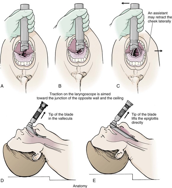
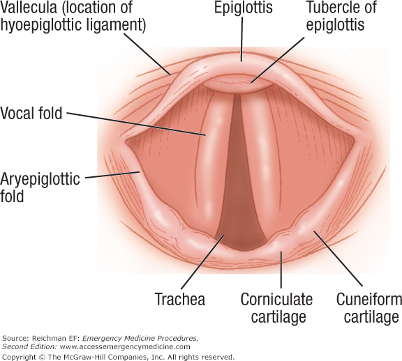
The vallecula is an important reference landmark used during intubation of the trachea. Intubation is then performed and tt position is checked by visualization of the carina through the tt and capnography.
The nasal fossa is bounded laterally by inferior middle and superior turbinate bones.
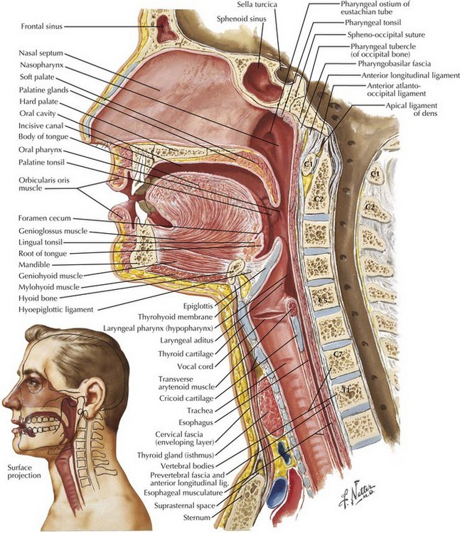
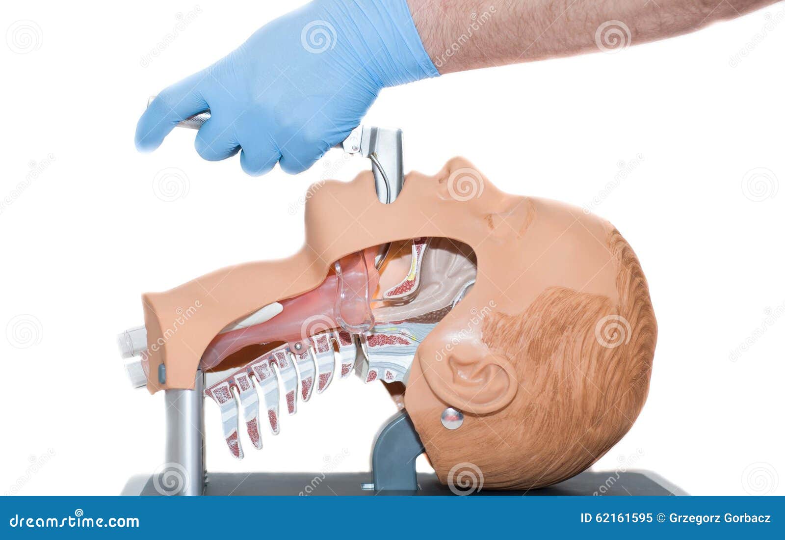
Intubation anatomy. Endotracheal intubation can be performed either orally or nasally although oral intubation is the more commonly used technique5the nasopharynx and oropharynx lead to the laryngopharynx hypopharynx. Anatomical abnormalities may affect only intubation only airway management or both. The ligaments of the larynx antero lateral view.
The larynx is a cartilaginous structure slung from the hyoid bone by the hyothyroid membrane. Nasotracheal intubation is an alternative approach to orotracheal intubation. Saliva is temporarily held in the valleculae to prevent initiation of the swallowing reflex.
These depressions serve as spit traps. The nasal fossae are divided by the midline cartilaginous septum and medial portions of the lateral cartilages fig. Anaesthesia is then induced using sevoflurane the cuff inflated and if necessary a neuromuscular blocking agent injected.
Try using search on phones and tablets. This section also focuses on the abnormal airways in obesity pregnancy children and neonate and patients with abnormal facial defects. At the base of the tongue the epiglottis separates the larynx from the laryngopharynx.
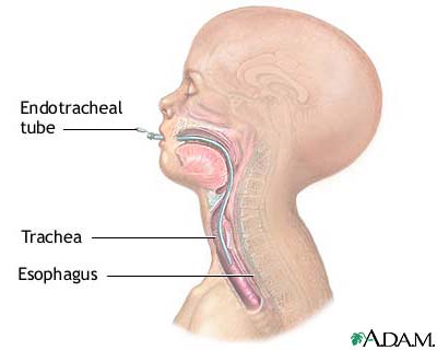
Tracheal intubation usually simply referred to as intubation is the placement of a flexible plastic tube into the trachea windpipe to maintain an open airway or to serve as a conduit through which to administer certain drugs. The two nasal fossae extend from the nostrils to the nasopharynx. The larynx is the key anatomical structure that needs to be identified when carrying out intubation.
If you cant recognize the vocal cords you will not be able to successfully intubate. It comprises of numerous separate cartilages held together with connective tissue. When first learning intubation a beginner often concentrates on memorizing the key laryngeal anatomy.
The epiglottic vallecula is a depression vallecula just behind the root of the tongue between the folds in the throat. This is important of course. Navigation best viewed on larger screens.
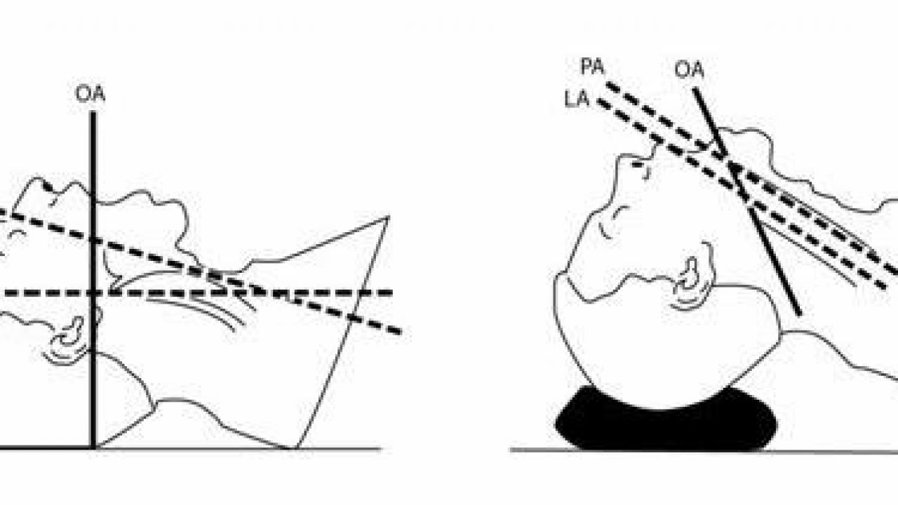
 Learning Intubation Head Position Effects Laryngeal View
 Anatomical Landmarks When Intubating
 Tracheal Intubation Clinical Gate
 Aime Airway
 Summit Medical Group
 Chapter 6 Essential Anatomy Of The Airway Emergency
 Chapter 19 Airway Management Morgan Mikhail S Clinical
 Endotracheal Intubation Medlineplus Medical Encyclopedia Image
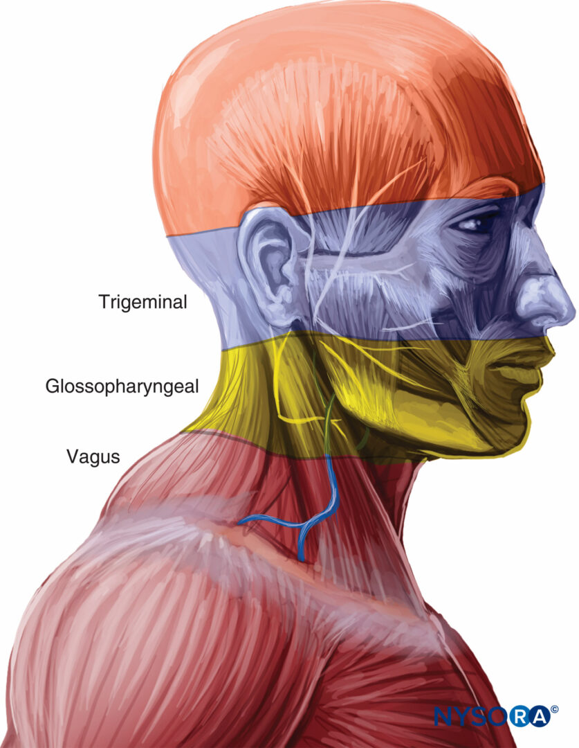 Regional And Topical Anesthesia For Awake Endotracheal

 Figure 3 29 From 3 Preparation For Awake Intubation
 How To Master Tracheal Intubation Acls Medical Training
 Chapter 6 Essential Anatomy Of The Airway Emergency
/intubation-021-5a299722e258f8003693b043.png) What Is Intubation And Why Is It Done
What Is Intubation And Why Is It Done
Tracheal Intubation And Endoscopic Anatomy Basicmedical Key
 Airway Procedures Resuscitation Harwood Nuss Clinical
 Barbra Villona On Twitter Nice Intubation Anatomy Graphic
 Intubation With Proper Sized Endotracheal Tube Medical Exhibit
:max_bytes(150000):strip_icc()/GettyImages-188057983-625125a51be848dfae5fbfa219f5a6f7.jpg) What Is Intubation And Why Is It Done
What Is Intubation And Why Is It Done
Airway Management
 Endotracheal Intubation Vs Esophageal Intubation Medical
 Airway Access The Unconscious Patient Stock Image Image Of
 Endotracheal Intubation Medical Illustration Human
Search Endotracheal Intubation Anatomy Of Pathway
 Naga Tracheal Intubation System Design Features And
 Chapter 122 Intubation And Airway Support Principles And
 Regional And Topical Anesthesia For Awake Endotracheal
 Functional Anatomy And Physiology Of Airway Intechopen


Posting Komentar
Posting Komentar