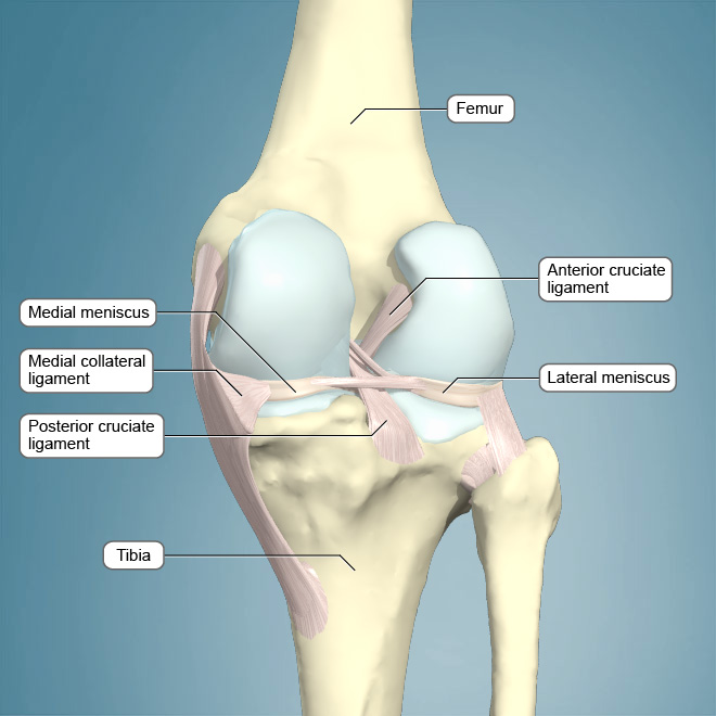
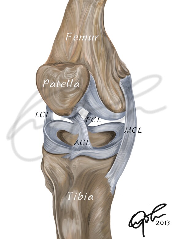
Ligaments of the knee. There are four knee ligaments thick bands of tough tissue that serve to maintain the stability of the knee joint.
Common Knee Injuries Orthoinfo Aaos
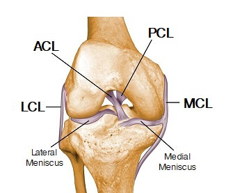
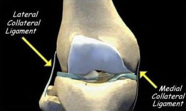
On the sides of the knee are the medial collateral ligament mcl and the lateral collateral ligament lcl.

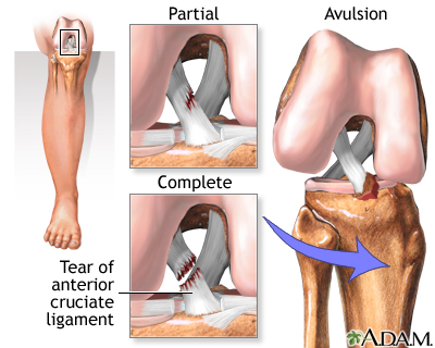
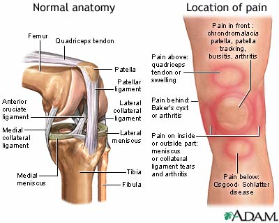
Knee ligaments anatomy. In knee joint anatomy knee ligaments are the main stabilising structures of the knee preventing excessive movements and instability. The function of ligaments is to attach bones to bones and give strength and stability to the knee as the knee has very little stability. Once stretched they tend to stay stretched and if stretched too far they snap.
The bones are held together by a joint capsule which consists of two distinct layers an outer layer of dense connective tissue and an inner membrane called the synovium which secretes a fluid to lubricate the joint. The knee includes four important ligaments all of which connect the femur to the tibia. Ligaments in the knee.
These two prevent sideways sliding of the knee joint ad also brace it against unusual movement. The medial collateral ligament on the inner side and the lateral collateral ligament on the outer side. The anterior cruciate ligament prevents the femur from sliding backward on the tibia or the tibia sliding forward on the femur.
Knee ligament impose limitations on the movement of the knee allowing it to concentrate forces of the muscles on extension and flexion. Knee anatomy share on pinterest the knee is the most complex joint in the human body. Tendons connect the knee bones to the leg muscles that move the knee joint.
Ligaments are tough fibrous connective tissues which link bone to bone made of collagen. Ligaments are strong tough bands that are not particularly flexible. The knee is a hinge joint that is responsible for weight bearing and movement.
Ligaments join the knee bones and provide stability to the knee. One ligament is on each side of the knee joint. The anterior cruciate ligament and posterior cruciate ligament provide front and back anterior and.
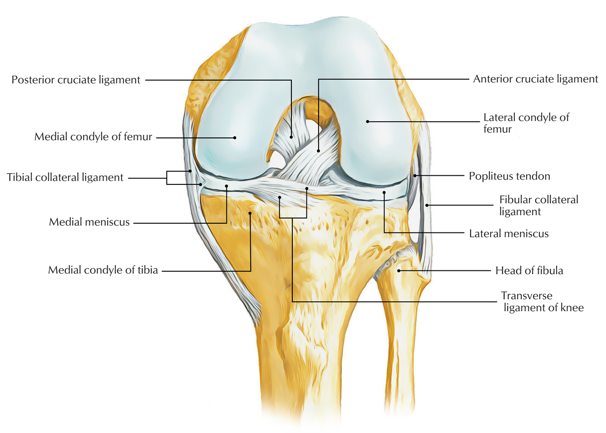
These are called the cruciate ligaments and consist of the anterior cruciate ligament and the posterior cruciate ligament. The medial meniscus situated on the inside of the knee.
Adolescent Sports Injuries Of The Knee Cleveland Clinic
 Anatomy Of The Knee Central Coast Orthopedic Medical Group
 Anatomy Of The Knee Baxter Regional Medical Center
 Sprains Of Knee Ligaments

Collateral Ligament Injuries Orthoinfo Aaos
 Anterior Cruciate Ligament Acl Injury Medlineplus Medical
 Anterior Cruciate Ligament Acl Tears For Parents
 Collateral Ligaments Of The Knee Joint Patellar Tendon
 Knee Joint Anatomy Motion Knee Pain Explained
 Biomechanics Functional Anatomy Behind Acl Tears Athletix
 Medial Collateral Knee Ligament Injury Background

 Human Knee Ligaments Printable Download Digital Illustration Medical Drawing Human Anatomy Knee Joint Knee Ligaments Anatomy Joint
 Anatomical Drawing Of The Ligaments In The Knee Download
 The Knee Anatomy Injuries Treatment And Rehabilitation
 Easy Notes On Ligaments Of The Knee Joint Learn In Just 3
 Knee Joint Picture Image On Medicinenet Com
 Amazon Com Kouber Anatomical Medical Knee Joint With
Acl Tears Pinnacle Orthopaedics
 14102 04b Tendons And Ligaments Of The Right Knee Anatomy
Graft Choices In Acl Surgery
 Collateral Ligament Cl Injury Aftercare Medlineplus
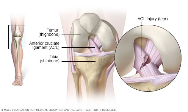 Acl Injury Symptoms And Causes Mayo Clinic


Posting Komentar
Posting Komentar