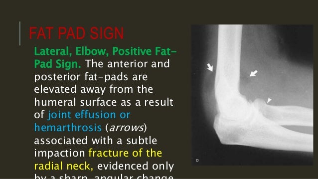
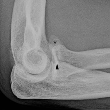
On an elbow x ray a fat pad sign suggests an occult fracture. Capitellum of the humerus with the ra.
 Imaging Of Elbow Fractures And Dislocations In Adults
Direct blow fall on an outstretched hand with flexed elbow avulsion fracture or stress fracture.
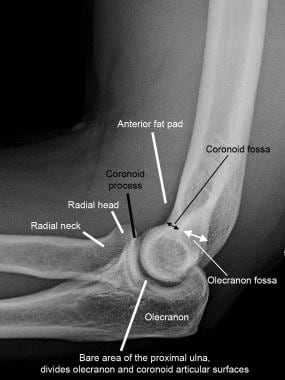
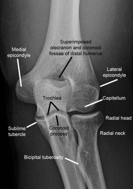
Elbow anatomy xray. On a normal elbow x ray only a small stripe of an anterior fat pad should be visible. The elbow is a complex synovial joint formed by the articulations of the humerus the radius and the ulna. No posterior fat pad should be seen.
A trivia quiz called elbow xray anatomy. The anterior fat pad protrudes more and looks pointy. The lucency on the radiograph which looks like a widened physis is due to cartilage ingrowth in the metaphysis.
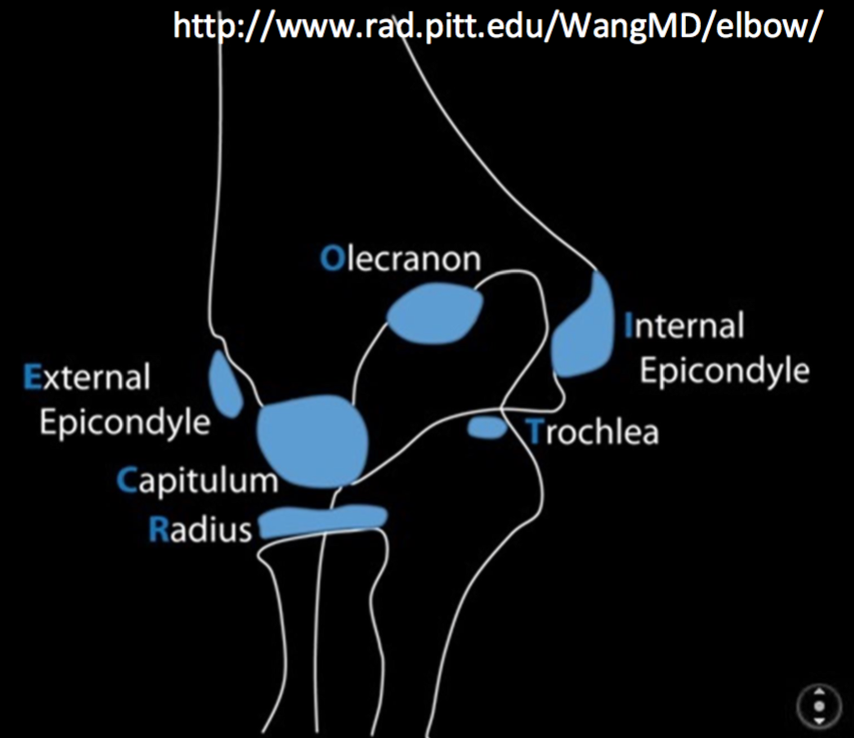
Continue with the mr. Injuries around the joint can produce a joint effusion which will displace the fat pads making them more visible. Gross anatomy articulations the elbow joint is made up of three articulations 23.
Both anterior and posterior fat pad signs exist and both can be found on the same x ray. Typically widely displaced due to unopposed pull of triceps. Use the mouse to scroll or the arrows.
Test your knowledge about elbow xray anatomy with this online quiz. Normal elbow x ray lateral 7 year old normal anterior fat pad. This is what is recognized as the sail sign.
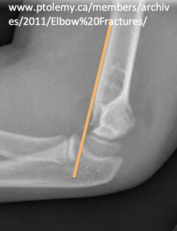
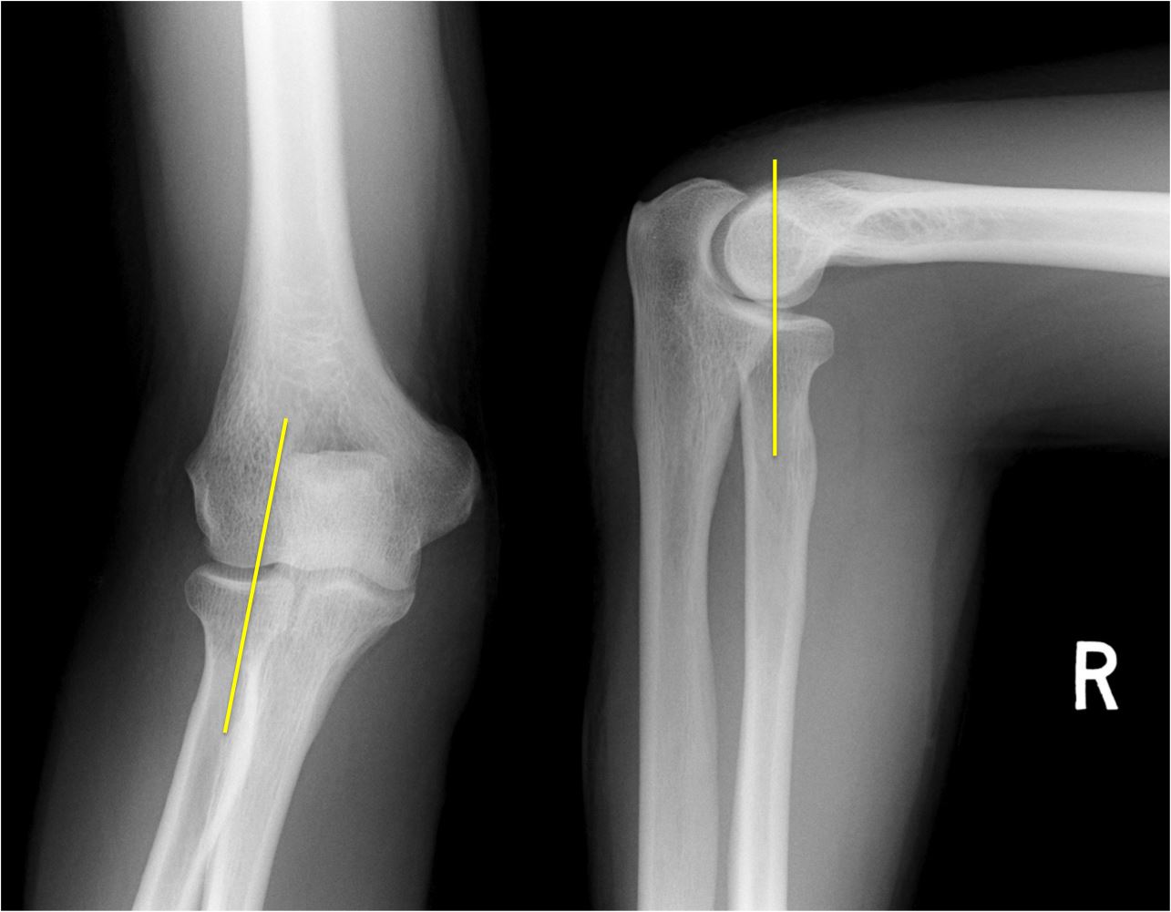
Copyright c 2005 2019 alex freitas md. The diagnosis is a little leaguers elbow which results from chronic stress injury. More than one third of the capitulum lies in front of the anterior humerus line.
It is caused by displacement of the fat pad around the elbow joint. Knee shoulder shoulder arthrogram ankle elbow wrist hip contact. Common represent 10 of all adult upper extremity fractures.
The posterior fat pad is not visible soft tissue of the triceps muscle is not separated from the posterior edge of the humerus.
 The Radiology Assistant Elbow Mri
 Joint Cubital Region Radiography Anatomy Humeroulnar
 The Elbow Mr Medical Imaging Anatomical Atlas
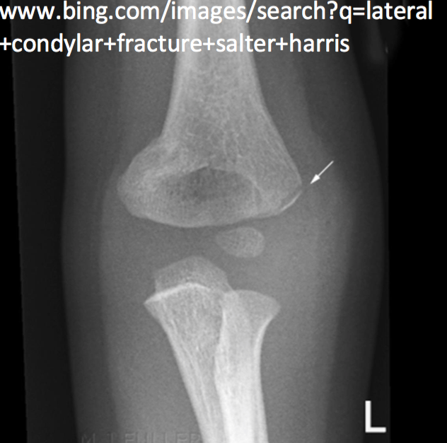
 The Radiology Assistant Elbow Fractures In Children
 Interpreting Elbow And Forearm Radiographs Taming The Sru
 Startradiology
 The Elbow
 Radiology In Ped Emerg Med Vol 1 Case 12
 Interpreting Elbow And Forearm Radiographs Taming The Sru
 Imaging Of Elbow Fractures And Dislocations In Adults
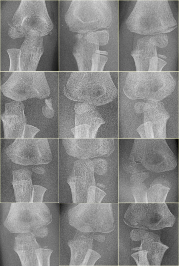
 Study Of Secondary Ossification Centers Of The Elbow In The
 Startradiology
 The Elbow
 The Radiology Assistant Elbow Fractures In Children
 Imaging Of Elbow Fractures And Dislocations In Adults
 Elbow Anatomy Pictures Bones Muscles Nerves
 Elbow Radiographic Anatomy Wikiradiography
 File X Ray Of Normal Elbow By Lateral Projection Jpg Wikipedia
 Normal Elbow Radiographs Radiology Case Radiopaedia Org
 Interpreting Elbow And Forearm Radiographs Taming The Sru
 Figure 3 From Three Dimensional Analysis Of Elbow Soft
 Startradiology
X Ray Of Elbow Joint
Xray Elbow Stock Photo Download Image Now Istock
 Anatomy Of The Elbow Ct Arthrography
 Film Critique Of The Upper Extremity Part 2 Elbow And Forearm
 Interpreting Elbow And Forearm Radiographs Taming The Sru
Elbow Radiographic Anatomy Wikiradiography
 Osteochondritis Dissecans Of Elbow Shoulder Elbow


Posting Komentar
Posting Komentar